Pluripotent stem cell models of Blau syndrome reveal
an IFN-g–dependent inflammatory response in macrophages
Blau
iPS
Abstract
Background: Blau syndrome, or early-onset sarcoidosis, is a juvenile-onset systemic
granulomatosis associated with a mutation in nucleotide-binding oligomerization domain
2 (NOD2). The underlying mechanisms of Blau syndrome leading to autoinflammation
are still unclear, and there is currently no effective specific treatment for Blau syndrome.
Objectives: To elucidate the mechanisms of autoinflammation in Blau syndrome, we
sought to clarify the relation between disease associated-mutant NOD2 and the inflammatory response in human samples.
Methods: Blau syndrome-specific induced pluripotent stem cells (iPSCs) lines were
established. To precisely evaluate the in vitro phenotype of iPSC-derived cells, the disease-associated NOD2 mutation of iPSCs was corrected using a CRISPR-Cas9 system. We also introduced the same NOD2 mutation into a control iPSC line. These isogenic iPSCs were then differentiated into monocytic cell lineages, and the status of NF-κB pathway and proinflammatory cytokine secretion were investigated.
Results: IFN-γ acted as a priming signal through the up-regulation of NOD2. In
iPSC-derived macrophages with mutant NOD2, IFN-γ treatment induced ligand-independent, NF-κB activation and proinflammatory cytokine production. RNA-seq analysis revealed
distinct transcriptional profiles of mutant macrophages both before and after IFN-γ treatment. Patient-derived macrophages demonstrated a similar IFN-γ-dependent inflammatory response.
Conclusions: Our data support the significance of ligand-independent autoinflammation
in the pathophysiology of Blau syndrome. Our comprehensive isogenic disease-specific iPSC panel provides a useful platform for probing therapeutic and diagnostic clues for the treatment of Blau syndrome patients.
Key words
Blau syndrome, nucleotide-binding oligomerization domain 2 (NOD2), nuclear factor kappa B (NF-κB), IFN-γ, disease-specific induced pluripotent stem cells
Abbreviations used
CARD15: Caspase recruitment domain-containing protein 15 ChIP: Chromatin immunoprecipitation
FDR: False discovery rate
GSEA: Gene set enrichment analysis iPSC: Induced pluripotent stem cell LPS: Lipopolysaccharide
MDP: Muramyl dipeptide NF-κB: Nuclear factor kappa B
NOD2: Nucleotide-binding oligomerization domain 2 PBMC: Peripheral blood mononuclear cell
PCA: Principal component analysis RNA-seq: RNA-sequencing
INTRODUCTION
Blau syndrome (OMIM: #186580), or early-onset sarcoidosis, is a hereditary juvenile-onset systemic granulomatosis1. Clinical symptoms appear before the age of 4 years and mainly affect the skin, joints, and eyes2. The symptoms are progressive and cause severe complications such as joint destruction and blindness3. The pathological findings of affected lesions include granuloma formation mainly composed of macrophages with other cells such as multinuclear giant cells and lymphocytes4, which is associated with local chronic inflammation. Although TNF-α antagonists are partially effective for controlling some symptoms, such as arthritis5, 6, there is no specific curative treatment at present for patients with Blau syndrome7.
Heterozygous mutations of nucleotide-binding oligomerization domain 2 (NOD2), or caspase recruitment domain-containing protein 15 (CARD15), were identified as responsible for the onset of Blau syndrome8. NOD2 protein is an intracellular pathogen recognition receptor, the ligand of which is known as muramyl dipeptide (MDP), found in bacterial cell walls9, 10. Upon recognition of the ligand, NOD2 leads to the activation of the nuclear factor kappa B (NF-κB) pathway, thereby causing the up-regulation of proinflammatory cytokines and chemokines9-13. However, the details regarding the
molecular mechanisms by which Blau syndrome-associated mutant NOD2 leads to autoinflammation and granuloma formation are still unclear.
mutations are gain-of-function or loss-of-function mutations is still argued. For example, in a model of HEK293 cells, ligand-independent auto-activation of NF-κB reporter activity was observed on the overexpression of Blau syndrome-associated mutant NOD2, indicating that Blau syndrome is caused by gain-of-function mutations of NOD214. Conversely, a knock-in mouse model described a reduction in the levels of proinflammatory cytokines responsive to NF-κB pathway upon stimulation with MDP, showing the possibility of loss-of-function15. However, since the mouse model did not show any phenotype of Blau syndrome in vivo, we recognized the need to investigate specific perturbations that abstract the unique function of mutant NOD2 using human samples.
Although NOD2 is mainly expressed in hematopoietic lineage cells, especially in monocytic cells13, the excessive production of proinflammatory cytokines or chemokines was not observed in peripheral blood mononuclear cells (PBMCs) of Blau syndrome patients16, 17 Whereas other autoinflammatory syndrome patients usually suffer from
periodic proinflammatory attacks, the clinical course of Blau syndrome is usually slow and progressive without specific episodes of febrile attacks, which hampers the evaluation of the disease activity of Blau syndrome. A lack of clinical parameters associated with disease activity makes it difficult to elucidate the mechanisms of this disease.
Substantial evidence supports a potential role of IFN-γ in the pathophysiology of Blau syndrome. Prominent IFN-γ expression was found in and around the granulomas of
biopsy specimens from Blau syndrome patients, indicating its commitment in the progression of granuloma4. IFN-γ is also described as a downstream mediator of NOD2-driven inflammation leading to uveitis18. In some patients, BCG vaccination was sometimes associated with the onset of this disease19, 20, showing the possibility that a BCG-associated immune response triggers a NOD2-dependent inflammatory pathway, which leads to flare up of Blau syndrome symptoms. Since IFN-γ is a major cytokine associated with BCG-mediated immune responses21, it may be involved in provoking a NOD2-mediate inflammatory response. Furthermore, IFN-γ, TNF-α, and other Toll-like receptor ligands act as priming signals to up-regulate NOD2 expression22-24. However, currently, there are no reports describing the direct effect of IFN-γ on Blau syndrome-associated immune cells.
Here we established Blau syndrome-patient-specific human induced pluripotent stem cell (iPSC) lines to develop in vitro disease models for elucidating the mechanisms of autoinflammation in Blau syndrome25. The iPSC model is free from the influence of
the in vivo cellular environment, such as medication or endogenous cytokines. Taking advantage of the nature of human pluripotent stem cells, in that their genome sequence can be edited relatively easily using the CRISPR-Cas9 genome editing system26, 27, we
established isogenic iPSC pairs sharing the same genomic backgrounds but different
NOD2 genotypes. In the present study, we corrected the NOD2 R334W mutation, which
In a similar manner, we introduced the NOD2 R334W mutation into an iPSC clone from a healthy donor. Using macrophages derived from these isogenic iPSC panels, we investigated the relationship between Blau syndrome associated-mutant NOD2 and the proinflammatory response.
METHODS Ethical matters
This study was approved by the Ethics Committee of Kyoto University, and informed consent was obtained from the patient’s guardians in accordance with the Declaration of Helsinki. Written informed consent was obtained from all patients. The peripheral blood mononuclear cells for iPSC generation were obtained from two independent Blau syndrome patients (Blau1, CIRA00232; and Blau2, CIRA00233).
Patients
Two Japanese boys with the NOD2-R334W-mutation diagnosed as Blau syndrome as previously described29, 30 were recruited to establish iPSCs. The patients were brothers.
Their father with the same NOD2-mutation was also diagnosed as Blau syndrome as previously described20, 30, 31. Each of the two patients had the similar clinical episode that erythematous papules appeared after BCG vaccination at 6 months of age. Thereafter one of the patients (Blau1) developed skin rash and arthritis without uveitis, then he had received treatment with non-steroidal anti-inflammatory drugs and methotrexate. Another patient (Blau2) developed skin rash without arthritis or uveitis, then he had not received any specific medication. PBMCs were obtained from another unrelated patient who had the same NOD2-R334W mutation to obtain monocyte-derived macrophages.
Primers
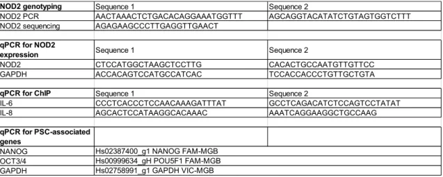
The primers used in this study are listed in Table E1.
Control iPSC lines
The human iPSC line 201B725 was kindly provided by Dr. Shinya Yamanaka (Kyoto University, Kyoto, Japan).
Establishment and maintenance of iPSCs
iPSCs were generated from blood cells as described previously32, 33. Briefly, blood cells were isolated from peripheral blood using a BD Vacutainer (BD BioSciences, San Jose, CA) in accordance with the manufacturer’s instructions. The PBMCs were cultured in StemSpan-ACF medium (StemCell Technologies, Vancouver, Canada) containing SCF (100 ng/mL), TPO (100 ng/mL), Flt-ligand (100 ng/mL), IL-6 (50 ng/mL), and IL-3 (10 ng/mL) for 5 days before transfection. SCF, TPO, Flt-ligand, IL-6, and IL-3 were purchased from R&D Systems (Minneapolis, MN). Episomal plasmids (3 µg) mixed with pCE-hOCT3/4, pCE-hSK, pCE-hUL, pCE-mp53DD, and pCXB-EBNA1 were transfected into 1-2 × 10⁶ cultured PBMCs using a Nucleofector 2b device with an Amaxa
Human CD34 cell Nucleofector kit. The electroporated cells were plated onto tissue culture plates coated with the Laminin511-E8 fragment iMatrix-511 (Nippi, Tokyo, Japan) containing StemSpan-ACF medium with the same cytokines as described above.
The culture medium was gradually changed to StemFit AK03 medium (Ajinomoto, Tokyo, Japan). Two to three weeks after the transduction, the individual colonies were isolated and expanded. The iPSCs were cultured on LN511E8-coated tissue culture plates with StemFit AK03 medium at 37 °C, with 5% CO2 and 21% O2, and passaged via
dissociation into single cells using TrypLE Select (Life Technologies, Gaithersburg, MD) as described previously32.
Characterization of established iPSCs
Chromosomal G-banding analyses were performed at the Nihon Gene Research Laboratory in Miyagi, Japan. For the teratoma formation assay, approximately 2 × 10⁶ cells were injected subcutaneously into the dorsal flank of immunocompromised NOD/scid/ccnull mice (Central Institute for Experimental Animals, Kawasaki, Japan). Masses were excised 9-14 weeks after injection and fixed with 4% paraformaldehyde in phosphate-buffered saline (-) (PBS [-]). Paraffin-embedded tissues were sliced and stained with hematoxylin and eosin. The slides were examined using the BIOREVO BZ-9000 microscope (Keyence, Osaka, Japan). A PlanApo 10×/0.45 objective (Nikon, Tokyo, Japan) and the BZ-II Viewer software program (KEYENCE) were used for image acquisition.
Surface markers on hematopoietic cells were evaluated with a MACSQuant Analyzer (Miltenyi Biotec, Bergisch Gladbach, Germany). The primary antibodies for the analysis were as follows: CD11b-FITC (Beckman Coulter, Brea, CA), CD45-FITC (BD Pharmingen, San Diego, CA), CD34-PE (Beckman Coulter), CD86-PE (BD Pharmingen), CD11c-APC (BD Pharmingen), and CD14-APC (Beckman Coulter). The dead cells were excluded by DAPI (Sigma-Aldrich, St Louis, MO) staining. The data analyses were performed using the FlowJo software program (TreeStar, Ashland, OR).
May-Giemsa staining
Cells were seeded onto glass slides by PLATINUMPRO (Matsunami, Osaka, Japan) and stained with May-Grunwald and Giemsa staining solution (Merck Millipore, Darmstadt, Germany) in accordance with the manufacturer’s instructions. The slides were examined using a BIOREVO BZ-9000 (Keyence). A PlanApo 40×/0.95 objective (Nikon) and the BZ-II Viewer software program (Keyence) were used for image acquisition.
Hematopoietic cell differentiation
Hematopoietic cell differentiation was performed in accordance with previously described protocols with some modifications34, 35. Briefly, 4 × 10²-1.6 × 10³ undifferentiated iPSCs were first disseminated onto a 6-well tissue culture dish coated with growth factor-reduced Matrigel in mTeSR1 medium. When individual colonies grew
to approximately 500 µm in diameter, differentiation was started by adding BMP4 (80 ng/mL, R&D systems). The cells were cultured under 37 °C, with 5% CO2 and 5% O2
during differentiation. On day 2, the medium was replaced by Essential 6 (Life Technology) containing VEGF (80 ng/mL), basic FGF (50 ng/mL), SCF (50 ng/mL), and SB431542 (2 µM). On day 4, the medium was replaced by Stemline 2 (Sigma-Aldrich) containing SCF (50 ng/mL), TPO (5 ng/mL), IL-3 (50 ng/mL), M-CSF (50 ng/mL), and Flt-3 ligand (50 ng/mL) for monocytic cell differentiation. On day 12, the cytokines were switched to the combination of M-CSF (50 ng/mL), Flt-3 ligand (50 ng/mL), and GM-CSF (25 ng/mL). BMP4, VEGF, SCF, TPO, IL-3, M-GM-CSF, GM-GM-CSF, and Flt-ligand were purchased from R&D Systems. Basic FGF, CHIR99021, was purchased from Wako Pure Chemical Industries (Osaka, Japan). SB431542 was purchased from Merck Millipore.
Sanger sequencing
Genomic DNA was extracted from the cells using the AllPrep DNA/RNA Mini Kit (Qiagen, Hilden, Germany) or PureLink Genomic DNA Kits (Invitrogen, Carlsbad, CA) and subjected to PCR amplification with primers for NOD2 genotyping (Table E1). After treatment with USB ExoSAP-IT (Affymetrix, Santa Clara, CA), the PCR products were used for Sanger sequencing. Sequencing was performed with the BigDye® Terminator v3.1 Cycle Sequencing Kit (Applied Biosystems, Carlsbad, CA), and the subsequent products were purified using the BigDye XTerminator® Purification Kit (Applied
Biosystems). The 3500xL sequencer (Applied Biosystems) was used for analyses. Data were analyzed using the CodonCode Aligner software program (CodonCode Corporation, Centerville, MA).
Generation of immortalized proliferating myeloid cell lines (iPS-MLs)
iPS-MLs were established as previously described36. In brief, lentiviral constructs encoding BMI1, cMYC, and MDM2 in the CSII-EF-RfA vector were kindly provided by Drs. Satoru Senju (Kumamoto University, Kumamoto, Japan) and Hiroyuki Miyoshi (RIKEN Bio Resource Center, Tsukuba, Japan). Floating hematopoietic cells differentiated into the monocytic lineage from iPSCs were collected on days 15 to 18. The cells were transfected with the lentiviral vectors and subsequently cultured in StemPro-34 serum-free medium containing 2 mM L-glutamine in the presence of M-CSF (50 ng/mL). For macrophage differentiation, the culture conditions were changed to RPMI-1640 medium (Sigma-Aldrich) supplemented with 10% fetal bovine serum (FBS) and M-CSF (100 ng/mL) for 7 to 9 days. For polarization of macrophages, IL-4 (20 ng/mL) or IFN-γ (20 ng/mL) was added for last 24 hours. We then collected the adherent macrophages by treating them with Accumax (Innovative Cell Technologies, San Diego, CA) and used the collected cells for subsequent experiments.
We corrected the mutant NOD2 gene using the CRISPR-Cas9 system. The guide RNA (gRNA) sequence was designed on the coding sequence of NOD2 in intron 3 (CCTTCAGTTATGTCAGCGTCCCC). gRNA vector and Cas9 vector were purchased from Takara Bio (Shiga, Japan). Clones maintained under feeder-free conditions were dissociated into a single cell. Vectors expressing gRNA and Cas9 were then introduced using a NEPA21 electroporator (Nepagene, Chiba, Japan). Two days after transfection, cells were incubated with puromycin (0.5-1 µg/mL) for 24-48 hours or neomycin (50 µg/mL) for 1 week. Surviving colonies were picked up 1 week after the start of selection, passaged and expanded several times, and frozen. The DNA was extracted from the cells and genotyped by Sanger sequencing as described above. Vectors expressing Cre recombinase were then introduced by HuGENE HD Transfection Reagent (Promega, Madison, WI) to excise the loxP-flanked drug selection cassette. For the construction of the C-terminal 3×FLAG sequence knocking-in allele, the gRNA sequence was designed on the coding sequence of NOD2 in exon 12 (CTTGCTTTGAAGTCTCCGGGAGG). Vectors were transfected, and cells were cultured as described above.
Quantitative PCR (qPCR)
RNA samples were prepared using AllPrep DNA/RNA Mini Kit (Qiagen) or RNeasy Mini Kit (Qiagen) and subjected to reverse transcription using a PrimeScript RT Master Mix (Takara Bio). All procedures were performed in accordance with the manufacturer’s
instructions. qPCR was performed on the StepOnePlus™ Real-Time PCR System (Applied Biosystems). For the detection of transgenes, DNA was subjected to qPCR, and SYBR Premix ExTaqII (Takara Bio) was used for detection. Relative quantification values are shown. For the quantification of the expression of PSC-associated genes (Fig.
E1C), qPCR was performed with cDNA and the Fast Advanced Master Mix (Applied
Biosystems). The data were processed using the ΔΔCt method, and the relative quantities (RQ) to the control 201B7 line are shown. The primer sequences are shown in Table E1.
Immunoblotting
Cell lysates were lysed for 30 min on ice in RIPA buffer (Wako Pure Chemical Industries) plus proteinase inhibitor cocktail (Nacalai Tesque, Kyoto, Japan). After being centrifuged at 15,000g for 20 min at 4 °C, the supernatants were collected, and dithiothreitol was added. The lysates were incubated at 95 °C for 5 minutes. The proteins were then separated by sodium dodecylsulfate-polyacrylamide gel electrophoresis (SDS-PAGE) and transferred to a PVDF membrane. The membranes were blocked with 3% nonfat milk in Tris-buffered saline (TBS) plus 0.1% Tween20. Immunoblotting was performed with FLAG M2 mouse monoclonal antibody (1:1000; Sigma-Aldrich) and HRP-conjugated anti-mouse IgG antibody (1:3000; Santa Cruz Biotechnology, Santa Cruz, CA) and detected using a LAS4000 (Fujifilm, Tokyo, Japan).
NF-κB Dual Luciferase assay
The iPS-MLs were transfected with pGreenFire NF-κB transcription reporter lenti vector (System Biosciences, Mountain View, CA), inserted with the Renilla gene under the CMV promoter as an internal control. The iPS-MLs were differentiated into macrophages as described above. The cells were cultured with or without 50 ng/mL of IFN-γ (R&D System) for 24 hours, with the addition of 10 µg/mL of MDP (Sigma-Aldrich) for the last 6 hours. The NF-κB activity was then measured using the Dual-Luciferase Reporter Assay System (Promega).
Immunofluorescence
The macrophages were seeded into multi-well glass-bottom dish (Matsunami) and cultured with or without 50 ng/mL of IFN-γ for 24 hours, with the addition of 10 µg/mL of MDP for the last 60 minutes. The cells were then fixed with 4% paraformaldehyde in PBS (-) for 30 minutes. Permeabilization and blocking were performed with blocking buffer (Blocking One [Nacalai Tesque] with 0.1% Tween20 [Santa Cruz Biotechnology]) for 30 minutes. The samples were incubated with anti-NF-κB p65 rabbit monoclonal antibody (1:400; Cell Signaling Technologies, Danvers, MA) in blocking buffer overnight at 4 °C, and subsequently incubated with Alexa Fluor 488 goat anti-mouse IgG (1:1000; Cell Signaling Technologies) for 30 minutes. The nuclei were stained with DAPI (1:100) in PBS (-). The cells were visualized using a FluoVIew10i confocal microscope
(Olympus, Tokyo, Japan) and analyzed using Image J software (NIH, Bethesda, MD).
Cytokine assay
The concentrations of cytokines in the culture supernatants were measured using LEGENDplexTM Multi-Analyte Flow Assay Kits (BioLegend, San Diego, CA) in accordance with the manufacturer’s instructions. Quantification was done with a MACSQuant Analyzer (Miltenyi Biotec). The iPS-MLs were differentiated into macrophages as described above. For stimulation, the cells were treated with IFN-γ (50 ng/mL) for 24 hours, and MDP (10 µg/mL) was added to the medium for the last 6 hours when indicated.
Isolation of PBMCs and macrophage differentiation
After receiving informed consent, peripheral blood was drawn from a patient and two healthy volunteers. PBMCs were isolated by Lymphoprep (STEMCELL Technologies). CD14-positive monocytic cells were sorted using EasySep Human CD14 Positive Selection Kit (STEMCELL Technologies). The cells were differentiated into macrophages as described above, and the differentiated macrophages were then subjected to an analysis.
After stimulation, the cells were cross-linked in 1% formaldehyde for 10 minutes at room temperature. The nuclear lysates were sonicated using a sonicator (Misonix, Farmingdale, NY). After being centrifuged at 15,000g for 30 min at 4 °C, the supernatants were collected. The clarified lysates were divided in half and treated overnight at 4 °C with 10 µg of anti-NF-κB p65 (C-20) rabbit polyclonal antibody (Santa Cruz Biotechnology) or Rabbit IgG polyclonal-Isotype control (Abcam, Cambridge, UK) pre-incubated with Dynabeads M-280 Sheep anti-Rabbit IgG (Thermo Fisher Scientific, Waltham, MA). The protein-DNA complexes were captured on a magnet and eluted with TE buffer containing 1% SDS at 65 °C. Following cross-link reversal and purification with MinElute PCR Purification Kit (Qiagen), quantitative PCR was performed as described above. The primer sequences are described in Table E1.
Cell viability assay
The cell viability was measured using a CountessTM Automated Cell Counter (Invitrogen,
Carlsbad, CA). For stimulation, the cells were treated with IFN-γ (50 ng/mL) for 24 hours, and MDP (10 µg/mL) was added to the medium for the last 6 hours when indicated.
RNA-sequence (RNA-seq) and data normalization
RNA-seq analysis was performed at Kazusa DNA Institute as previously described37. RNA-seq libraries were prepared using a SureSelect Strand Specific RNA Library
Preparation kit (Agilent Technologies, Santa Clara, CA). Sequencing was performed on an Illumina HiSeq1500 using a HiSeq Rapid SBS kit v2 (Illumina, San Diego, CA) in a 50-base single-end mode. The mRNA profiles were compiled using the Cufflinks (ver. 2.2.1) software program and expressed as fragments per kilobase of exon model per million mapped fragments (FPKM). For the analysis, the data sets filtered by the criterion requiring FPKM >0 in at least one sample were first subjected to variance-stabilizing transformation and interquartile range filtering. Then, upper-quartile (UQ) normalization followed by remove unwanted variations (RUV) normalization regarding each three line of treated and untreated samples as a replicated group (RUVs), modeling k = 1 factors of unwanted variation was performed to remove unwanted variations38, 39. UQ and RUVs normalization was conducted using the open-source R package Deseq2 and RUVSeq, the source code of which is freely available through the Bioconductor Project.
Gene Set Enrichment Analysis (GSEA)
GSEA (http://software.broadinstitute.org/gsea/index.jsp) was used to estimate the biological signatures. Normalized data sets were applied for each analysis using Molecular Signatures Database v5.1 (Hallmark gene sets, size-filters set at 15-500).
GSE accession numbers
accession number GSE98454.
Statistics
All of the statistical analyses were performed using the Excel, R and Prism software programs. The values are presented as the mean ± S.E.M or S.D., as indicated in the Figure Legends. The statistical analysis was done using either Student’s t-test or the Wilcoxon rank sum test.
RESULTS
Generation of Blau syndrome patient-specific iPSCs and associated isogenic iPSC panels
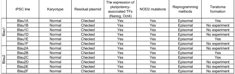
We first established and characterized multiple iPSC lines from 2 Blau syndrome patients with a heterozygous R334W (1000C>T) NOD2 mutation (referred to as Blau1A-1F and Blau2A-2F, see Table E2). Blau-iPSCs were confirmed to carry the corresponding heterozygous point mutation on exon 4 of the NOD2 gene, showed typical pluripotent stem cell like-morphology, maintained a normal karyotype, and expressed comparable levels of pluripotency-associated genes (NANOG and OCT3/4) to a control iPSC line (Table E2). We selected representative iPSC clones from both patients (Blau1E and Blau2B) and named them Blau1-R334W (Blau1E) and Blau2-R334W (Blau2B). The data of these representative clones for the evaluation of iPSC-quality are shown in Figure
E1A-E1C. The pluripotency of Blau-iPSC clones including Blau1-R334W and
Blau2-R334W was confirmed using a teratoma formation assay (Table E2 and Figure E1D). To understand the pathophysiology derived from Blau syndrome-associated mutant
NOD2 in a strictly controlled isogenic background, we next corrected the mutation in the
Blau1-R334W and Blau2-R334W iPSC clones using a CRISPR-Cas9 mediated targeting26, 27 (referred to as Blau1-corrected and Blau2-corrected, respectively, Figure
1A). In the corrected clone (Blau1-corrected and Blau2-corrected), the inserted allele was
validated by genomic PCR (Figure 1B and E1E), and the T to C correction of NOD2 on exon 4 was confirmed via the Sanger sequencing (Figure 1C and E1F). In a similar manner, we also introduced a heterozygous NOD2-R334W mutation into control 201B7-iPSCs (referred to as 201B7-WT for parental control 201B7, and 201B7-R334W for mutated 201B7, Figure E1G-E1I). To investigate the expression of endogenous NOD2, we generated 201B7-WT iPSCs with 3× FLAG-tagged NOD2 in the C-terminal using the CRISPR-Cas9 system (referred to as 201B7-WT-NOD2-FLAG, Figure E1J and E1K).
Differentiation of iPSCs into a monocytic lineage
Since monocytic lineage cells, especially macrophages, express NOD2, produce inflammatory cytokines robustly, and play a pivotal role in the formation of granulomas, we first differentiated iPSCs into a monocytic lineage using a previously reported protocol35. We tracked the expression of NOD2 mRNA during the differentiation and
found the monocytic lineage cells derived from iPSCs expressed NOD2 on day 12 of differentiation (Figure 1D). The iPSC-derived monocytes were then further differentiated into macrophages and stimulated with IFN-γ or IL-4 in order to elicit polarization into M1 or M2 macrophages35. Of note, the iPSCs-derived macrophages treated by IFN-γ showed especially high expression of NOD2, as previously described22-24 (Figure 1D). IFN-γ treatment up-regulated the expression of NOD2 protein (Figure 1E), thus indicating a priming role of IFN-γ in the NOD2-NF-κB signaling pathway.
Given these observations, we next investigated the in vitro function of NOD2 in iPSC-derived macrophages. One of the obstacles facing disease iPSC studies is that directed differentiation from iPSCs is often time- and labor-intensive, whereas the functional analysis often shows high variance. To overcome these issues and stably obtain a scalable number of mature monocytic cells from engineered iPSC clones, immortalized proliferating myeloid cell lines (iPS-MLs) were established from iPSC-derived monocytes by introducing three transgenes: cMYC, BMI1, and MDM236. The established
iPS-MLs showed a monocytic appearance (Figure E2A) and expressed CD33 but only expressed CD14 slightly (Figure E2B), indicating their characteristics of immature monoblast-like status. When the iPS-MLs were differentiated into macrophages (iPS-ML-derived macrophages, referred to as iPS-MPs), the expression of macrophage surface cell markers CD14, CD68, and CD11b were up-regulated (Figure E2C). These iPS-MPs demonstrated an adherent cell appearance, and IFN-γ treatment gave them an
M1-macrophage-like elongated, tightly attached appearance (Figure E2D).
NF-κB is similarly activated in iPS-MPs with wild-type and mutant NOD2
The activation of NOD2 by its ligands such as MDP is involved in the immune response induced by NF-κB activation9, 11, 13. To investigate the activation of NF-κB in cells with wild-type NOD2 and NOD2 mutations, we next tracked the nuclear translocation of NF-κB p65, a subunit of NF-NF-κB transcription complex, as a parameter of NF-NF-κB activation (Figure 2A-2C). In untreated iPS-MPs, NF-κB p65 localized mostly in the cytosol. Upon stimulation with MDP, NF-κB p65 remained in the cytosol for a shorter time course (1 to 5 minutes after stimulation) and translocated into the nucleus after 30 minutes to 1 hour. With MDP treatment after 6 hours, NF-κB p65 was returned into the cytosol. Overall, the response of NF-κB to MDP stimulation was similar in cells with wild-type and mutant NOD2.
IFN-γ induces ligand-independent NF-κB activation on NOD2-mutated iPS-MPs
To investigate the role of mutant NOD2 on immune response, we first investigated the NF-κB transcription activity in iPS-MPs using a luciferase reporter system. Interestingly, IFN-γ priming significantly increased the luciferase activity in mutant iPS-MPs without secondary stimulation by MDP (Figure 3A). The degree of NF-κB activation in mutant iPS-MPs by IFN-γ increased in a dose- and time-dependent manner (Figure E3A and
E3B). Both wild-type and mutant iPS-MPs were able to react to MDP at comparable
levels of NF-κB activation (Figure 3A). The time course of the degree of NF-κB activation in both the wild-type and mutant iPS-MPs by MDP peaked at 4 hours after stimulation and then started to decrease, which was earlier than observed with IFN-γ (Figure E3C). The activation of NF-κB in cells treated with IFN-γ and MDP, was higher than that in cells that were only treated with MDP in both wild-type and mutant iPS-MPs (Figure 3A), suggesting a priming role of IFN-γ in the NOD2-NF-κB signaling pathway. The activation of NF-κB signaling with lipopolysaccharide (LPS), which is mainly mediated by the Toll-like receptor signaling pathway40, was not significantly different between wild-type and mutant clones (Figure E3A).
To evaluate the NF-κB activation in each cell, we next investigated the nuclear translocation of NF-κB p65. Some of the IFN-γ-primed cells in the mutant clone showed NF-κB p65 nuclear translocation without MDP (Figure 3B), suggesting a ligand-independent activation state. The immunofluorescent density of the nuclear-translocated NF-κB p65 in mutant iPS-MPs was significantly increased compared to that in wild-type cells (Figure 3C). In contrast, untreated cells demonstrated no obvious spontaneous nuclear translocation of NF-κB in either wild-type or mutant iPS-MPs (Figure 3B and
3C). MDP treatment increased NF-κB activity at comparable levels between wild-type
and mutant iPS-MPs (Figure 3B and 3C), as mentioned above in Figure 2.
pathway in mutant iPS-MPs, which may associate with an overproduction of proinflammatory cytokines.
IFN-γ induces proinflammatory cytokine production on NOD2-mutated iPS-MPs by increasing the binding of NF-κB p65 to their promoter regions
To evaluate the consequence of NF-κB activation, we next measured the production of proinflammatory cytokines and chemokines (Figure 4A-4C). When untreated, the production of proinflammatory cytokines from both wild-type and mutant clones was negligible. IFN-γ priming significantly increased the production of common NF-κB-responsive cytokines such as IL-6, IL-8, and TNF-α in mutant iPS-MPs without secondary MDP stimulation, consistent with the status of NF-κB pathway. No marked differences in cell viability were noted with each stimulation, regardless of the genotype or type of stimulation (Figure E4), excluding the effect of cell death on the cytokine profiles. On the other hand, despite the comparable levels of NF-κB activity, the production of these cytokines with MDP in mutant iPS-MPs was significantly lower than those from wild-type iPS-MPs. No significant secretion of other cytokines such as IL-1β, IL-10, or IL-12p70 was observed in either wild-type or mutant iPS-MPs under these conditions (Figure E3C-E3E).
To confirm that the increased cytokine production on IFN-γ-primed mutant iPS-MPs was associated with the NF-κB activation, a chromatin immunoprecipitation (ChIP)
assay with NF-κB p65 protein was performed. IFN-γ priming induced the ligand-independent recruitment of NF-κB p65 to the promoter specific regions of IL-6 and IL-8 in mutant iPS-MPs (Figure 4D and 4E). We referenced the NF-κB activation, proinflammatory cytokine production, and binding to the promoters using another isogenic pair of iPS-MPs to evaluate the consistency (Blau1-corrected and Blau1-R334W,
Figure E5A-E5F) and confirmed that all isogenic pairs including Blau1 showed
consistent results regarding the response to IFN-γ. Overall, our data indicate that the production of proinflammatory cytokines by IFN-γ-primed iPS-MPs with mutant NOD2 is at least partially induced through the ligand-independent activation of the NF-κB pathway.
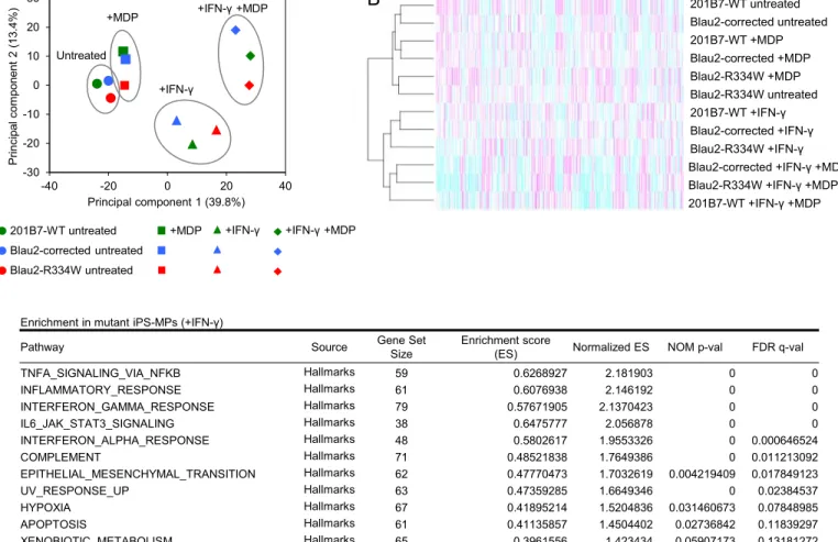
RNA-seq analysis reveals the involvement of inflammatory response in IFN-γ-primed iPS-MPs with a Blau syndrome-associated NOD2 mutation
The above data revealed the relevance of the IFN-γ-induced NF-κB activation and proinflammatory cytokine production, indicating difference in the biological status between mutant and wild-type untreated iPS-MPs in evoking an inflammatory response. To clarify the involvement of this response, we performed an RNA-sequencing (RNA-seq) analysis to evaluate the changes in the global transcriptional profiles of iPS-MPs. To assess the global effects of genotype and treatment on the transcriptome of iPS-MPs, we first performed a principal component analysis (PCA) plot from the normalized gene
counts of samples with each treatment. The transcriptome clustered predominantly by treatment, while the inter-clonal difference was minimal (Figure 5A, for the process of normalization, see Figure E6A-E6C). A hierarchical clustering analysis also demonstrated that untreated and IFN-γ-treated samples grouped into distinct clusters (Figure 5B). In order to extract the differently regulated pathways between mutant and wild-type iPS-MPs, we next performed a gene set enrichment analysis (GSEA)41. We confirmed that IFN-γ treatment significantly enriched IFN-γ response pathway in all clones compared to the untreated state (Figure E6D and E7E). In mutant iPS-MPs upon IFN-γ treatment, several inflammatory pathways, such as “TNFA_SIGNALING_VIA_NFKB” and “INFLAMMATORY_RESPONSE” (False discovery rate (FDR) <0.25) were significantly enriched when compared to wild-type iPS-MPs (Figure 5C). Interestingly, we noticed that similar inflammatory pathways were already up-regulated in mutant iPS-MPs, even in the untreated state (Figure 5D). These data support the idea that untreated mutant iPS-MPs are already in a distinct biological state for evoking an inflammatory response, which is maintained even after IFN-γ priming, leading to an enhanced inflammatory response, such as via proinflammatory cytokine production.
Blau syndrome patient’s primary macrophages primed by IFN-γ caused ligand-independent NF-κB activation and proinflammatory cytokine production
Although the iPSC-derived isogenic monocytic cell line pairs enabled us to precisely evaluate the in vitro phenotype, there remains a possibility that the long-term in vitro culture and introduction of transgenes might alter the cellular phenotype. To exclude this possibility, we investigated whether or not the in vitro phenotypes could be recapitulated with primary monocyte-derived macrophages (Mo-MPs). For this, Mo-MPs were obtained from the PBMCs of a Blau syndrome patient with a NOD2-R334W mutation. IFN-γ treatment up-regulated the endogenous expression of NOD2 in Mo-MPs from the control (Figure 6A). Both the control- and patient-derived Mo-MPs demonstrated no obvious spontaneous nuclear translocation of NF-κB when untreated (Figure 6B and 6C). Similar to the observation with iPS-MPs, some of the IFN-γ-primed Mo-MPs from the patient showed NF-κB p65 nuclear translocation without MDP, and the degree of NF-κB p65 nuclear translocation was significantly larger in the patient-derived Mo-MPs than in those of the control (Figure 6B and 6C). MDP stimulation promoted the nuclear translocation of NF-κB p65 in both the control and patient, but the degree was smaller in the patient’s Mo-MPs than in those of the control.
We then evaluated production of proinflammatory cytokines from Mo-MPs. IFN-γ priming significantly increased the production of IL-6, IL-8, and TNF-α from the patient-derived Mo-MPs in a ligand-independent manner (Figure 6D-6F). Collectively, IFN-γ treatment activated NF-κB pathway and induced production of proinflammatory cytokines from patient-derived macrophages. These data were compatible with those
DISCUSSION
In this study, we demonstrated that IFN-γ-primed human macrophages with Blau syndrome-associated mutant NOD2 cause the ligand-independent activation of NF-κB transcription and production of proinflammatory cytokines such as IL-6 and IL-8. One of the main roadblocks to studying Blau syndrome-associated NOD2 mutants has been the lack of a proinflammatory response in mutant cells in vitro despite autoinflammatory symptoms in vivo. Since both patient-derived cells and a knock-in mouse model also showed reduced proinflammatory responses, the argument remains as to whether the Blau syndrome-associated NOD2 mutations are gain-of-function or loss-of-function. In the present study, we identified IFN-γ as a specific stimulant inducing a pro-inflammatory response in both iPSC- and patient-derived primary macrophages. Of note, humans with complete IFN-γ receptor deficiency, whether IFN-γR1 or IFN-γR2, do not display any mature granuloma42, indicating the essential role of IFN-γ in the formation of granulomas. Our data provide important insights into the role of NOD2 in IFN-γ-dependent granuloma formation. Although the involvement of IFN-γ signaling has been implicated in the pathology of BS, there have been no reports proving the direct effect of IFN-γ on the function of mutant NOD2. Our findings revealed a previously undefined pathway for activating NOD2-mutated macrophages.
RNA-seq data indicated that untreated macrophages with mutant NOD2 are already in a primed biological state to evoke an inflammatory response. While untreated
macrophages did not activate the NF-κB pathway due to a shortage of auto-activated mutant NOD2 protein, the up-regulation of mutant NOD2 expression by IFN-γ may cause auto-activation of NOD2, leading to subsequent NF-κB activation. However, since the relationship between IFN-γ-induced prionflammatory cytokine production and granuloma formation is still largely unknown, further studies will be required to gain a comprehensive understanding of the pathophysiology of Blau syndrome.
A response to MDP subsequently reduced the level of proinflammatory cytokine production in mutant iPS-MPs, which is consistent with the previous findings in the mouse model15 and in the patients’ PBMCs17. While MDP treatment did not lead to reduced cytokine production in the primary Mo-MPs in our study, one of the fundamental problems in the functional analysis of human primary cells is the large degree of variance, even among healthy controls, which is shown in Figure 6D-6F. Cell-derived iPSCs share the same genomic backgrounds as primary blood cells, but the NOD2 genotypes differ between isogenic pairs, which enables us to exclude any interpersonal variance. The loss of response to MDP in cytokine production may be associated with the pathogenesis of Blau syndrome, because granulomas are sometimes caused by the inability to eliminate foreign bodies and pathogens43. In contrast to the findings of the cytokine-production
assay, MDP stimulation activated NF-κB pathway in both mutant and wild-type clones, and tended to enhance the recruitment of the NF-κB p65 subunit to promoter regions of
paradoxical response remain to be elucidated, our data indicate the possible existence of an alternative suppressive pathway activated by mutant NOD2 or another pathway through MDP for producing proinflammatory cytokines independent of NOD2.
Based on our observation from iPS-MPs and patient-derived MoMPs, blocking the IFN-γ signaling in Blau syndrome patients can be a potential treatment for managing chronic inflammation. For example, the Janus kinase inhibitors tofacitinib, baricitinib, and ruxolitinib are already clinically available. However, before this concept can be applied in a clinical setting, a number of issues remain to be solved. First, we do not have direct evidence to prove that the IFN-γ pathway is activated in Blau syndrome patients. For this, further studies investigating the primary samples of patients, including blood and pathological specimens, will be required to confirm the up-regulation of IFN-γ signatures. Second, since there are other priming signals, such as TNF-α, LPS, and other Toll-like receptor ligands, known to up-regulate the expression of NOD222-24, IFN-γ-dependent priming of macrophages may not be the principal condition for evoking autoinflammation. In the current study, we did not evaluate the effects of these signals because of the technical difficulty for distinguishing the Toll-like receptor- or TNF receptor-dependent NF-κB activation from the NOD2-dependent one. However, IFN-γ usually does not transduce signals activating the NF-κB pathway, enabling us to investigate the role of IFN-γ as a priming signal. Third, we must consider other effects of IFN-γ associated with a substantial immune response and other signal pathways. To avoid
these issues, we must suppress the specific pathways induced by mutant NOD2 when applying this therapy in treatments.
In conclusion, by investigating the functions of macrophages under controlled conditions using a comprehensive isogenic iPSC panel with a Blau syndrome-associated
NOD2 mutation, we were able to perform strictly controlled cellular assays and found
that IFN-γ provokes a series of aberrant inflammatory responses in mutant macrophages. Since we mainly focused on the NOD2-dependent NF-κB-pathway activation using macrophages in this study, further studies exploring alternative NOD2-dependent pathways or with other cell types are required to clarify the comprehensive pathophysiology of Blau syndrome. Phenotypes related to genotypes other than R334W should be also evaluated. Through the Blau syndrome study, we expect that our iPSC model will also provide an opportunity for elucidating the previously unknown mechanisms of granuloma formation in other diseases.
ACKNOWLEDGEMENTS
We thank the patients and family for their cooperation. We also thank Drs. Megumu K. Saito, Tatsutoshi Nakahata, Yuri Kawasaki, Akira Niwa, Fumiko Honda-Ozaki, Kazuki Kobayashi, Mitsujiro Osawa, Ayako Nagahashi, Katsunori Semi, Akitsu Hotta, Isao Asaka, Yasuhiro Yamada (Center for iPS Cell Research and Application, Kyoto University, Kyoto, Japan), Naotomo Kambe (Kansai Medical University, Hirakata, Japan), Ryuta Nishikomori, Toshio Heike (Kyoto University Graduate School of Medicine, Kyoto, Japan), Hiroyuki Matsue (Chiba University Graduate School of Medicine, Chiba, Japan) for the contribution to this project. We are grateful to Drs. Satoru Senju (Kumamoto University, Kumamoto, Japan) and Hiroyuki Miyoshi (RIKEN Bio Resource Center, Tsukuba, Japan) for providing lentiviral vectors. We are grateful to Ryunosuke Ikeda and Tomoyo Ukai for his technical assistance. We also thank Yoshinori Hasegawa and Osamu Ohara for supporting RNA-seq analysis. We would also like to thank Harumi Watanabe for administrative assistance.
This work was supported by the grant for the Core Center for iPS Cell Research of Research Center Network for Realization of Regenerative Medicine from the Japan Agency for Medical Research and Development (AMED) [A.H., T.N. and M.K.S.]; the Program for Intractable Diseases Research utilizing Disease-specific iPS cells of AMED (15652070) [I.A., T.N. and M.K.S.]; Practical Research Project for Allergic Diseases and Immunology (Research on Allergic Diseases and Immunology) of AMED (14525046)
[M.K.S.]; Practical Research Project for Rare/Intractable Diseases of AMED (15634527) [N.K., R.N., T. H., T.N. and M.K.S.]; Research Project for Practical Applications of Regenerative Medicine from AMED [M.K.S.] and the Japan Society for the Promotion of Science (JSPS) KAKENHI, grant numbers 13389802 [M.K.S.] and 14431432 [R.N., T.H. and M.K.S.].
REFERENCES
1. Blau EB. Familial granulomatous arthritis, iritis, and rash. J Pediatr 1985; 107:689-93.
2. Hetherington S. Sarcoidosis in young children. Am J Dis Child 1982; 136:13-5. 3. Fink CW, Cimaz R. Early onset sarcoidosis: not a benign disease. J Rheumatol
1997; 24:174-7.
4. Janssen CE, Rose CD, De Hertogh G, Martin TM, Bader Meunier B, Cimaz R, et al. Morphologic and immunohistochemical characterization of granulomas in the nucleotide oligomerization domain 2-related disorders Blau syndrome and Crohn disease. J Allergy Clin Immunol 2012; 129:1076-84.
5. Milman N, Andersen CB, Hansen A, van Overeem Hansen T, Nielsen FC, Fledelius H, et al. Favourable effect of TNF-alpha inhibitor (infliximab) on Blau syndrome in monozygotic twins with a de novo CARD15 mutation. Apmis 2006; 114:912-9.
6. Otsubo Y, Okafuji I, Shimizu T, Nonaka F, Ikeda K, Eguchi K. A long-term follow-up of Japanese mother and her daughter with Blau syndrome: Effective treatment of anti-TNF inhibitors and useful diagnostic tool of joint ultrasound examination. Mod Rheumatol 2014:1-5.
7. Wouters CH, Maes A, Foley KP, Bertin J, Rose CD. Blau syndrome, the
prototypic auto-inflammatory granulomatous disease. Pediatr Rheumatol Online J 2014; 12:33.
8. Miceli-Richard C, Lesage S, Rybojad M, Prieur AM, Manouvrier-Hanu S, Hafner R, et al. CARD15 mutations in Blau syndrome. Nat Genet 2001; 29:19-20.
9. Girardin SE, Boneca IG, Viala J, Chamaillard M, Labigne A, Thomas G, et al. Nod2 is a general sensor of peptidoglycan through muramyl dipeptide (MDP) detection. J Biol Chem 2003; 278:8869-72.
10. Inohara N, Ogura Y, Fontalba A, Gutierrez O, Pons F, Crespo J, et al. Host recognition of bacterial muramyl dipeptide mediated through NOD2. Implications for Crohn's disease. J Biol Chem 2003; 278:5509-12.
11. Inohara N, Koseki T, Lin J, del Peso L, Lucas PC, Chen FF, et al. An induced proximity model for NF-kappa B activation in the Nod1/RICK and RIP signaling pathways. J Biol Chem 2000; 275:27823-31.
12. Abbott DW, Wilkins A, Asara JM, Cantley LC. The Crohn's disease protein, NOD2, requires RIP2 in order to induce ubiquitinylation of a novel site on NEMO. Curr Biol 2004; 14:2217-27.
13. Ogura Y, Inohara N, Benito A, Chen FF, Yamaoka S, Nunez G. Nod2, a Nod1/Apaf-1 family member that is restricted to monocytes and activates NF-kappaB. J Biol Chem 2001; 276:4812-8.
14. Kanazawa N, Okafuji I, Kambe N, Nishikomori R, Nakata-Hizume M, Nagai S, et al. Early-onset sarcoidosis and CARD15 mutations with constitutive nuclear factor-kappaB activation: common genetic etiology with Blau syndrome. Blood 2005; 105:1195-7.
15. Dugan J, Griffiths E, Snow P, Rosenzweig H, Lee E, Brown B, et al. Blau syndrome-associated Nod2 mutation alters expression of full-length NOD2 and limits responses to muramyl dipeptide in knock-in mice. J Immunol 2015; 194:349-57.
16. Martin TM, Zhang Z, Kurz P, Rose CD, Chen H, Lu H, et al. The NOD2 defect in Blau syndrome does not result in excess interleukin-1 activity. Arthritis Rheum 2009; 60:611-8.
17. Son S, Lee J, Woo CW, Kim I, Kye Y, Lee K, et al. Altered cytokine profiles of mononuclear cells after stimulation in a patient with Blau syndrome. Rheumatol Int 2010; 30:1121-4.
18. Holly L, Rosenzweig, Tatsushi K, Martin TM, Planck SR, Davey MP,
Rosenbaum JT. Nucleotide oligomerization domain-2 (NOD2)-induced uveitis: dependence on IFN-gamma. IOVS 2009; 50:1739-45.
19. Osborne GE, Mallon E, Mayou SC. Juvenile sarcoidosis after BCG vaccination. J Am Acad Dermatol 2003; 48:S99-102.
20. Okafuji I, Nishikomori R, Kanazawa N, Kambe N, Fujisawa A, Yamazaki S, et al. Role of the NOD2 genotype in the clinical phenotype of Blau syndrome and early-onset sarcoidosis. Arthritis Rheum 2009; 60:242-50.
21. Black GF, Weir RE, Floyd S, Bliss L, Warndorff DK, Crampin AC, et al. BCG-induced increase in interferon-gamma response to mycobacterial antigens and efficacy of BCG vaccination in Malawi and the UK: two randomised controlled studies. The Lancet 2002; 359:1393-401.
22. Rosenstiel P, Fantini M, Brautigam K, Kuhbacher T, Waetzig GH, Seegert D, et al. TNF-alpha and IFN-gamma regulate the expression of the NOD2 (CARD15) gene in human intestinal epithelial cells. Gastroenterology 2003; 124:1001-9. 23. Totemeyer S, Sheppard M, Lloyd A, Roper D, Dowson C, Underhill D, et al.
IFN- Enhances Production of Nitric Oxide from Macrophages via a Mechanism That Depends on Nucleotide Oligomerization Domain-2. The Journal of Immunology 2006; 176:4804-10.
24. Lee KH, Biswas A, Liu YJ, Kobayashi KS. Proteasomal degradation of Nod2 protein mediates tolerance to bacterial cell wall components. J Biol Chem 2012; 287:39800-11.
25. Takahashi K, Tanabe K, Ohnuki M, Narita M, Ichisaka T, Tomoda K, et al. Induction of pluripotent stem cells from adult human fibroblasts by defined factors. Cell 2007; 131:861-72.
26. Ding Q, Regan SN, Xia Y, Oostrom LA, Cowan CA, Musunuru K. Enhanced efficiency of human pluripotent stem cell genome editing through replacing TALENs with CRISPRs. Cell Stem Cell 2013; 12:393-4.
27. Mali P, Yang L, Esvelt KM, Aach J, Guell M, DiCarlo JE, et al. RNA-guided human genome engineering via Cas9. Science 2013; 339:823-6.
28. Rose CD, Pans S, Casteels I, Anton J, Bader-Meunier B, Brissaud P, et al. Blau syndrome: cross-sectional data from a multicentre study of clinical, radiological and functional outcomes. Rheumatology (Oxford) 2015; 54:1008-16.
29. Ikeda K, Kambe N, Satoh T, Matsue H, Nakajima H. Preferentially inflamed tendon sheaths in the swollen but not tender joints in a 5-year-old boy with Blau syndrome. J Pediatr 2013; 163:1525 e1.
30. Ikeda K, Kambe N, Takei S, Nakano T, Inoue Y, Tomiita M, et al. Ultrasonographic assessment reveals detailed distribution of synovial inflammation in Blau syndrome. Arthritis Res Ther 2014; 16:R89.
31. Kanazawa N, Okafuji I, N K, R N, M N-H, S N, et al. Early-onset sarcoidosis and CARD15 mutations with constitutive nuclear factor- κB activation: common genetic etiology with Blau syndrome. Blood 2005; 105:1195-7.
32. Nakagawa M, Taniguchi Y, Senda S, Takizawa N, Ichisaka T, Asano K, et al. A novel efficient feeder-free culture system for the derivation of human induced pluripotent stem cells. Sci Rep 2014; 4:3594.
33. Okita K, Yamakawa T, Matsumura Y, Sato Y, Amano N, Watanabe A, et al. An efficient nonviral method to generate integration-free human-induced pluripotent stem cells from cord blood and peripheral blood cells. Stem Cells 2013; 31:458-66.
34. Niwa A, Heike T, Umeda K, Oshima K, Kato I, Sakai H, et al. A novel serum-free monolayer culture for orderly hematopoietic differentiation of human pluripotent cells via mesodermal progenitors. PLoS One 2011; 6:e22261. 35. Yanagimachi MD, Niwa A, Tanaka T, Honda-Ozaki F, Nishimoto S, Murata Y,
et al. Robust and highly-efficient differentiation of functional monocytic cells from human pluripotent stem cells under serum- and feeder cell-free conditions. PLoS One 2013; 8:e59243.
36. Haruta M, Tomita T, Yuno A, Matsumura K, Ikeda T, Takamatsu K, et al. TAP-deficient human iPS cell-derived myeloid cell lines as unlimited cell source for dendritic cell-like antigen-presenting cells. Gene Therapy 2013; 20:504-13. 37. Tanaka S, Suto A, Iwamoto T, Kashiwakuma D, Kagami S, Suzuki K, et al.
Sox5 and c-Maf cooperatively induce Th17 cell differentiation via RORgammat induction as downstream targets of Stat3. J Exp Med 2014; 211:1857-74.
38. Risso D, Ngai J, Speed TP, Dudoit S. Normalization of RNA-seq data using factor analysis of control genes or samples. Nat Biotechnol 2014; 32:896-902. 39. Peixoto L, Risso D, Poplawski SG, Wimmer ME, Speed TP, Wood MA, et al. How data analysis affects power, reproducibility and biological insight of RNA-seq studies in complex datasets. Nucleic Acids Res 2015; 43:7664-74.
40. Akira S, Takeda K. Toll-like receptor signalling. Nat Rev Immunol 2004; 4:499-511.
41. Subramanian A, Tamayo P, Mootha VK, Mukherjee S, Ebert BL, Gillette MA, et al. Gene set enrichment analysis: a knowledge-based approach for interpreting genome-wide expression profiles. Proc Natl Acad Sci U S A 2005; 102:15545-50.
42. Bustamante J, Boisson-Dupuis S, Abel L, Casanova JL. Mendelian susceptibility to mycobacterial disease: genetic, immunological, and clinical features of inborn errors of IFN-gamma immunity. Semin Immunol 2014; 26:454-70.
43. Vinay Kumar AKA, Jon C. Aster. Granulomatous inflammation. In: Robbins and Cotran Pathologic Basis of Disease, Professional Edition, 9e 2014:352-3.
FIGURE LEGENDS
Figure 1. Establishment of an isogenic iPSC panel with Blau syndrome-specific clones. A, A schematic illustration of the stepwise gene correction of the NOD2 R334W
mutation using the CRISPR-Cas9 system. The arrows indicate the PCR primers used in (B, E1E). B, The validation of the corrected allele by genomic PCR. C, Representative chromatogram of Sanger sequencing of NOD2. D, The quantitative RT-PCR validation of
NOD2 expression during differentiation into macrophages derived from iPSCs. Data are
presented as the means ± S.D (n = 3). E, Western blotting of NOD2-FLAG protein in iPS-MPs treated with IFN-γ (50 ng/mL) for 24 hours. Data are from at least 3 independent experiments.
Figure 2. The time course of the nuclear translocation of NF-κB p65 in iPS-MPs by MDP. A, The representative confocal images of immunofluorescence staining of NF-κB
p65 (green). The cells were treated with MDP (10 µg/mL) for the indicated times. The cells were stained with DAPI for the visualization of the nucleus (blue). Scale bars = 40 µm. B-C, Quantitation of the nuclear translocation of NF-κB p65 by immunofluorescence staining. The mean fluorescence intensity of NF-κB p65 (green) merged with the nucleus (blue) in each cell was plotted. The bars indicate the mean ± S.D (n = 36-80). Data are from at least 3 independent experiments.
Figure 3. IFN-γ-dependent NF-κB activation on iPS-MPs with Blau syndrome-associated mutant NOD2. A, NF-κB activation as evaluated using a NF-κB luciferase
reporter system. Cells were treated with IFN-γ (50 ng/mL) for 24 hours, with the addition of MDP (10 µg/mL) for the last 6 hours. The relative fold-change from the untreated condition (set as 1) is shown. Data are presented as the means ± S.E.M (n = 3). *p < 0.05. An unpaired t-test was used for the statistical analysis. B-C, Evaluation of the nuclear translocation of NF-κB p65. (B), The representative confocal images of immunofluorescence staining of NF-κB p65 (green). The cells were treated with IFN-γ (50 ng/mL) for 24 hours, with the addition of MDP (10 µg/mL) for the last 60 minutes. The cells were stained with DAPI for the visualization of the nucleus (blue). Scale bars = 40 µm. (C), Quantitation of the nuclear translocation of NF-κB p65 at the single-cell level. The mean fluorescence intensity of NF-κB p65 (green) merged with the nucleus (blue) in each cell was plotted. The bars indicate the means ± S.D (n = 20-35). NS, not significant; **p < 0.01. A Wilcoxon rank sum test was used for the statistical analysis. Data are from at least 3 independent experiments.
Figure 4. IFN-γ induces proinflammatory cytokine production on NOD2-mutated iPS-MPs by increasing the binding of NF-κB p65 to their promoter regions. A-C,
Secretion of proinflammatory cytokines IL-6 (A), IL-8 (B), and TNF-α (C). The cells were treated with IFN-γ (50 ng/mL) for 24 hours, with the addition of MDP (10 µg/mL) for the
last 6 hours. Data are presented as the means ± S.E.M (n = 3). NS, not significant; *p < 0.05; **p < 0.01. An unpaired t-test was used for the statistical analysis. D-E, Quantitative PCR analysis of chromatin immunoprecipitation samples (ChIP-qPCR) using anti-NF-κB p65 antibody. The binding to the promoter region of IL-6 (D) and IL-8 (E) was evaluated. The cells were treated with IFN-γ (50 ng/mL) for 24 hours, with the addition of MDP (10 µg/mL) for the last 60 minutes. Data are presented as the means ± S.D (n = 3). NS, not significant; *p < 0.05; **p < 0.01. An unpaired t-test was used for the statistical analysis. Data are from at least 3 independent experiments.
Figure 5. RNA-seq analysis of iPS-MPs. A, Principal component analysis based on the
normalized RNA-seq data. Each symbol indicates each clone with each treatment. B, Hierarchical clustering and heat map of the normalized RNA-seq data. The peak transcript level is magenta, and the trough is cyan. C-D, Gene set enrichment analysis (GSEA). The gene sets of significantly (FDR<0.25) enriched in mutant iPS-MPs with each treatment (treated with IFN-γ (C), or untreated (D)) in Hallmarks are shown (size-filtered by 15-500).
Figure 6. IFN-γ-dependent NF-κB activation and proinflammatory cytokine production in patient-derived Mo-MPs. A, Quantitative RT-PCR validation of NOD2
The relative fold-change from the untreated condition (set as 1) is shown. Data are presented as the means ± S.D (n = 3). B-C, Evaluation of the nuclear translocation of κB p65. (B), The representative confocal images of immunofluorescence staining of NF-κB p65 (green). The cells were treated with IFN-γ (50 ng/mL) for 24 hours, with the addition of MDP (10 µg/mL) for the last 60 minutes. The cells were stained with DAPI for visualization of the nucleus (blue). Scale bars = 40 µm. (C), Quantitation of the nuclear translocation of NF-κB p65 at the single-cell level. The mean fluorescence intensity of NF-κB p65 (green) merged with nucleus (blue) in each cell was plotted. The bars indicate the means ± S.D (n = 18-34). NS, not significant; *p < 0.05; **p < 0.01. A Wilcoxon rank sum test was used for the statistical analysis. D-F, Secretion of proinflammatory cytokines IL-6 (D), IL-8 (E), and TNF-α (F). The cells were treated with IFN-γ (50 ng/mL) for 24 hours, with the addition of MDP (10 µg/mL) for the last 6 hours. Data are presented as the means ± S.E.M (n = 3). NS, not significant; *p < 0.05. An unpaired t-test was used for the statistical analysis. Data are from 2 independent experiments.
Figure E1. Characterization of iPSCs. A, Morphology of iPSCs. Scale bars = 100 µm. B, Karyotype analysis of representative clones. C, Quantitative RT-PCR validation of the
expression of pluripotent stem cell-associated genes NANOG and OCT4. D, Representative images of the teratoma formation assay. The arrow indicates melanin. Scale bars = 100 µm. E, The validation of the corrected allele by genomic PCR in
Blau1-R334W iPSCs. F, Representative chromatogram of Sanger sequencing of NOD2. G, A schematic illustration of the stepwise gene knock-in of NOD2 R334W mutation into control 201B7 iPSC clones using the CRISPR-Cas9 system. The arrows indicate the PCR primers used in (H). H, Validation of the knocked-in allele by genomic PCR. I, Representative chromatogram of Sanger sequencing of NOD2. J. A scheme for introducing the 3 × FLAG tag into the C-terminus of NOD2 using the CRISPR-Cas9 system. The arrows indicate PCR primers used in (E). K, The validation of the knocked-in allele by genomic PCR.
Figure E2. Characterization of iPS-MLs and iPS-MPs. A, The representative
May-Giemsa images of MLs. Scale bars = 40 µm. B, Flow cytometric analysis of iPS-MLs. The white area shows the isotype control. C, Flow cytometric analysis of untreated iPS-MPs. The white area shows the isotype control. D, The representative May-Giemsa images of iPS-ML-macrophages. Scale bars = 40 µm. The cells were treated with IFN-γ (50 ng/mL) for 24 hours.
Figure E3. Response of iPS-MPs to IFN-γ. A-B, Dose- and time-dependence of
NF-κB activation by IFN-γ evaluated using a NF-NF-κB luciferase reporter system. (A), iPS-ML-macrophages were treated with the indicated dose of IFN-γ for 24 hours or LPS (100 ng/mL) for 6 hours (n = 3). (B), iPS-MPs were treated with IFN-γ (50 ng/mL) for
the indicated time periods. MDP (10 µg/mL) was added for the last 60 minutes, when indicated (n = 3). The relative fold-change from the untreated condition (set as 1) is shown. C, Time course of NF-κB activation by MDP evaluated using a NF-κB luciferase reporter system. iPS-MPs were treated with MDP (10 µg/mL) for the indicated time periods (n = 3). The relative fold-change from the untreated condition (set as 1) is shown. D-F, Secretion of cytokines IL-1β (D), IL-10 (E), and IL-12p70 (F). The cells were treated with IFN-γ (50 ng/mL) for 24 hours, with the addition of MDP (10 µg/mL) for the last 6 hours (n = 3). A-F, Data are presented as the means ± S.E.M. Data are from at least 3 independent experiments.
Figure E4. Cell viability of iPS-MPs. The cells were treated with IFN-γ (50 ng/mL)
for 24 hours, with the addition of MDP (10 µg/mL) for the last 6 hours (n = 3). Data are presented as the means ± S.D. Data are from at least 3 independent experiments.
Figure E5. Ligand-independent NF-κB activation and proinflammatory cytokine production on iPS-MPs with Blau syndrome-associated mutant NOD2. A, NF-κB
activation as evaluated using a NF-κB luciferase reporter system. Cells were treated with IFN-γ (50 ng/mL) for 24 hours, with the addition of MDP (10 µg/mL) for the last 6 hours. The relative fold-change from the untreated condition (set as 1) is shown. Data are presented as the means ± S.E.M (n = 3). B-D, Secretion of proinflammatory
cytokines IL-6 (B), IL-8 (C), and TNF-α (D). The cells were treated with IFN-γ (50 ng/mL) for 24 hours, with the addition of MDP (10 µg/mL) for the last 6 hours. Data are presented as the means ± S.E.M (n = 3). E-F, Quantitative PCR analysis of chromatin immunoprecipitation samples (ChIP-qPCR) using anti-NF-κB p65 antibody. The binding to the promoter region of IL-6 (E) and IL-8 (F) was evaluated. The cells were treated with IFN-γ (50 ng/mL) for 24 hours, with the addition of MDP (10 µg/mL) for the last 60 minutes. Data are presented as the means ± S.D (n = 3). A-F, *p < 0.05; **p
< 0.01. An unpaired t-test was used for the statistical analysis. Data are from at least 3
independent experiments.
Figure E6. RNA-seq analysis of iPS-MPs. A, Principal component analysis based on
the RNA-seq data before normalization. Each symbol indicates each clone with each treatment. B-C, Box plots of relative log expression for unnormalized (B) and normalized (C) counts. The bottom and top of the box indicate the first and third quartiles, respectively; the inside line indicates the median. D-E, Gene set enrichment analysis (GSEA). The gene sets of significantly (FDR<0.25) enriched in IFN-γ-treated iPS-MPs from all clones (D) and untreated (E) are shown (size-filtered by 15-500).
TABLES
Table E1. List of primers
NOD2 genotyping Sequence 1 Sequence 2
NOD2 PCR AACTAAACTCTGACACAGGAAATGGTTT AGCAGGTACATATCTGTAGTGGTCTTT
NOD2 sequencing AGAGAAGCCCTTGAGGTTGAACT qPCR for NOD2
expression Sequence 1 Sequence 2
NOD2 CTCCATGGCTAAGCTCCTTG CACACTGCCAATGTTGTTCC
GAPDH ACCACAGTCCATGCCATCAC TCCACCACCCTGTTGCTGTA
qPCR for ChIP Sequence 1 Sequence 2
IL-6 CCCTCACCCTCCAACAAAGATTTAT GCCTCAGACATCTCCAGTCCTATAT
IL-8 AGCACTCCATAAGGCACAAAC AAATCAGGAAGGCTGCCAAG
qPCR for PSC-associated genes NANOG OCT3/4 GAPDH Hs02387400_g1 NANOG FAM-MGB Hs00999634_gH POU5F1 FAM-MGB Hs02758991_g1 GAPDH VIC-MGB
Table E2. Characterization of the Blau iPSCs.
TFs: Transcription Factors
iPSC line Karyotype Residual plasmid
The expression of pluripotency-associated TFs
(Nanog, Oct4)
NOD2 mutations Reprogrammingmethods Teratomaformation
Blau1A Normal Checked Yes Yes Episomal Yes
Blau1B Normal Checked Yes Yes Episomal No experiment
Blau1C Normal Checked Yes Yes Episomal No experiment
Blau1D Normal Checked Yes Yes Episomal No experiment
Blau1E Normal Checked Yes Yes Episomal Yes
Blau1F Normal Checked Yes Yes Episomal No experiment
Blau2A Normal Checked Yes Yes Episomal No experiment
Blau2B Normal Checked Yes Yes Episomal Yes
Blau2C Normal Checked Yes Yes Episomal Yes
Blau2D Normal Checked Yes Yes Episomal No experiment
Blau2E Normal Checked Yes Yes Episomal No experiment
Blau2F Normal Checked Yes Yes Episomal No experiment
Bl au 1 Bl au 2
iPSC s Day4 Day8Day1 2 Mac roph ages Mac roph ages +IF Nγ Mac roph ages +IL-4 0 50 100 150 200 250 R el at iv e N O D 2 m R N A le ve l 4.2 kbp 2.0 kbp B C D A 4.2 kbp 2.0 kbp 2.0 kbp Ex.3 Ex.4 Final Ex.3 PGKneo 5’ arm 3’ arm LoxP pA Targeting allele Ex.4 mutation
Ex.3 PGKneo Ex.4
Immediate Targeting vector Blau2-R334W R/W R/R Blau2-corrected Cre + Inserted ー Figure 1 E NOD2-FLAG β-actin
Untre ated 1 min 5 min 30 m in 1 ho ur 2 ho ur 6 ho ur 24 h our 0 20 40 60 80 N F -κ B p 65 in th e nu cl eu s F lu or es ce ns e in te ns ity / pi xe l + MDP Untre ated 1 min 5 min 30 m in 1 ho ur 2 ho ur 6 ho ur 24 h our 0 20 40 60 80 N F -κ B p 65 in th e nu cl eu s F lu or es ce ns e in te ns ity / pi xe l + MDP Untreated +MDP
1 min 5 min 30 min 1 hour 24 hour
B A Figure 2 C 201B7-WT 201B7-R334W 2 hour 6 hour 201B 7-WT 201B 7-R33 4W
201B 7-WT 201B 7-R33 4W
Untreated +IFN-γ +MDP +IFN-γ +MDP
NF-κB p65 DAPI Merge NF-κB p65 DAPI Merge B A C NF -κB p6 5 in th e nu cleu s Figure 3
IL6 IL8
D E
A B C