SPECTROPHOTOMETRIC IDENTIFICATION
OF FUCOSE
Teiiti NARASAKI
(Laboratory of Agricultural Products
Technology) and Sin'itir6 KAWAMURA
(Laboratory
of Biqlogical Chemistry)
(Received July
2,1960)
KAWAMURA and NARASAKICI) found the presence of a methylpentose as one of the constituents of hemicel- lulose I31 from broad-bean seeds This methylpentose, isolated by paper chromatography and cellulose col- umn chromatography, was assumed to be fucose by spectrophotometric observations of various color reac. tionsC2 3)
,
determination of Rf .on paper chromatog~am(~ 2, , and synthesis of 2,g-dinitxophenylhydrazoneby the solvent diffusion method (2).
It was deemed useful to apply characteristic spectrophotometry in order t o identify a minute amount of a sugar separated by such means as paper chromatography. In the course of such studies the auhors discover
-
ed that the phenol-sulfuric acid reactiod4) could identify fucose and rhamnose, the two methylpentoses relatively widely distributed in nature as the polysaccharide constituentsEXPERIMENTAL
1
Spectrophotometric examinations of some color reactions of fucose and rhamnose.The spectrophotometer used was of the Beckman
DU
model of the Shimadzu Co , Ltd , Ky8to The light source was a tungsten lamp. The photoelectron multiplier IP28 a n d the quartz cells of lomm widthwere used 1 2
1.1.
The specific cysteine-sulfuric acid reac- 1.0tion
0.8
The sugar solutions of about 25 Y/ml were used for this reaction CG) The time of heating was 10 0.6 min The absorption spectra (Fig 1 - A ) were o b t a i n 0.4
ed after allowing the reaction mixture to stand for 0 2
24 hours at the r oom temperature Both fucose and
I hamnose showed strong absorption at 400mth charac-
8
5
0 teristic to methylpentoses. The spectra were en-2
0::
tixely similar for the two sugaxs$
0 8
1
2.
The thioglycolic acid-sulfuric acid reac- 0 6tion
0 4 As shown in Fig 1 - B , the two methylpentoses showed almost the same spectra again with maxi- 0 2 mum absorptions at 400mp i n this reaction ( 6 ) 0
300 420 540 300 420 540
1
3 The Molisch reaction as revised by Wave length, rnpFigure 1 Absorption spectra of fucose and rham-
Dische. nose in A) cysteine-sulfuric acid, B) thio
The sugar solutions of about 50 Y/ml were used glycolic acid, C) 1-naphthol, and D) orcinoI reactions
The classical reaction with 1-naphthol was examin- fucose ,
.
. . . r hamnoseOLIVE 香川大学学術情報リポジトリ
Tech. Bull Fac. Agr Kagawa Univ. ed according to DISCHE ('). The spectra (Fig 1-C)
l o r
were again very similar for fucose a n d rhamnose with the maximum absorption a t 565mp
1
4
The orcinol-sulfuric acid reaction 0 8 .I
4 8By this method@) only ketoses develope color by
!
:
very short heating, but aldoses also do by longer 0.6
heating. Fig 1 - D shows almost the same absorp.
2
tion spectra for the two methylpentoses of about $'
0.450 Y/ml by 15-min heating
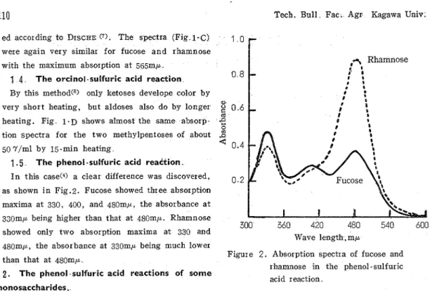
1.5
The phenol -sulfuric acid reaction I n this case(" a clear difference was discovered, as shown i n Fig.2. Fucose showed three absorption maxima a t 330, 400, and 480mjh, the absorbance a t 330n-p being higher than that a t 480mp. Rhamnose300 360 420 480 540 600
showed only two absorption maxima a t 330 and
Wave length, m p 480mp, the absorbance at 330mp being much lower
Figure 2. Absorption spectra of iucose and than that a t 480mp
I hamnose in the phenol -sulfuric
2.
The phenol sulfuric acid reactions of someacid r eaction monosaccharides.
The phenol-sulfuric acid reaction of DUBOIS et al. C4) was applied to eigth common monosaccharides in about 5 0 ~ / m l solutions Hexoses showed high absorption maxima at 490mp, and pentoses showed those a t 480mp; both groups of sugars showed lower absorption maxima at 330mp. Their spectra were similar to the spectrum for rhamnose shown in Fig 2.
The absorbnaces for various sugars as well as the ratio of the absorbance a t 330mp to that at 480 or 490 m p are given in Table I
Table
I
Characteristics of common monosaccharides in the phenol-sulfuric acid reactionSugars
Absorbance a t Absorbance a t hmax. in longer wave region
I
I
330mp (*)I
Xmax, , m p1
Absorbance (B)I
D-
GlucoseI
0.5281
490/
0 8101
0.65 I I 1'
D-
Mannose 0.346 L -A1 abinose 0 294 D-Ribose 0 298 D-Xylose 0 354 D - GalactoseThus this ratio higher t h a n 1 would show the presence of fucose
0 402
1
4901
0 770I
0 523.
The effect of heating time in the phenol-sukfuric acid reactionIn some color reactions of sugars the heating time affects so largely the absorption spectra of the reac- tion products that the qualitative and quantitative analyses become thereby possible (3") By the original method of DEBOIS e t a1 (I), the heating was not controlled, but i t was carried out by utilizing the heat produced by dropwise addition of sulfuric acid into the aqueous medium Now an experiment was made to see the effect by varying the heating time on this reaction
Vol,
12,
No.1 (1960)
Sulfuric acid was added under ice cooling T h e reaction mixture was kept for 2, 8 , or 32 min i n a boiling-water bath The spectra are shown in Fig.
3.
As the heating time was lengthened, the sbsorb- ance a t 330ml~ increased, while that a t 480mp did not so much increase In the case of fucose longer heating than 8 min led to the disappearance of the maximum a t 480mp Anyhow no conditions were found to be more effective in identifying fucose from rhamnose than the conditions of the original method,4.
Detection of fucose as a constituent of hemicellulose Bl from broad - bean seeds.The hydrolyzate of the hemicellulose B1 was puri- fied in respect to the methylpentose fraction step by step, and the following four samples, A , B, C , and D, were pr epar ed :
A
- -
the hydrolyzate was chromatog~aphed on alarge sheet of paper (TByB No 51A, 40cmx 40cm) with phenol-water (4:l) as the solvent,
B
--
sample A was chromatographed with butanol-acetic acid-water (4 : 1 : 2) as the solvent,
C
- -
sample B was again chromatographed with phenol-water as the solvent, andD
- -
sample C was chr omatogr aphed on a cellulose column with butanol-ethanol -water (4 : 1 : 5 , the upper layer).
The results of the phenol -sulfuric acid reaction of these four sample solutions are shown in Table
II
Fig. 4 shows the absorption spectra for the samples A and D (spectra for the other inter medi- ate samples are omitted for simplicity sake).
Fig.4 and Table
II:
show instantly that the unknown sugar isolated and purified is fucose This paper confirms the former that the hemi- cellulose B1 from broad bean seeds contains fucose as a constituent(This paper was presented b e f o ~ e the 170th meet- ing of the Kansai Branch of the Agr icultural Chem-
300
360
420
480
540
600
Wave length, m p
Figure 3 Effect of heating time on the absorp- tion spectra of fucose a n d rhamnose in the phenol-sulfur ic acid reaction
.
A
0,
300
360
420
480
540
600
Wave length, m p
Figure 4. Absorption spectra of t h e samples A and D in the phenol -sulfuric acid reac- ical Society of ,Japan on June 4, 1960, a t the tion
Table
II.
Phenol-sulfuric acid reaction of methylpentose fractions separated from the hydrolyzate of hemicellulose B1 of b r o a d - b e a n z e d s .Samples
1
Absorbance a t 330mp (A)I
Absorbance a t 48Omp ( 5 )I
A 1 50
..
542 0.63 0..474 0..
406 0,,98A
B C D 0.458 0.382 -1
0 , 7 8 1.20-
0.342 0.374 0.398OLIVE 香川大学学術情報リポジトリ
112
Tech Bull. Fac. A ~ I.
Kagawa Univ. Mukogawa Women's University, Nisinomiya )LITERATURE CITED
(1) KAWAMURA, S , NARASAKI,
T.
: Bull Agr.
C h m . ,2 8 , 350
(1956)
Chem Soc Japan,
2 2 ,
436 (1958)
(5) DISCHE,z . ,
SHEIILES, L B : J . B Z O ~ .(2) ---, - : Nzpfion Ndgez -kagaku Chem.,
115,
595 (1948).
Kaishi,
33, 817 (1959)
(6)
GIBBONS,M
N : A n a l y s t ,80,
268 (1955)
(3) NARASAKI, T : T e c h . Bull Fac Agr (7) DISCHE, Z . : Mzkrochemie,
7
,
33
(1929)
Kagawa U n z v . ,11
,265 (1959)
(8)
B R ~ ~ C K N E R ,B
J : Bzochem1
,6 0 , 200
(4) DUBOIS, M . , GILLES, K . A . , HAMILTON,
(1955).
J K , REBERS, P A , S M I T H , F : Anal