緒 言
特発性内頚動脈解離は比較的稀な疾患であり2,9)一般 的に内科的治療を中心とした保存的治療が優先される6,8). しかし,解離部の狭窄・閉塞や動脈瘤様の拡張を呈し, 症状の悪化を呈する症例も多く報告されている2,10,13,14,17,20). 本邦においては,内頚動脈解離は欧米に比し,頻度が低 いことが知られているが,若年性脳梗塞の原因でもあり4), 急性期の診断,治療は重要である20).今回我々は内科的 治療に抵抗し,急性期にステント留置術を施行し良好な 結果を得た1例を経験したので報告する.症例呈示
66歳,男性.左側の一過性黒内障,右上下肢の脱力発 作による歩行不安定を主訴に救急外来を受診した.初診 時,意識清明,失語症および構語障害はなく,右上下肢 の若干の筋力低下および感覚障害を認めるものの立位保持, 独歩は可能であった.既往歴にfibromuscular dysplasia, Marfan症候群などの結合織疾患はなかった.また症状 出現4時間前に1時間程度のゴルフスウィングをしてい た.1年前に眩暈に対して行った脳MRI,MRAでは明 らかな異常所見は認めなかった.入院時,脳MRIでは脳 梗塞巣は認めなかったが,MRAで左内頚動脈の頭蓋内特発性内頚動脈解離の1例:症例報告
竹内昌孝1) 石黒朋子2) 阿波根朝光2) 小西善史3) 松前光紀1)Spontaneous dissection of the internal carotid artery: case report
Masataka TAKEUCHI1) Tomoko ISHIGURO2) Asamitsu AWANE2)
Yoshifumi KONISHI3) Mitsunori MATSUMAE1) 1) Department of Neurosurgery, Tokai University School of Medicine 2) Department of Neurosurgery, Kasai Cardiology & Neurosurgery hospital 3) Department of Neurosurgery, Kyorin University School of Medicine
●Abstract●
Objective: We report a case in which acute carotid stenting was performed for spontaneous dissection of the internal carotid artery (IC). Case: A 66-year-old male developed sudden amaurosis fugax and right hemiparesis and came to us. Cerebral angiography revealed irregularities of the carotid wall, stasis of the contrast agent, and delayed cerebral blood flow from the cervical carotid bifurcation to the C5 portion of the IC. Based on these observations, IC dissection was diagnosed. After hospitalization, we initiated bed rest and medical therapy. Two days after onset, increased infarcts on MRI and a decrease of hemispheric blood flow on SPECT due to possible embolization secondary to thrombus formation were observed. We determined that active treatment was indicated and acute stenting was performed on the same day. Stenting was performed under local anesthesia and distal protection. Implementation of two Driver stents and 1 Wallstent RP improved the IC morphology and cerebral blood flow. Although sensory disturbance remained in the right lower limb, the patient was ambulatory and was discharged on Day 11.
Conclusion: Minimally invasive percutaneous carotid stenting is a useful alternative for cases presenting with spontaneous IC dissection that exhibit resistance to medical therapy.
●Key Words●
carotid artery stenting, endovascular surgery, progressive stroke, spontaneous dissection of the internal carotid artery
(Received September 16, 2009:Accepted December 30, 2009)
1)東海大学付属病院 脳神経外科 2)葛西循環器脳神経外科病院 脳神経外科 3)杏林大学 脳神経外科
への血流低下を認めた.入院後,直ちにヘパリンナトリ ウム持続点滴を10,000単位/日で開始し,active clotting time(ACT)を200秒に維持するように投与量を増減し た.また,抗血小板薬(アスピリン100 mg・シロスタゾ ール200 mg/日)の内服を開始した.第1病日に,右上 下肢の脱力発作が頻回となり,脳MRI拡散強調画像(以 下DWI)にて左大脳半球に塞栓性と考えられる新たな 梗塞巣の出現を認めた(Fig. 1).脳血管撮影を施行し たところ,左総頚動脈分枝部よりC5 portionにかけ, long tapered irregular stenosisおよび造影剤の停滞を認 め(Fig. 2),また,頭蓋内血流の遅延がみられた.対 側内頚動脈および後方循環からの側副血行は乏しかっ た.脳血流SPECTにおいて,左大脳半球に有意な血流 低下を認めた.3D-CTAでは解離病変に特徴的なintimal
flapがcervical portion-petrous portion移行部のみに存在 したが,MRA のsource imageではcrescent signはなく, 正確な解離病変範囲の同定は困難であった.第2病日に, 右上下肢脱力発作と失語症の悪化がみられ,また頭位挙 上や体位変換にて意識障害が出現した.DWIにて梗塞 巣の増多を認めた(Fig. 1).以上より,塞栓性の進行 性脳卒中で大脳半球血流低下を伴う症候性特発性内頚動 脈解離と診断し,内科的治療の限界と判断し,緊急で経 皮的内頚動脈ステント留置術を施行した.
血管内治療
局所麻酔下,右大腿動脈アプローチにより,コアキシ ャ ル シ ス テ ム に て 8Frガ イ デ ィ ン グ カ テ ー テ ル Launcher(Medtronic, Santa Rosa, CA, USA)を左総頚 Fig. 1 Diffusion-weighted images (upper row) show ischemic lesions in the left watershed region on動脈に留置した.ヘパリン7,000単位を静注し,ACT
310秒であることを確認した.ロードマッピング下に
Guardwire Plus(Medtronic, Santa Rossa, CA, USA) を慎重にcavernous portionに誘導した.その際,解離部 通過前後の血管撮影を比較し,偽腔を通過していないこ とを確認した後,内頚動脈の血流遮断試験を行った.10 分間の血流遮断では,新たな神経学的異常の出現を認め なかった.血流遮断下に,術前3D-CTAにてintimal flap が描出されたcervical portion-petrous portion移行部に, stent遠位端が遠位正常血管にかかるようにDriver stent
4mm~30 mm(Medtronic, Santa Rosa, CA, USA)を,
近 位 側 へ の 落 下 に 注 意 し な が ら 9 気 圧(nominal pressure)で,10秒間拡張して留置した.3D-CTAにて 解離部遠位正常血管が4.1 mmであったため,4mm径の stentを選択した.ThrombusterⅢ7Fr(カネカメディ ッ ク ス, 大 阪 ) に て 血 液 吸 引 後,distal balloon protectionを解除した.血管撮影でステント近位部に狭 窄の残存と血流の停滞を認めたため,新たなDriver stent 4mm~30 mmを 誘 導 し,Guardwire Plusに よ る distal balloon protection下に,先に留置したステントと ステントエッジをオーバーラップするように9気圧,10
秒間にて拡張,留置した.Protectionを解除して再度,血 管撮影を行うと,狭窄部は消失したもののステント近位 側に若干の造影剤の停滞を認めたため,再度protection 下 に Wallstent RP 6 mm ~24 mm(Boston Scientific, Natick, MA, USA)を誘導し,その遠位側を,2本目 Driver stentに被せるようにして留置した.結果的には
3個のステントがpetrous portion-cervical portion移行
部 か ら 近 位 側 へ 連 続 し た 形 で 留 置 さ れ た(Fig. 3). ThrombusterⅢ7Frにて各ステント留置毎に合計3回 (180 ml)の血液吸引をしたが,明らかな血栓は認めら れなかった.治療直後の血管撮影では内頚動脈の血流の 停滞は消失し(Fig. 4),頭蓋内血流の改善を認めた.
術後経過
術後,ヘパリンは自然中和とし,アルガトロバン 180 mg/日を2日間使用し,アスピリンとシロスタゾー ルを継続とした.術後のDWIに新たな脳梗塞巣は認め られず,早期リハビリテーションを開始した.術前より みられた右上下肢の脱力発作や失語症は術後直ちに消失 した.第7病日の血管撮影では内頚動脈病変部の血流の 停滞はなく,頭蓋内血流も良好に保たれていた.第11病 Fig. 2 Left common carotid artery angiograms on admission (A:AP view andB lateral view). Long tapper stenosis (arrows) and stasis of the contrast agent are observed from the C5 portion.
日に右上下肢の感覚障害は若干残存するものの,独歩に て自宅退院となった.術後1年の時点では再狭窄はなく, 感覚障害は軽快しmRS1で経過している.
考 察
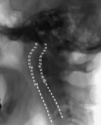
頚部内頚動脈解離の発生率は,Schievinkらにより2.6 人/10万人/年と報告されている18).内頚動脈解離全体で は頭蓋外41%,頭蓋内47%,両者合併が12%であり,本 邦において頚動脈解離は椎骨脳底動脈解離と比較し低率 で,その比率は約1:4で椎骨脳底動脈解離が多い19). しかし,近年MRI,MRAおよび超音波検査などが簡便 に施行されるようになり,日常診療において遭遇する疾 患として念頭に置く必要がある7,15).頚動脈解離の症状 として,川口らは,片麻痺が73%,頭痛や頚部痛が50% と報告し9),Houserらはそれらに加え不完全Horner徴候 が57%で認められたとした7).不完全Horner徴候は本疾 患に特徴的な症状であり,顔面の発汗障害を伴わないの は内頚動脈周囲の交感神経叢のみ障害され,外頚動脈周 囲の交感神経叢は温存されるためと説明している15).わ れわれの症例では,左黒内障,右上下肢の脱力発作とい った左内頚動脈領域の虚血症状にて発症した. 内頚動脈解離は,頭頚部に強い回転応力がかかる頭頚部 Fig. 3 The deployed stents are indicated on the intraoperativeimage. Two Driver stents, (= = =) ( 4mm ~ 30mm) proximal to the C5 portion are shown. Due to slight stasis of the contrast agent on the proximal side, Wall stent RP (- - -) ( 6mm ~ 24mm) is additionally deployed.
Fig. 4 Postoperative angiograms (A:AP view and B:lateral view). Stenosis and stasis of the contrast agent have disappeared.
の打撲や穿通創による外傷性内頚動脈解離,fibromuscular dysplasia,Marfan症候群などの基礎疾患を有する内頚 動脈解離,また基礎疾患がなく原因不明である特発性内 頚動脈解離に分類される.しかし,咳やテニス,スイミ ング等のスポーツ後に続発した例も知られ10),頚部の小 さな回旋や過伸展が関与している可能性も示唆されてい る.本症例では症状出現4時間前にゴルフスウィングを 1時間程度しており,入念な現病歴の聴取も診断に際し て必要不可欠と考える.治療は,安静,抗凝固・抗血小 板療法などの内科的治療が第1選択である.今回の症例 においてもヘパリン持続点滴にて,ACT 200秒に維持 し,抗血小板薬を2剤併用しており,内科的治療は適切 であったと考えている.脳血管撮影などの画像所見の改 善や臨床症状消失など治癒率は外傷性解離に比し特発性 解離は高いとされる8).しかし,Houserらは特発性頚動 脈解離の約80%の症例で完全寛解もしくは改善を認める ものの,20%では狭窄の進行や完全閉塞に至る例が存在 すると報告している7).また,外傷性解離に認められる 動脈瘤様の拡張は寛解しにくいとされる. 診断には脳血管撮影が有用であり,その所見に解離部 位より数cm遠位より始まる狭窄像であるstring sign, tapering occlusion,頭蓋内循環時間の延長などが挙げら れる.String signは最も高頻度でみられるが,この所見 は頭蓋底部でとどまることが多く,その先の頭蓋内へ進 展することはない.内科的治療に抵抗性で進行性経過を たどる内頚動脈解離に対する血行再建術を目的とした外 科的治療として,病変部の切除とgraftを用いた再建術3),
gradual intramural dilatation,EC-IC bypassなどが報告 されているが16,20),近年,より低侵襲なステントを用い た血行再建の報告も散見される1,11,12,13,17).本症例におい ても,抗凝固・抗血小板療法による内科的治療を選択し たが,進行性に症状の増悪を認めたため積極的な治療が 必要と判断した.解離病変部位を考慮すると直達術は困 難なこと,頭位挙上や体位変換による症状の増悪もある ことからhemodynamic compromiseの状態であり,低侵 襲的な局所麻酔下にて可能なステント留置術を選択し た.ステント留置術に関しては,急性閉塞やステントの distal migrationなどが問題としてあげられる.今回の症 例では,proximal protection可能なガイディングカテー テルの準備ができず,Guardwire plusをロードマッピン グ下に解離病変遠位にまで誘導しdistal balloon protection を行った.幸い遠位塞栓を来すことなく治療することが できたが,狭窄部に壁在血栓を伴っている可能性もあり, proximal protection下での誘導がより安全な手技であっ たものと考えられた.また,Guardwire plusが偽腔を通 過することにも十分な注意が必要である.今回,われわ れはバイプレーン,ロードマッピング下にGuardwire plus をその先端の動きに注意を払いながら誘導し,解離部通 過前後の血管撮影にて真腔への誘導を確認したが, intravascular ultrasoundを使用すれば,真腔の確保や解 離範囲の同定,偽腔内血栓の状態などのより的確な確認 ができたものと思われた. 解離病変に対しての複数本のステント留置が必要な場 合,近位側から留置すべきか遠位側から留置すべきかに ついて定見は無い.長い病変では近位側から留置するこ とによりentry pointを先に抑えることによって遠位側の 留置本数や範囲を少なくすることができる可能性がある が,一方で先に留置したステントのためにより遠位側へ のステント留置が妨げられる可能性がある.本症例では 解離病変の遠位側の頭蓋底部分に高度の狭窄を認めたた め,血管径,柔軟性および通過性を考慮して遠位側から 手前に向かってDriver stentを留置した.その後,内腔 が広く誘導が容易で近位側の解離血管壁に対するstent strutの 影 響 の 少 な いclosed cell-typeのself-expanding stentであるWallstent RPを留置した.これによって解 離部血管壁の十分な圧着,偽腔の閉塞が得られ,十分な 血流改善を認めたものと思われる. ステント留置後の長期予後としては,再狭窄が問題と なる.動脈硬化に起因する内頚動脈狭窄症に対するステ ント留置術については,SAPPHIREの追跡調査では治 療3年後の再狭窄率2.4%と良好であった5).解離病変に 対するステント留置での長期予後に関して,Marioらは 12例の経験で24ヵ月の経過では何ら問題なかったと報告 している13).しかし未だ十分な知見は得られておらず, 本症例についても今後の注意深い経過観察が必要である と考える.
結 語
一過性脳虚血発作にて発症し,内科的治療に抵抗し, 進行性の症状増悪を呈した特発性内頚動脈解離の1例を 経験した.本疾患に対する治療は内科的治療が主体であ るが,十分な経過観察が必要であり,進行性の経過をた どる例では,低侵襲のステント留置術は選択肢のひとつ として有用であり,今後,長期予後を含めた症例数の蓄 積が必要である.文 献
1) Ansari SA, Gregory Thomson B, Gemmete JJ, et al: Endovascular treatment of distal cervical and intracranial dissection with the neuroform stent: Neurosurg 62:636- 646, 2008.
2) 青木信彦:非外傷性頚部内頚動脈解離の2例:血管撮影 所見を中心として.Brain Nerve 35:361-366,1983. 3) Ehrenfeld WK, Wylie EJ: Spontaneous dissection of the
internal carotid artery. Arch Surg 111:1294-1301, 1976. 4) Goldstein LB, Gray L, Hulette CM: Stroke due to recurrent
ipsilateral carotid artery dissection in a young adult. Stroke 26:480-483, 1995
5) Gurm HS, Yadav JS, Pierre Fayad, et al: Long-term results of carotid stenting versus endarterectomy in high-risk patients: N Engl J Med 358:1572-1579, 2008. 6) 濱野利明,高塚勝哉,西村洋,他:特発性頚部内頚動脈
解離の2例:脳卒中 12:251-259,1990.
7) Houser OW, Mokri B, Sundt TM, et al: Spontaneous cervical cephalic arterial dissection and its residuum: angiographic spectrum. AJNR5:27-34, 1984
8) 岩室康司,中原一郎,東登志夫,他:自然軽快した特発 性頭蓋内内頚動脈解離の 1 例. No Shinkei Geka 36:625- 631, 2008. 9) 川口哲朗,朝田雅博,長尾朋典,他:頚部内頚動脈の特 発 性 解 離 性 動 脈 瘤: 2 症 例 報 告 と 文 献 的 考 察.No Shinkei Geka 12:1395-1400, 1984.
10) Kawajiri K, Kiyama M, Hayazaki K: Spontaneous dissection in the common carotid artery: Neurol Med Chir (Tokyo) 35:373-376, 1995.
11) Liu AY, Paulsen RD, Marcellus ML, et al: Long-term
outcomes after carotid stent placement for treatment of carotid artery dissection. Neurosurg 45:1368-1374, 1999. 12) Malek AM, Higashida RT, Phatouros CC, et al: Endovascular
management of extracranial carotid artery dissection achieved using stent angioplasty. AJNR 21:1280-1292, 2000.
13) Mario Fava, Luis Meneses, Soledad Loyola, et al: Carotid artery dissection: endovascular treatment. Report of 12 patients: Catheter Cardiovasc Interv 71:694-700, 2008. 14) Miyamoto S, Kikuchi H, Karasawa J, et al: Surgical
treatment for spontaneous carotid dissection with impending stroke. J Neurosurg 61:382-386, 1984.
15) Mokri B, Sundt TM, Houser OW, et al: Spontaneous internal carotid dissection, hemicrania, and Horner's syndrome. Arch Neurol 36:677-680, 1979.
16) Morgan MK, Sekhon LH: Extracranial-intracranial saphenous vein bypass for carotid or vertebral artery dissections: a report of six cases. J Neurosurg 80:237- 246, 1994.
17) Sam Heye, Geert Maleux, Rik Vandenberghe, et al: Symptomatic internal carotid artery dissecting pseudoaneruysm: endovascular treatment by stent-graft. Cardiovasc Intervent Radiol 28:499-501, 2005.
18) Schievink WI, Mokri B, Whisnant JP: Internal carotid artery dissection in a community. Rochester Minnesota, 1987-1992. Stroke 24:1678-1680, 1993.
19) 高木誠:脳動脈解離.Brain Nerve 58:963-970, 2006. 20) 宇野昌明,上田伸,新野清人,他:頚動脈解離に対する
治療方法と長期予後の検討.No Shinke Geka 25:417-423, 1997. JNET 4:27-32, 2010