(Original) Matsumoto Shigaku 22 : 13--25, 1996
key words : Japanese macaque-corticotomy-orthopedic force-strain gauge-Class Il malocclusion
Effect of Corticotomy on Maxillary Anterior Bone Segment Retraction
Induced by Orthopedic Force
YOSHIYASU YOSHIKAWA NORIMASA OKAFUJI YUJI ASHIZAWA
and Tosmo DEGUCHI
DePartment of Orthodontics, Matsumoto Dental College (Chief : Prof T. Deguchi)
Summary
The dry skulls of two adult female Japanese macaques were used in this study of bone strains produced by corticotomy combined with orthopedics. The directions and magni-tudes of the principal strains were measured using an eight-pair triaxial strain gauge rosette.
While compressive strains were seen in the control study (orthopedic force alone before corticotomy), tensile strains were seen in the experimental study (orthopedic force after corticotomy) on the frontzygomatic suture, the lateral side of the zygomaticomaxillary suture of the zygomatic bone, and on the maxillary bone. Corticotomy, combined with the posterior transfer of the anterior segment, had the following effects : in the control study, traction was diffused though the maxillofacial structures, while in the experimental study, it did not readily diffuse because the compact bone was separated. This resulted in favorable conditions for the posterior transfer of the anterior maxillary segment between the maxilla proper and the maxillary alveolar bone which are connected only by trabecular bone. These results demonstrate the usefulness of combining a corticotomy with retractien of the maxillary anterior segment by orthopedic forces.
A clinical case of Class II malocclusion is presented to illustrate what can be accom-plished with treatment by corticotomy. The treatment objectives were achieved with improvement in the patient's profile and in the function and esthetics of the dentition.
Introduction
Anterior maxillary osteotomy is a well-established surgical procedure for use in the correction of anterior skeletal or dental maxillary prognathism in adults. Daniel et ali). reported that the main advantage of the procedure is the relatively short treatment time.
The reported complications associated with the early applications of this technique included devitaiization of teeth, loss of part or all of the osteotomized segment, and relapse2). With current techniques, however, no major problems involving the teeth, bone or soft tissue loss are reported3), although minor complications such as devitalization still occur. Leibold et al`). reported that 50%
14 Yoshikawa, et al, : Effect of Corticotomy on IVIaxillary Anterior Bone Segment Retraction Induced by Orthopedic Force
of the non-responsive teeth were maxillary canines. The relatively high positions of their apices and their cornerstone positions in the mobilized segments apparently make the canines the teeth most vulnerable to surgical trauma.
Corticotomy was advocated by KOIe5), who reported more rapid movement of teeth since the main resistance to movement arises from cortical bone rather than from the thin trabecula of t]he spongiosa. Corticotomy of the maxillary and mandibular bones facilitates orthodontic displacement of the alveolar processes of a single tooth or of a group of teeth. This displacement saves time and reduces the risk of relapse. There are both experimenta16-'9) and clinicaliO"-'2) reports describing this method.
Nishimotoi3) previously performed corticotomies in conjunction with maxillary anterior bone segment retraction in Japanese macaque monkeys (A4acaca fuscata) and conducted evaluative cephalometric, dental cast, and histopathologic studies of the related sutures. The present study was undertaken to obtain strain gauge measurements of the macaque dry skulls following the applica-tion of the technique in his study.
A clinical case of Class II malocclusion treated by corticotomy is also presented.
Materials and Methods
1. Materials
The two adult female Japanese macaque dry skulls were used in thi$ experiment. Their dental development appears to be advanced for their chronological ages (at least 9.5 years) as determined by Kirino et al. (Yoshikawa6)). Since bone is an elastic materiali`), the same skull was used for both the control (before corticotomy) and experiment (after corticotomy) measurements, and the other skull was used for temperature compensation in the strain measurement procedure.
2. Anchoring of Skull
One skull was fixed with acrylic resin to a stainless steel pipe imbedded in a block of dental stone. The sagittal and occlusal planes were set vertically and horizontally, respectively (Fig. 1). The other dry skull was not anchored.
3 . Strain gauge
The directions and magnitudes of the principal strains were measured with trixial rosette strain
gauges (KFG-1-120-D17-11LIM2S, Kyowa Electric Instruments Co., Ltd., Tokyo, JAPAN) cement-ed to the bone. Prior to attachment the 16 strain gauges (Fig. 1), the surface of each skull was
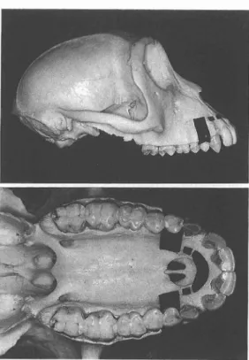
degreased in acetone and allowed to dry overnight in a desiccator. The gauges were cemented to the skull with a cyano-acrylate monomer adhesive (Type CC-33A, Kyowa Electronic Instruments Co., Ltd., Tokyo JAPAN). A Wheatstone bridge circuit recording device was used to measure the strains. Since a strain gauge may be affected by its own heating and the ambient temperature, a dummy strain gauge, having the same gauge factor, was cemented to the second skull. With this arrangement, the sensitivity of the gauges could be increased without being influenced by the temperaturei5). Figure 2 shows the bilateral placement of the strain gauge Pairs 1 to 8. Pair 7 was placed on the medial side of the zygomatic arch.
4. Data Processing
The strains produced by the orthopedic traction were recorded on a personal computer (PC -9801, NEC, Tokyo JAPAN) via a strain amplifier (Universal Digital Measuring System, UCAM -5B, Kyowa Electronic Instruments Co., Ltd., Tokyo JAPAN) and a scanner (Universal Scanning Box, USB-51A, Kyowa Electronic Instruments Co., Ltd., Tokyo JAPAN) which was connected to
Mkptts\. 22(1) 1996
Figure 1. Testing apparatus showing skulls and recording equipment.
15
i"
;Jif!XN
Figure 2. Strain gauge locations on the skull. Pair 7 was positioned on the mediai side of the zygomatic arch.
each of the 16 gauges. The following formulae were used for the rosette analysisi6) :
Maximum principal strain
Ei=
t (ea+Ec+ 2{(Ea-Eb)2+(Eb-E,)2})
Minimum principal strain
E2=-
lllJ (Ea+Ec- J2{(EaNEb)2+(eb-Ec)2})
Direction of maximum principal strain ÅíaÅrE, e=S tanra (2 EEbi-eEa,- Ec)
eaÅqEc 0=-li- tanri (2 EEbi-EEa,; Ec) +goo
where Ea, Eb, and Ec are the output strains and 0 is the direction of the maximum principal strain
s
in the countercloclÅqwise direction from the E.-axis. The direction of the minimum principal strainis perpendicular to the maximum principal strain. 5 , Braces and Traction Method
An alginate impression vL'as taken of the skull and an acrylic resin headgear, resembling a helmet, was constructed. The force module was positioned in place with the headger, Acrylic resin was then applied to fix the module. A resin splint was constructed using the same method as for the headgear (Fig. 3). In this study, micro-load cells (LTS-l KA, Kyowa Electronic Instruments Co., Ltd,, Tokyo, JAPAN) were used to accurately measure forces. A strain amplifier was used to measure forces with output readings with E Å~10-6 equivalent strain. In the calibration record, a value which corresponds to 1X10⁻⁶ equivalent strain, multiplied by the reading, can be found. The desired Ioad value was defined as (output from a strain amplifier Å~ 10'6) Å~ (calibration factor, gVlÅ~lo-6).
On the right side, the rated output was 3917Å~ 10-6 strain/1 kgf from the calibration sheet. The output from the strain amplifier was-334Å~10u6 strain when it was unloaded and 1115Å~ 10-6 strain when it was loaded. Thus, on the right side:
16 Yoshika"'a, e't ag. i Effect of Corticotomy on Maxillary Anterior Bone Segment Retraction Induced bs' Orthopedlc Force
Figure3. Testing apparatus showing loading equipment and strain gauges.
l kgf: 3917=x: 1115 -(- 334) x-- 370 gram.
Similarly. on the Ieft side, the rated output was 3878Å~10-6 strain/1 kgf, The unloaded output "ras 68Å~10-6 strain, while the loaded reading was 1476Å~10L6 strain. Therefore, on the left side :
1 kgf: 3878 = x: 1476 - 68 x = 363 gram.
Thus, the traction forces were 370 gram on the right and 363 gram on the left at 20 degrees upvLTard from the occlusal plane. The strain data were taken three times and averaged.
6. Corticotomy
After both maxillary first premolars were extracted, control data were obtained. Then, a corticotomy was perforrned in the same manner as in the previous studyi3) (Fig. 4). Several 2 mm deep holes were made through the cortical bone with a # 8 round bur, and connected with a # 261 fissure bur. Then the cortical bone was cut labio-lingually along the holes. Because the Japanese macaque has a shallow palate and because space for moving the anterior bone segment was needed, some parts of the maxilla rnesial to the incisive fossa were cut off. The bone at the extracted first
premolars was removed to provide room for moving the anterior segment, The.g.e procedures were perforrned under air cooling xxrith a three-way syringe. The experimental study was then performed to examine the effect of cor"ticotomv.
Results
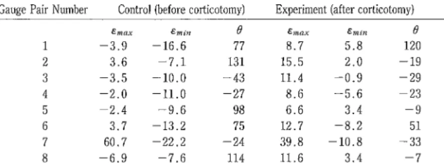
In the control stud}r (Fig, 5). high compressiv•e strain was found in the frontozygomatic suture (Pair 1, Table 1). but less strain was seen in the inferior region of the frontozygomatic suture (Pair 2). Along the z}rgornaticomaxillary suture (Pairs4 and 6), the zygomatic bone (Pair3), and the maxillary bone adjacent to the canine apex (Pair 5). the compressive strains were also high. The highest compressive strain was found in the medial zygomatic arch and at the temporozygomatic suture (Pair 7). Lateral to the suture (Pair 8), high compressive strain was found.
After corticotomy (Fig. 6), the strain inagnitudes and directions showed obvious alteration. The compressive strains became tensile at the following locations : the frontozygomatic suture (Pair 1) ; the maxillary bone adjacent to the canine apex (Pair 5);the lateral aspects of the zygomatic arch suture (Pair 8). For Pair 7, the magnitude was markedly decreased.
R[}dsdvs'i`E` 22(1) 1996 17
Ngure 4. Region of corticotomy a[)uccal gual sides) (From Nishimotoi3))
and
Iin-/
.tJt)-,-Å~
1ox 1o'6 strain
compressten
tenslon
traction force
18 Yoshikawa, et aL : Effect of Corticotomy on Maxillary Anterior Bone Segment Retraction Induced by Orthopedic Force
Table 1. Maximum, and minimum principal strain and the direction of maximum principal strain
Gauge Pair Number Control (before corticotomy) Experiment (after corticotomy)
1 2 3 4 5 6 7 8 Emax -3.9
3.6
-3.5 -2.0 -2.43.7
60.7-6.9
Emin -16.6 -7.1 -10.0 -11.0 -9.6 -13.2 -22.2 -7.6 e77
131-43
-27
98
75
-24
114 emax 8.7 15.5 11.4 8.6 6.6 12.7 39.8 11.6 Emin 5.8 2.0 -O.9 -5.6 3.4 -8.2 -10.8 3.4 e 120-19
-29
-23
-9
51
-33
-7
Emax, maximum principle strain ; E.i., mmimum principle strain ; direction of maximum principle strain ; data with negative sign represent compression strain (X 10-6) and those with positive sign represent tension strain (X 10-6)." 1ox1o'6 strain
compresslontenslon
-Figure 6. Strain distribution in the experimental skull.
traction force
Case Presentation
A clinical case of Class II malocclusion is presented to illustrate what can be accomplished with treatment by corticotomy.
History and general clinical background
The female patient was 34 years old at the start of treatment. Her general health was excellent and there were no conditions present that would in any way contraindicate treatment. The oral tissue appeared to be healthy and the dentition in a good state of repair, with no caries lesions present. No detrimental habits were noted. Her mother showed maxillary protrusion. The patient's profile was protrusive and the lips strained on closure. An excessive vertical
tE}Agts`,iP=" 22(l) 1996 19
growth pattern was evident. Her overjet was 11 mm and overbite was 4 mm (Figs. 7 and 8). Diagnosis
The occlusion was classified as Angle Class II with maxillary protrusion. There were no apparent tissue pathoses. Cephalometrically, marked protrusion of the maxillary dentures was evident. The Angle Classification was Class II with an ANB re!ationship of 7C. The maxillary mcisors were very protrusive and forward of the bony base, as indicated by the Ul to FH position of 1280 (Table 2, and Figs 8, 12). The panoramic radiograph confirmed the presence of all permanent teeth with the exception of the upper and lower right wisdom teeth (Fig. 11).
Diagnostic summary
The following clinical findings were noted : Angle Class II ; Skeletal II (ANB 70) ; Maxillary protrusion ; and Prognathic profile with protruded lip position.
Treatment objective
Although the Wassmund-Wunderer method was considered for the initial treatment plan, it was very difficult for the patient to remain in the hospital for the extended period of time required, as she had three children. Corticotomy and orthopedic force method was chosen after consultations and obtaining informed consent. This method was selected because it is safer and requires less time in the hospital than the Wassumund-Wunderer method and because it was anticipated that the time spent wearing the head gear equipment and the total treatment period would be less than that required for conventional orthodontic treatments.
t
"
Figure 7. Pre-treatment facial
xad
",-photographs.mrl
20 Yoshikawa, et al. : Effect of Corticotomy on Tvlaxillary Anterior Bone Segment Retraction Induced by Orthopedic Force Table 2. Cephalometric rneasurements in the patient
measurement
SNA
SNB
ANB
IMPA
FMA
FMIA
UI-FH
UI-SN
Y-Axis Interincisal Occlusal PI U-Lip to E-line L-Lip to E-linenorm
82
79
3
96 29 55 110 104 65 126 11o
2
pre-treatrnent 89 82 7 97 30 53 128 127 70 105 5 54
post-treatment86
78
6
98
31
51
98
97
71
133 153
o
Treatment stages
A full-banded edgewise, Roth type of appliance with pretorqued, preangulated O.022 Å~O.029 inch bonded brackets was used to treat this malocclusion. The treatment stages described below were undertaken.
1 . All of the teeth of both the maxillar and mandibular dentition were bonded or banded except the maxillary first premolars. Leveling of the arches and correction of rotations of each tooth were accomplished with super elastic wires. Leveling was performed by O.O17Å~O.025 inch stainless steel wire before corticotomy operation.
2. After application of general anesthesia, the corticotomy operation was performed in the maxilla. The Neiman incision was performed and the location of the basal margin of the piriform aperture was confirmed. The horizontal corticotomy procedure was then performed on the buccal and lingual sides for two- or three-mm width using a cross cut tapered fissure bur. The vertical corticotomy was performed at the site of the first premolars after extraction of these teeth for eight-mm width using bone chisel.
The patient used a transpalatal bar, J-hook and face bow combination head gear. During the period of use of this equipment, the extraction space was half closed within 50 days after the operation. The maxillary anterior segment was retracted.
3 . 0ne year after the operation, occlusion, interdigitation, and finishing were completed. Progress of treatment
The patient was seen at four-week intervals during the treatment. No appliance breakage was noted ; the patient's cooperation and oral hygiene were excellent at all times and her progress was satisfactory.
Treatment results
The facial esthetics improved considerably, and the occlusion and soft-tissue response were good. The maxillary protrusion-type profile was reduced as a result of the retraction of th.e maxillary incisors (Fig. 9 and 10). The dental occlusion and interdigitation were acceptable. Canine rise was good, with no interferences, and canine protection and anterior guidance were considered to be adequate for future protection of the dentition. The panoramic radiograph showed good approximation of the roots in the extraction sites. No root resorption was evident in either the upper or lower anterior segment (Fig. 11). The FMA angle was slightly opened (Fig. 12).
wtISiigi+#- 22(1) 1996
Figure 9,
21
Post-treatment facial photographs.
Figure 10, Post-treatrnent intraoral photographs.
Figure 11. Panoramic radiographs. Pre-treatment (Left) and post-treatment (Right).
Secondary Treatment
All brackets and bands were removed, and the patient was instructed to wear a custom-made tooth positioner with face bow for one year during sleep. Final records were taken immediately after removal of the bands. The patient was observed monthly for the first six months. Retention time is completed and she has shown no relapse.
Final evaluation of the clinical case
We believe that, in the clinical patient with maxillary protrusion, the satisfactory occlusion, ANB change, tooth position, and facial changes justified the corticotomy and orthopedic therapy.
22 Yoshikawa, et al, : Effect of Corticotomy on Maxillary Anterior Bone Segment Retraction Induced by Orthopedic Force
g2•s
695 1275 65 e ( (L
Figure 12. Tracing of cephalometric radiograph at pre-treatment (Left) and superimposition of the pre-treatment and post-treatment tracings on SN-plane registered at sella. Solid line at pre-treatment and doted line at post-treatment (Right).
The treatment objectives were achieved with improvement in the patient's profile and .in the function and esthetics of the dentition.
Discussion
In the study of dynamic bone morphology, the following techniques have been used : photoelas-ticityi7''8), stress coati9'20}, holography interference2i'22), and measurement with strain gauges6•i5•2iA-27). The strain gauge method has provided excellent results in elucidating medical and dental biome-chanics. Nakanishii5), Suzukii6), and Shirai23) performed triaxial strain gauge experiments to analyze the deformations of the maxillary complex under orthodontic and orthopedic loads. Similarly, Fukui2`) and Hata25} studied the orthodontic and orthopedic effects induced by a chin cup appliance, and Hylander26•2') tested competing hypotheses by means of strain gauges. However, in few studies have corticotomies been investigated with strain gauges6"). Yoshikawa6) reported that strain values were decreased at all suture sites after corticotomy, but vertical strains could not be measured because single strain gauges were used.
Compressive strains were noted in our control study (before corticotomy), in opposition to the tensile strains observed in the experimental study (after corticotomy), for the frontozygomatic suture, the zygomaticornaxMary suture of the zygomatic bone, and on the maxillary bone. The greatest tensile and compressive strains in the control study were found in the zygomatic arch suture (Pair 7), and the lateral aspect of that suture (Pair 8) showed high compressive strain. These results conflict with those of previous studies.
Suzukii6) found that the curvature of the temporal part of the zygomatic arch was increased by backward traction force. This implies that there are tensile strains on the lateral side of the zygomatic arch and compressive strains on the medial side of the arch6'i6}. The zygomatic arch and temporozygomatic suture demonstrated the highest strains but these strains were -absorbed by increased curvature of the zygomatic. arch6'i6). In contrast, the present results suggest that there is a large tensile strain on the medial side of the zygomatic arch (Pair 7) but small compressive strains
?Z}IKvae}k 22(I) 1996 23
on the lateral side of the zygomatic arch (Pair 8) in control skull. In experimental skull, the strain readings showed decreased tension on the medial side of the zygomatic arch (Pair 7), however, the pattern is the same as the control skull. These data indicate the tendency for the curvature of the temporal part of zygomatic arch to increase in experimental skull (after corticotomy). This can be attributed to the smaller appliance (covered only anterior segment) used in this study than the other studies (covered all maxillar).
Nishimotoi3), in a study of the histopathological changes following the application of the same procedure in living animals, found enlargement of the suture spaces in both the control (no cor-ticotomy) and, to a lesser extent, in the experimental (with corcor-ticotomy) animal. Similar results were obtained by Drosch128) who studied the effect of heavy continuous orthopedic forces on young squirrel monkeys. He found vigorous cell activity in the sutures after one month, when the width of the sutures had enlarged two-to three-fold. Elder and Tuenge29) reported the effects of high-pull force application on the sutures in Rhesus monkeys. They found, in contrast to our results for the suture surfaces, that bone resorption occurred at the zygomaticomaxillary and zygoMaticotemporal sutures. When heavy loads were placed on the monkey skulls, the sutures demonstrated immediate high strains. After corticotomy, these strains were decreased. In living animals, expanded sutures -were noted 30-70 days Iater.
Corticotomy, combined with the posterior transfer of the anterior segment, seemed to have the following effects: in the control study (before corticotomy), traction was diffused through the maxillofacial structures via the sutures, while in the experimental study (after corticotomy), the traction did not diffuse as readily because the compact bone was separated. This results in favorable conditions for the posterior transfer of the anterior maxillary segment between the maxilla proper and the maxillary alveolar bone, which are connected only by trabecular bone. These results demonstrate the usefulness of combining a corticotomy with orthopedic retraction of the maxillary anterlor segment.
Acknowledgments
The authors wish to thank Dr. Kiyofumi Furusawa, Oral Maxillofacial Surgery Department II, Matsumoto Dental College, performed the surgical procedures.
This work was supported by a grant from the Ministry of Education, Science and Culture of Japan (02670934).
References
1 ) Daniel H. T., White R. P., and Proffit W. R. (1971) Anterior maxillary osteotomy in dental treatment. JADA, 83 : 338-343.
2 ) Westwood R. M. and Tilson H. B. (1975) Complications associated with maxillary osteotomies. J. Oral Surgery, 33 : 104-115.
3 ) Epker B. N. (1977) A modified anterior maxillary osteotomy. J. Max. Fac. Surg. 5 : 35-38.
4 ) Leibold D. G., Tilson H. B. and Rank K. R. (1971) A subjective evaluation of the re-establishment of the neurovascular supply of teeth involved in anterior maxillary osteotomy procedures. Oral Surg. 32 : 531 -534.
5 ) K61e H. (1959) Surgical operations on the alveolar ridge to correct occlusal abnormalities. Oral Surg. Oral Med. Oral Pathol. 12 : 515-529.
6) Yoshikawa Y. (1987) Effect of corticotomy on maxillary retraction induced by orthopedic force. Matsumoto Shigaku, 13 : 292-320. (in Japanese with English summary)
24 Yoshikawa, et at, : Effect of Corticotomy on Maxillary Anterior Bone Segment Retraction Induced by Orthopedic Force
Shikwa Gakuho, 82 : 219-252. (in Japanese with English summary)
8) Matsuda Y. (1989) Effect of two stage corticotomy on maxillary protraction. J. Japan Orthodontic Society, 48 : 506-520. (in Japanese with English summary)
9 ) Dttker J. (1975) Experimental animal research into segmental alveolar movement after corticotomy. J. Max. Fac. Surg. 3: 81-84.
Ie) Generson R. M. and Stratigos G. T. (1978) Combined surgical and orthodontic management of anterior open bite using Corticotomy. J. Oral Surg. 36 : 216-219.
11) Lines P. A. (1975) Adult rapid maxillary expansion with corticotomy. Am. J. Orthod. 67 : 44-56. I2) Fitzpatrick B. N. (1980) Corticotomy. Aust. Dent. J. 25 : 255-258.
13) Nishimoto M. (1992) An experimental study on maxillary anterior bone segment retraction by dic force. J. Fukuoka Dent. Coll. 19 : 171-194. (in Japanese with English summary)
14) Emata T. (1976) The analysis of strain and displacement, produced by the Kloehn type face bow in the dry skull of the macaca irus, J. Japan Orthodontic Society, 35 : 130ul49. (in Japanese with English summary)
15) Nakanishi Y. (1973) Effect of headgear traction on the Human facial skeleton: A study with strain gauges, J. Osaka Dent. Univ. 7: 7-30.
16) Suzuki T. (1982) Experimental study of the deformity of maxillary complex loaded by orthopedic forces. -measurement by three axial strain gauges-. Shikwa Gakuho, 82 : 469-502. (in Japanese with English summary)
17) Chacanas S. J., Caputo A. A. and Davis J. C. (1976) The effects of orthodontic forces on the craniofacial complex utilizing cervical and headgear appliances, Am. J. Orthod. 69 : 527-539.
18) Ayala C.P., De Alba J.A. and Caputo A.A., Chaconas S.P. (1980) Canine retraction with J hook headgear, Am. J. Orthod. 78 : 538-547.
19) Evans F. G. and Lissner H. G. (1948) "Stresscoat" deformation studies of the femur under static vertical loading. Anat. Rec. 100 : 159T- 190.
20) Evans F. G., Lissner H. G. and Pedersen H.E. (1948) Deformation studies of the femur under dynamic vertical loading. Anat. Rec. 101 : 225-241.
21) Iijima M. (1988) Action patterns of orthopedic forces on the maxillary complex utilizing holography interface and strain gauge methods. J.Japan Orthodontic Society, 47 : 127-144. (in Japanese with English summary)
22) Iijima M., Fujimoto K., Matsui S., Kubodera T. and Kobayashi K. (1986) Study of mechanical reaction of Human dry skull to extra-oral anchorage appliance as measured by holography interface and strain gauge methods. Bull, Josai Dent. Univ. 15 : 41-49. (in Japanese with English summary)
23) Shirai H. (1990) The experimental study of the effects of J-hook type headgear on the craniofacial ' complex. Nichidai Kokuu Kagaku, 16 : 274-293. (in Japanese with English summary)
24) Fukui T. (1971) Effect of the chin cap on the Human mandible based upon the strain distribution. J. Osaka Dent. Univ. 5 : 109-128,
25) Hata S. (1983) Orthopedic effects on the craniofacial complex induced by chin cap appliance. Kyushu shikagakaishi, 37 : 506-535. (in Japanese with English summary)
26) Hylander W. L. (1984) Stress and strain in the mandibular symphysis of primates : A test of competing hypotheses. Am. J, Phys. Anthr. 64:1-46.
27) Hylander W. L. and Johnson K, R. (1992) Strain gradients in the craniofacial region of primates. In Davidovitch Z. (ed.). The Biological Mechanisms of Tooth Movement and Craniofacial Adaptation, 559 -569. The Ohio State University College of Dentistry, Columbus, Ohio.
28) Droschl H. (1975) The effect of heavy orthopedic forces on the sutures of the facial bones. Angle Orthodont. 45 : 26-33.
29) Elder J.R. and Tuenge R.H. (1974) Cephalometric and histologic changes produced by extraoral high-pull traction to the maxilla in Macaca mulatta, Am. J. Orthod 66 : 599-617.
zaJ!tsitse}g 22(1) 1996 25 Oi'en : XfiasI:.h ltMLN t: Å}t'fi MR ntu"tsreg5 di fkhptfu va C: "b' Ct 6 = YV+= F S - di stMC: eaV6Mnde
sJIIeff, maeeaniE, figere=, Nmtwde
(M}pttsJi( • dvNkeIE)2 ma6D pt ÅrÅqbreX= ,trs '/-v"ivoutmewtges igMvNv(, u 7veu ts Q -EvwaasJY-fJtJ igetHl LfoLLvwaEiltru]E
ts ag zz o th fi pt tz Hi aE V-or' ly tzc ea -e- 6 61F sOtue ig fi . k. nN U-S" JY O" E fi fl O va[J fi e*, 8 ma OE wt JK F v h/ tr" - .ti igm L N -c fi . tc . ko re m, *t wa -(s• eutwa nB lic, rJ ut eea, pt. nn owa ptH Lywa waao srk ailm t6- : as rrL vÅéE -E izlz Se LN v( v*, EE wt V-9`' jCk ig t7N. bt) fo a) vc su L vC , u 7vf = F E - 6r fjt re L fo =wt ec Sb' LN v( , eI -)
KbVy-e'lkAtptwt-9-6-*rbKhj4Åq62Ltc. -9-tsibtb, -ltsve.ptEiltiilJEgneesOpaJtiptivffekSb•Åë]-6uiVfu
F E-ambMeMtaoat 5 va;4%6ktc. ntufizzo=7vf= F s -igT}MLtsuNStuaeefo-uN-(v*, eegl"
thN'`&ft 6Dl#AzzigAJi Lw({XXt Z5 d E tzex -) "(swagetmtzcinAtrtwL'(Li6. C2LtzcSifLw( u ivf= F E -ig STLMLfoscwt-(}•el:twvat!Ng•th:bjma}ISthL-CLN K,kbt)ell, rvua2 LtwtL.-C, l1ODtweIJi])b:at{twLtzCÅq Åq tst b,tu*ntE a) Jg -6-D k rdg . ( u N 6 LsM. aH 8 hsve. nH a) ma ig ca twe L O F.7 tr= fo- L N (Lywa ntu fi tszz zz Er pahpt et " -}Å} 6 O L L 'C 7fi imi f" A] 'e 6 ig o 8 ig % 6 thL fo. rk tc, Zsc X21 )5r ms JE L -( Rff ts ee ut, pmL)cA i5r rek7 y pt