INTRODUCTION
Galectins are a family of carbohydrate-binding proteins characterized by a conserved amino acid sequence defined by structural similarities in their carbohydrate-binding domain and affinity for β-galactoside-containing glycoconjugates. It was re-ported that galectins play a significant role in apopto-sis, several of which, such as 1 and galectin-9, were implicated in the promotion of apoptosis of immune and melanoma cells, respectively, whereas galectin-7 induces apoptosis of colon cancer cells. In contrast, galectin-3 was reported to be an antitotic molecule, inhibiting Fas-induced T-cell
apop-tosis as well as epithelial cell apopapop-tosis induced by staurosporine, cisplatin, genistein, and anoikis (1). Galectin-3 is isolated as a 32 kDa monomer, and consists of three distinct structural domains : the carboxyterminal domain, which appears as the carbohydrate-recognition domain, the N-terminal domain, comprising the 20 NH2-terminal residues,
and the R domain, composed of Pro-Gly-Tyr-rich re-peating motif (2). Galectin-3 contains the Asp-Trp-Gly-Arg (NWGR) motif in the C-terminal domain, and it doesn’t exist in other galectins. The NWGR motif is critical for the anti-apoptotic function of galectin-3 (3).
Galectin-3 is expressed in normal and neoplastic cells (4). Galectin-3 is localized not only in intracel-lular space such as the cytoplasm or the nucleus but also in extracellular space such as the cell surface or the extracellular matrix (5).
In certain types of cancers, the expression of galectin-3 is positively correlated to tumor
progres-ORIGINAL
Serum level of galectin-3 in human bladder cancer
Manabu Sakaki, Natsuo Oka, Ryoichi Nakanishi, Kunihisa Yamaguchi,
Tomoharu Fukumori, and Hiro-omi Kanayama
Department of Urology, Institute of Health Biosciences, The University of Tokushima Graduate School, Tokushima, Japan
Abstract : We examine serum level of galectin-3 in patients with bladder cancer. We used serum samples of 67 patients with urological diseases and classified these patients into bladder cancer group (n=43) and control group (n=24). Galectin-3 concentration was meas-ured by ELISA (Human Galectin-3 Assay Kit, IBL). And we selected the patient with high serum galectin-3 concentration (Urothelial Carcinoma, G3, pT3a pN0M0), we performed immunohistochemical staining with the VECTASTAIN ABC (Avidin Biotinylated enzyme Complex) system. Median value of serum galectin-3 concentration was 1068 pg/ml (range 551-2028) in the cancer group vs 584 pg/ml (range 259-1262) in controls. Serum galectin-3 concentration of the bladder cancer patients was statistically higher than that of controls (p 0.0005). There was no apparent correlation in serum galectin-3 concentration with the clinico-pathological features such as stage and grade. Higher expression of galectin-3 was observed in bladder cancer tissue than in normal bladder tissue. We suggest the meas-urement of serum galectin-3 is useful for diagnosis of bladder cancer. J. Med. Invest. 55 : 127-132, February, 2008
Keywords : galectin-3, serum, bladder cancer
Received for publication November 26, 2007 ; accepted January 9, 2008.
Address correspondence and reprint requests to Manabu Sakaki. M.D., Department of Urology, Institute of Health Biosciences, The University of Tokushima Graduate School, Kuramoto-cho, Tokushima 770-8503, Japan and Fax : +81-88-633-7160.
sion, such as gastric cancer (6), liver cancer (7), thyroid cancer (8). In other types of cancers, it is inversely correlated to tumor progression, such as head and neck cancer (9), uterine sarcoma (10), breast cancer (11), pancreas cancer (12).
In bladder cancer, galectin-3 mRNA levels were increased in most tumors compared with normal urothelium (13). However, the clinical relevance of this finding is not fully understood. We examined serum level of galectin-3 in patients with bladder cancer.
PATIENTS AND METHODS
Measurement of serum galectin-3 concentration We studied serum samples of 67 patients who were admitted for urological diseases at our insti-tution between 1996 and 2006. We classified these 67 patients into the cancer group (n=43) and the control group (n=24). Control group consisted of patients with 21 urolithiasis, 1 varicocele and 2 stress incontinence. Clinical data are described in Table 1.
All serum samples were stored at -80"!until this study. All samples were returned to room tempera-ture before use. Serum galectin-3 concentration was measured by ELISA in accordance with the manu-facture’s instructions (Human Galectin-3 Assay Kit, IBL). Measurable range of this ELISA is from 117.19 to 7500 pg/ml. This ELISA kit has been de-scribed and validated elsewhere.
Immunohistochemical staining
After radical cystectomy, both bladder cancer tis-sue and normal bladder tistis-sue were embedded in OCT compound and stored at -80"!. We selected the patient with high serum galectin-3 concentra-tion, and cut these tissue in thickness of 5
microme-ters at -18"!and put section on the slide. We per-formed hematoxylin eosin staining, and chose the section by which a core is stained clearly. The good bladder cancer tissue and normal bladder tissue could be obtained. The histopathological diagnosis of selected bladder cancer tissue was urothelial car-cinoma (UC), G3, pT3a pN0M0.
Immunohistochemical staining was performed with the VECTASTAIN ABC (Avidin Biotinylated enzyme Complex) system. The brief procedure was as follows. Sections were air dried, and fixed with acetone. We rinsed sections in tap water and washed in buffer. After incubating sections for 30 minutes with diluted normal blocking serum, the primary antibody (rat monoclonal anti-Gal-3 antibody, TIB 166) was incubated for 60 minutes at room tem-perature. Following incubation for 30 minutes with diluted biotinylated secondary antibody solution, we added VECTASTAIN ABC Reagent and waited for 30 minutes at room temperature. We incubated sec-tions in peroxidase substrate solution until desired stain intensity develops. Cancer tissue and normal tissue were stained for the same hour at the same time. The expression of galectin-3 was evaluated us-ing light microscopy at
!
400 magnification and the results interpreted according to the intensity of im-munoreactive product in the bladder cancer cells. The immunohistochemical results were evaluated as : weak, less than 20% positive cells with weak in-tensity ; moderate, 20-50% positive cells with mod-erate intensity ; and strong, more than 50% positive cells with strong intensity.Statistical analysis
Statistical analysis was performed using
SPSS-II"". To compare results of measurement of serum
galectin-3 concentration in two groups, we used Mann-Whitney test. Differences were considered significant at p!0.05.
RESULTS
Serum level of galectin-3
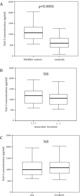
Serum galectin-3 concentration was increased in the cancer group. Median value of serum galectin-3 concentration was 1068 pg/ml (range 551-2028) in the cancer group vs. 584 pg/ml (range 259-1262) in controls (p!0.0005, Fig. 1A). We divided 43 blad-der cancer patients into 33 non muscle invasive group and 10 muscle invasive group, and examined these samples. There was no significant difference
Table 1. Patient’s clinical data
cancer group controls
n 43 24
mean age (range) 72 (45-83) 60 (28-85) sex ratio M/F 34/9 12/12 non muscle invasion 33 -muscle invasion 10
-G1 2
-G2 21
-in serum galect-in-3 concentration between the two groups (Fig. 1B). We also divided these 43 patients into 20 patients with G3 and 23 patients with G1 or G2. There was no apparent correlation in serum level of galectin-3 between the two groups (Fig. 1C).
Based on data from the patients with bladder cancer, receiver operating characteristic (ROC) curve was obtained. The ideal cutoff value of serum galectin-3 concentration was recommended to be 586 pg/ml. Using this cutoff value, the sensitivity and specific-ity were 97.7% and 54.2%, respectively.
Immunohistochemical staining of galectin-3 We selected one patient with high serum galectin-3 concentration, and studied immunohistochemical staining of galectin-3 in bladder cancer tissue and normal bladder tissue. The histopathological diag-nosis of selected bladder cancer tissue was UC, G3, pT3a pN0M0. Higher expression of galectin-3 was observed in bladder cancer tissue than in normal bladder tissue. The cytoplasm was stained strongly in bladder cancer tissue, but almost no cytoplasm was stained in normal tissue (Fig. 2). We performed immunostaining of galectin-3 to other three sam-ples (UC, G3, pT2b or pT3a pN0M0). We evaluated all bladder cancer tissue as strong, and all normal tissue as weak according to our criteria. There was no correlation between the level of serum galectin-3 and degree of staining. The pattern of staining was same as the above.
DISCUSSION
Galectin-3 is a member of the galectin gene fam-ily that is expressed at elevated levels in a variety of neoplastic cell types and has been associated with cell growth, cellular adhesion process, cell prolif-eration, transformation, metastasis, and apoptosis (14-20). Galectin-3 has both anti-apoptotic and pro-apoptotic function depending on cell’s differentia-tion status and tissue type (21). It was reported that A
B
C
Fig. 1. Analysis of serum galectin-3 concentration ; Serum galectin-3 concentration of the cancer group was statistically higher than that of controls (A). There was no apparent corre-lation in galectin-3 concentration between non muscle invasive group and muscle invasive group (B). No significant difference was seen in galectin-3 concentration between G3 and G1 or G2 by Mann-Whitney test (C).
A
A BB
Fig. 2. Immunohistochemical staining of galectin-3 ; Black arrow indicates nucleus and white arrow indicates cytoplasm. Higher expression of galectin-3 was observed in bladder cancer tissue (B) than in normal bladder tissue (A). The cytoplasm was stained strongly in bladder cancer tissue, but almost no cyto-plasm was stained in normal tissue (
!
400). We evaluated blad-der cancer tissue as strong, and normal tissue as weak accord-ing to our criteria.the Asp-Trp-Gly-Arg (NWGR) motif (3) and phos-phorylation (22) are critical for the anti-apoptotic function of galectin-3. Fukumori, et al . mentioned that secreted extracellular galectin-3 was related to pro-apoptotic function of galectin-3, but its mecha-nisms are quite complicated and there are many un-answerable questions (23).
The expression of galectin-3 is up-regulated in various types of cancer. Several reports have indi-cated its involvement in carcinogenesis (24, 25). One possible reason for this is the anti-apoptotic activity of galectin-3. Some reports have proposed mechanisms by which galectin-3 protects cells from apoptosis. Takenaka, et al. have shown that nuclear export of galectin-3 is important for its anti-apoptotic activity (26).
Galectin-3, which has been reported to be ex-pressed in the nucleus, in the cytoplasm and on the cell surface, can be secreted into the stroma (5). It has been shown to be up-regulated in some cancers such as thyroid carcinoma, hepatocellular carcinoma and lymphoma, and down-regulated in others in-cluding breast, uterine and pancreas cancer (27).
Expression pattern of galectin-3 is altered in many types of cancers. Therefore, several attempts to use galectin-3 expression as a diagnostic indicator are under development. Inohara, et al. demonstrated that expression of galectin-3 in fine needle aspirates could be a diagnostic marker for thyroid cancer (28). Aron, et al. mentioned that galectin-3 was strongly expressed in smears of papillary thyroid carcinoma. However, since it is also expressed in a variety of benign lesions, its role as a pre-surgical marker for differentiating benign from malignant thyroid nodules is limited (29). Nakamura, et al. reported that strong expression of galectin-3 in colorectal cancer correlated with cancer progression, liver metastasis, and poor prognosis for patients (30). Shimamura, et al. studied decreased expression of galectin-3 was associated with advanced stage, tumor de-differentiation, and metastasis in ductal adenocarcinoma of the pancreas (12).
The expression of galectin-3 is down-regulated in some of urological cancers, suggesting that galectin-3 has a tumor suppressive role in urologi-cal organs. The mechanism by which galectin-3 promotes cancer progression has been reported. However, little is known about tumor suppressive functions of galectin-3 (31).
In prostate cancer, Van den Brule, et al. have re-ported galectin-3 was usually not expressed or de-creased compared with the normal glands (32). In
renal cell carcinoma, it has been shown galectin-3 expression was significantly higher in low-grade conventional (clear cell) RCCs, indolent chromo-phobe RCCs (33, 34).
On the contrary, Cindolo, et al. have reported that increased galectin-3 mRNA expression com-pared to basal levels of normal bladder samples was observed in many bladder cancer samples with no apparent correlation with the clinico-pathological features such as stage and grade (13). We thought if the role of galectin-3 could be solved clinically, it was useful for diagnosis, prognostic prediction and selection of anticancer therapies. Thus we tried to measure the serum level of galectin-3 in bladder can-cer patients.
We used ELISA kit to measure serum galectin-3 concentration in patients with bladder cancer. To our knowledge, this is the first report using ELISA kit to measure serum galectin-3 concentration in pa-tients with bladder cancer. We demonstrated that serum galectin-3 concentration in bladder cancer pa-tients was statistically higher than normal control patients (p!0.0005).
However, there was no statistical difference in either histological grade or pathological stage with bladder cancer patients. Those results are consistent with these as Cindolo, et al. has reported in vitro.
In immunohistochemical staining of galectin-3, the expression of galectin-3 was apparently higher in bladder cancer tissue than in normal bladder tissue.
A standard method for diagnosis of bladder cancer is urine cytology and cystoscopy. However, cystoscopy is invasive and expensive. Several uri-nary markers for bladder cancer have been inves-tigated. Nuclear matrix protein-22 (NMP22), blad-der tumor antigen (BTA) and urine cytology are major urinary markers for diagnosis of bladder cancer. Sensitivity and specificity were previously reported with 85% and 91.3% for NMP22, 67% and 80.8% for BTA, 44% and 100% for urine cytology (35). However, Poulakis, et al. have demonstrated the specificity of urinary markers obviously became low with pyuria in cystitis and urolithiasis (36). Con-trary to urinary markers, serum level of galectin-3 is not affected by pyuria. For that reason we con-sider that the measurement of serum galectin-3 will be helpful to diagnosis of bladder cancer, if blad-der cancer is clinically suspected.
As the results of this study, the possibility that galectin-3 will be useful in diagnosis of bladder can-cer was suggested. However, there was no
signifi-cant difference in the stage and grade in our study. Further studies are needed to characterize the role of galectin-3 in bladder cancer. We will examine the level of urine galectin-3, and evaluate the utility through a combination of serum galectin-3 and urine markers including urine galectin-3. The combined use of these markers might be able to improve ac-curacy of diagnosis of bladder cancer.
In conclusion, the serum galectin-3 concentra-tion of the bladder cancer patients was statistically higher than that of controls. This result suggests that the measurement of serum galectin-3 concen-tration is helpful to diagnose bladder cancer.
REFERENCES
1. Oka N, Nakahara S, Takenaka Y, Fukumori T, Hogan V, Kanayama HO, Yanagawa T, Raz A : Galectin-3 inhibits tumor necrosis factor-related apoptosis-inducing ligand-induced apoptosis by activating Akt in human bladder carcinoma cells. Cancer Res 65 : 7546-7553, 2005
2. Vereecken P, Zouaoui Boudjeltia K, Debray C, Awada A, Legssyer I, Sales F, Petein M, Vanhaeverbeek M, Ghanem G, Heenen M : High serum galectin-3 in advanced melanoma : preliminary results. Clin Exp Dermatol 31 : 105-109, 2006
3. Akahani S, Nangia-Makker P, Inohara H, Kim HR, Raz A : Galectin-3 : a novel antiapoptotic molecule with a functional BH1 (NWGR) do-main of Bcl-2 family. Cancer Res 57 : 5272-5276, 1997
4. Takenaka Y, Fukumori T, Raz A : Galectin-3 and metastasis. Glycoconj J 19 : 543-549, 2004 5. Wang JL, Werner EA, Laing JG, Patterson
RJ : Nuclear and cytoplasmic localization of a lectin-ribonucleoprotein complex. Biochem Soc Trans 20 : 269-274, 1992
6. Lotan R, Ito H, Yasui W, Yokozaki H, Lotan D, Tahara E : Expression of a 31-kDa lactoside-binding lectin in normal human gastric mucosa and in primary and metastatic gastric carcino-mas. Int J Cancer 56 : 474-480, 1994
7. Hsu DK, Dowling CA, Jeng KC, Chen JT, Yang RY, Liu FT : Galect3 expression is in-duced in cirrhotic liver and hepatocellular car-cinoma. Int J Cancer 81 : 519-526, 1999 8. Xu XC, el-Naggar AK, Lotan R : Differential
ex-pression of galectin-1 and galectin-3 in thyroid tumors. Am J Pathol 147 : 815-822, 1995
9. Choufani G, Nagy N, Saussez S, Marchant H, Bisschop P, Burchert M, Danguy A, Louryan S, Salmon I, Gabius HJ, Kiss R, Hassid S : The levels of expression of galectin-1, galectin-3, and the Thomsen-Friedenreich antigen and their binding sites decrease as clinical aggres-siveness increases in head and neck cancers. Cancer 86 : 2353-2363, 1999
10. Schwarz G Jr, Remmelink M, Decaestecker C, Gielen I, Budel V, Burchert M, Darro F, Danguy A, Gabius HJ, Salmon I, Kiss R : Galectin fingerprinting in tumor diagnosis. Dif-ferential expression of galectin-3 and galectin-3 binding sites, but not galectin-1, in benign vs. malignant uterine smooth muscle tumors. Am J Clin Pathol 111 : 623-631, 1999
11. Castronovo V, Van Den Brûle FA, Jackers P, Clausse N, Liu FT, Gillet C, Sobel ME : De-creased expression of galectin-3 is associated with progression of human breast cancer. J Pa-thol 179 : 43-48, 1996
12. Shimamura T, Sakamoto M, Ino Y, Shimada K, Kosuge T, Sato Y, Tanaka K, Sekihara H, Hirohashi S : Clinicopathological significance of galectin-3 expression in ductal adenocarci-noma of the pancreas. Clin Cancer Res 8 : 2570-2575, 2002
13. Cindolo L, Benvenuto G, Salvatore P, Pero R, Salvatore G, Mirone V, Prezioso D, Altieri V, Bruni CB, Chiariotti L : Galectin-1 and galectin-3 expression in human bladder transitional cell carcinomas. Int J Cancer 84 : 39-43, 1999 14. Gong HC, Honjo Y, Nangia-Makker P, Hogan
V, Mazurak N, Bresalier RS, Raz A : The NH2
terminus of galectin-3 governs cellular compart-mentalization and functions in cancer cells. Cancer Res 59 : 6239-6245, 1999
15. Herrmann J, Turck CW, Atchison RE, Huflejt ME, Poulter L, Gitt MA, Burlingame AL, Barondes SH, Leffler H : Primary structure of the soluble lactose binding lectin L-29 from rat and dog and interaction of its noncollagenous praline-, glycine-, tyrosin-rich sequence with bacterial and tissue collagenase. J Biol Chem 268 : 26704-26711, 1993
16. Yang RY, Liu FT : Galectins in cell growth and apoptosis. Cell Mol Life Sci 60 : 267-276, 2003 17. Raz A, Lotan R : Endogenous
galactoside-binding lectins : a new class of functional tu-mor cell surface molecules related to metasta-sis. Cancer Metastasis Rev 6 : 433-452, 1987 18. Barondes SH, Cooper DN, Gitt MA, Leffler H :
Galectins. Structure and function of a large fam-ily of animal lectins. J Biol Chem 269 : 20807-20810, 1994
19. Perillo NL, Marcus ME, Baum LG : Galectins : versatile modulators of cell adhesion, cell pro-liferation, and cell death. J Mol Med 76 : 402-412, 1998
20. Rabinovich GA : Galectins : an evolutionarily conserved family of animal lectins with multi-functional properties ; a trip from the gene to clinical therapy. Cell Death Differ 6 : 711-721, 1996
21. Nakahara S, Oka N, Raz A : On the role of galectin-3 in cancer apoptosis. Apoptosis 10 : 267-275, 2005
22. Yoshii T, Fukumori T, Honjo Y, Inohara H, Kim HR, Raz A : Galectin-3 phosphorylation is required for its anti-apoptotic function and cell cycle arrest. J Biol Chem 277 : 6852-6857, 2002 23. Fukumori T, Takenaka Y, Yoshii T, Kim HR, Hogan V, Inohara H, Kagawa S, Raz A : CD29 and CD7 mediate galectin-3-induced type II T-cell apoptosis. Cancer Res 63 : 8302-8311, 2003 24. Takenaka Y, Inohara H, Yoshii T, Oshima K, Nakahara S, Akahani S, Honjo Y, Yamamoto Y, Raz A, Kubo T : Malignant transformation of thyroid follicular cells by galectin-3. Cancer Lett 195 : 111-119, 2003
25. Yoshii T, Inohara H, Takenaka Y, Honjo Y, Akahani S, Nomura T, Raz A, Kubo T:Galectin-3 maintains the transformed phenotype of thy-roid papillary carcinoma cells. Int J Oncol 18 : 787-792, 2001
26. Takenaka Y, Fukumori T, Yoshii T, Oka N, Inohara H, Kim HR, Bresalier RS, Raz A : Nu-clear export of phosphorylated galectin-3 regu-lates its antiapoptotic activity in response to che-motherapeutic drugs. Mol Cell Biol 24 : 4395-4406, 2004
27. Califice S, Castronovo V, Van Den Brule F : Galectin-3 and cancer. Int J Oncol 25 : 983-992, 2004
28. Inohara H, Honjo Y, Yoshii T, Akahani S, Yoshida J, Hattori K, Okamoto S, Sawada T, Raz A, Kubo T : Expression of galectin-3 in fine-needle aspirates as a diagnostic marker
differ-entiating benign from malignant thyroid neo-plasms. Cancer 85 : 2475-2484, 1999
29. Aron N, Kapila K, Verma K : Utility of galectin-3 expression in thyroid aspirates as a diagnostic marker in differentiating benign from malig-nant thyroid neoplasms. Indian J Pathol Mi-crobiol 49 : 376-380, 2006
30. Nakamura M, Inufusa H, Adachi T, Aga M, Kurimoto M, Nakatani Y, Wakano T, Nakajima A, Hida JI, Miyake M, Shindo K, Yasutomi M : Involvemant of galectin-3 expression in colorec-tal cancer progression and metastasis. Int J Oncol 15 : 143-148, 1999
31. Oka N, Takenaka Y, Raz A : Galectins and urological cancer. J Cell Biochem 91 : 118-124, 2004
32. van den Brûle FA, Waltregny D, Liu FT, Castronovo V : Alteration of the cytoplasmic/ nuclear expression pattern of galectin-3 corre-lates with prostate carcinoma progression. Int J Cancer 89 : 361-367, 2000
33. Francois C, van Velthoven R, De Lathouwer O, Moreno C, Peltier A, Kaltner H, Salmon I, Gabius HJ, Danguy A, Decaestecker C, Kiss R : Galectin-1 and galectin-3 binding pattern ex-pression in renal cell carcinomas. Am J Clin Pathol 112 : 194-203, 1999
34. Young AN, Amin MB, Moreno CS, Lim SD, Cohen C, Petros JA, Marshall FF, Neish AS : Expression profiling of renal epithelial neo-plasms : a method for tumor classification and discovery of diagnostic molecular markers. Am J Pathol 158 : 1639-1651, 2001
35. Eissa S, Swellam M, Sadek M, Mourad MS, El Ahmady O, Khalifa A : Comparative evalu-ation of the nuclear matrix protein, fibronectin, urinary bladder cancer antigen and voided urine cytology in the detection of bladder tu-mors. J Urol 168 : 465-469, 2002
36. Poulakis V, Witzsch U, De Vries R, Altmannsberger HM, Manyak MJ, Becht E : A comparison of urinary nuclear matrix protein-22 and bladder tumour antigen tests with voided urinary cy-tology in detecting and following bladder can-cer : the prognostic value of false-positive re-sults. BJU Int 88 : 692-701, 2001