TUMSAT-OACIS Repository - Tokyo University of Marine Science and Technology (東京海洋大学)
Research for determination factors of fish
life span
学位授与機関
東京水産大学
学位授与年度
2004
RESEARCH FOR DETERMINATION FACTORS
OF FISH LIFE SPAN
2004
GRADUATE SCHOOL OF FISHERIES
TOKYO UNIVERSITY OF FISHERIES
DOCTORAL COURSE OF AQUATIC BIOSCIENCES
= L'- ' -'='1"f A; ',*', t / _ i' ;' - 4 ...,
1'
-<
0049062
+ :
Reiko NAGASAKA
CONTENTS
ABSTRACT
ACKNOWLEDGEMENTS
CHAPTER I. fNTRODUCTIONAND GENERAL BACKGROUNDS
Section1.General background Section2。The pu荘)ose and briefsummaries ofthis researchCHAPTER IL OXIDATIVE STRESS OF CARP RED BLOOD CELLS
CHAPTER III. APOPTOSIS n“AYU BRAm AND LIVER
Section1.Elevated levels ofoxidative DNAdamage activate p53and caspases in brain ofayu with aging Section2。Enhanced oxidative damages and apoptosis in aging ayu liver DisucussionCHAPTER IV EFFECTS OF CALORIC RESTRICTION ON
POS㍗SPAWMNG DEATH OFAYU
CHAPTERVl ROLES OF LEPTlN IN POST−SPAWNING DEATH OF AYU
(PlθooglOSSμSα1∫’Vθ1’S)CHAPTER VL GENERAL DISCUSSION
REFERENCES
i
iv1
3
31 3554
57
70
81 83 110126
134
ABSTRACT
Some fish species show parental death shortly after their first spawning. The well-known
exanrples are ayu (Plecoglossus altivelis) which dies in only one year. Although the mechanisms for such a short life span are still unclear, there have been proposed some
hypotheses. Since it is shown clearly that ayu produced ROS higher than other species, it is
supposed that high ROS production strongly related in aging advances, resulting in
shortened life span. Homeostasis disturbances by maturation, debility for exhaustingenergy of spawning and decreasing of feeding activities during spawning and after
spawning are also considered to be factors which ayu dies in only one year. Along these hypotheses, this study dealed with carp as a model fish with long life span and ayu as a model fish with short life span and was performed for disclosing whether 1) influence ofthe oxidative stress on biomembrane, 2) apoptosis related factors, 3) caloric restriction, and/or 4) feeding activity would be relevance to short life span or not. Clarification of fish life span determination factors will also contribute to the stable fish eulture techniques including 'programmed' fish culture on the basis of mechanism elucidations.
This thesis is composed of five chapters. Chapter I deals with introduction and general diseussion. It was given for gaining insight into aging and senescence studies. The free
radical theory of aging, telomere theory of aging, progranuned cell death, p53 tumor
suppressor protein induced apoptosis and caloric restriction were reviewed
comprehensively. Regulatory peptides and control of food intake were also described.In Chapter II, it was investigated the influence of partial oxidative stress on penueability
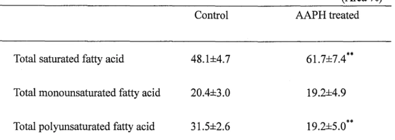
and fluidity of nucleated fish red blood cells for simulating nucleated somatic cells. Peroxide value indicating lipid hydroperoxide level in nucleated red blood cells of common carp (Cyprinus carpio) increased with increasing body size. It was detected that
oxidation of nucleated red blood cells led to the degraded PUFA compositions and
accelerated the permeability of calcein and ATP in the nucleated red blood cells restrictedly
oxidized with AAPH treatment. Using fluorescence probes, PC3P, it was found that
oxidative stress reduced the membrane fluidity of nucleated red blood cells. It was also
observed that AAPH had no significant influence on the osmotic fragility and
electrophoretic profiles of red blood cell proteins. These results suggest that partial
oxidative-stress, even if failure to fragment the membrane, may affect membrane
permeability of fish nucleated red blood cells for an important energy molecule, ATP. It is well known that ayu (Plecoglossus altivelis) die after spawning and the life span is
only one year. One possible cause is that enhanced oxidative stress might induce DNA
damage and subsequent DNA repair systems as phosphorylated p53 in ayu, Ieading to
apoptosis and relating to their short life span. Telomeres, the non-coding sequences at the ends of chromosomes, shortening of telomeres can induce cell cycle arrest and apoptosis.
Chapter 111 was, then, addressed to the p53 and its phosphorylation in ayu brain, the oxidative DNA damage by measuring the levels of 8-0HdG and the induction of apoptosis by measuring the levels of caspase-9/6, -3 with aging in brain and liver. It was also
investigated that age related changes in telomere length in the ayu. The findings indicate that oxidative stress activated caspase-9/6, -3 in brain and liver, and activated p53 through
the phosphorylation of p53 and p53 with aging in ayu brain. There was no significant change in telomere length through life span. It was suggested that the age-related of
apoptosis might be involved in increasing of DNA damage and mutations in brain and liver,
and could partially explain the short life span of ayu.
The effeets of caloric restriction on post-spawning death of ayu were investigated in
Chapter IV. Caloric restriction is the only established intervention that extends life span in mamlnals, insects and nematodes. One of the hypotheses suggested that most of the effects of CR on aging may be due to reduced oxidative stress at the cellular level. It was known that ayu produced ROS higher than other fish and that the 1ife span of ayu is only one year.
It was attempted to quantify age-associated changes of the degree of attenuation on
oxidative damage and honuonal homeostasis in CR. The oxidative DNA damage by
measuring the levels of 8-0HdG and the induction of apoptosis by measuring the levels of caspase-9/6, -3 with aging in brain and liver were surveyed. Changes in maj or sexual
honuones were also investigated. Caspase activities in brain and liver were reduced by CR,
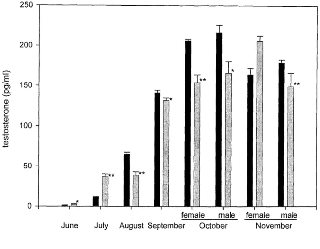
although CR was no influence to DNA damage level. Plasma testosterone levels of CR ayu
were significantly higher and progesterone and 1 7 -estradiol levels were lower than the control ayu. However, Iife span of ayu was not prolonged by CR. These results suggest that there would be factors detenuining life span of ayu other than CR and apoptosis.
Chapter V deals with roles of leptin in post-spawning death of ayu. It is well known that ayu dye after spawning and the life span is only one year. The deterntinants for such a short
life span are probably involved in spawDing and some accompanied changes in hormonal
homeostases. It is one of the accompanied changes that feeding activity of ayu decreases
during spawning and after spawning. Then, it was investigated the relationships among leptin and ghrelin, they are regulators for food intake, and other maj or honnones, 1 7 P-estradiol and prolactin. Leptin levels were significantly higher during spawning,
associated with decrease in appetite. Leptin levels were also synchronized with levels of 1 7 -estradiol and prolactin. Ghrelin levels were no significant difference. Therefore, one possible explanation for decrease in appetite during ayu spawning is that the alteration of 1 7 -estradiol homeostasis induced the secretion of leptin. The inability to reduce the leptin level into the basal after spawning would be in part responsible for a short life span
of ayu.
In conclusion, it was revealed that the mechanisms governing life span of ayu were through at least two pathways. One is apoptosis induced by oxidative stress with aging,
This pathway is probably, however, an alleyway, since CR could afford to down-regulate
apoptosis pathway but did not extend the life span of ayu. Another is decreasing appetite
during and after spawning induced by leptin in ayu. Reproduction induced physiological
ACKNOWLEDGMENTS
The successful completion of this dissertation was made possible by the help of many
persons.
First of all, I would like to express my appreciation to my adviser. Professor Nobuaki
Okamoto for his advice. I also gratefully acknowledge Associate Professor Hideki Ushio for his excellent guidance and encouragement throughout my research term. I owe deep
gratitude to Professor Hideo Fnkuda for his kind advice and for reviewing my dissertation.
I would like to extend my thanks to Associate Professor Toshiaki Ohshima and Professor
Shuichi Satoh for their useful suggestions sinee my Master 's course and for encouraging
me in the Doctor's course. My sincere gratefulness goes to Professor Takeshi Akita,
Professor Sonoe Muramatsu and Professor Shuichi Kitada for all kindness during my
study.
I sincerely wish to thank Mr. Hitoshi Kubota and all members at Tochigi Prefectural
Fisheries Experimental Station for their hospitality and help in conducting the feeding trial.
I am grateful to Associate Professor Kenj i Fukunaga (Kansai University), Mr. Makoto Furuhashi and Mr. Hiroaki Hirayama (Kyoto Marine High School) for their kind advice and help. Special thanks are given to Dr. Akiko Nagasaka (Yamanashi University), Ms.
Midori Nagasaka, Saki-Q chang and my lovely family for their kind advice during my
difficult times.I also wish to thank my colleagues in the Laboratory of Fish Physiology for their friendships during my research term. I would like to thank members of the Laboratory of Marine Biochemistry, Laboratory of Food Preservation, Laboratory of Fish Nutrition and
Fish Pathology for their kindness and help.
CHAPTER I
It is mentioned that the control system for maintaining the constancy ofthe homeostases of animals is always working. Although it is becoming clear that these are controlled by the
network of immunity systems, nervous systems and intemal secretion systems, the
regulation mechanisms in fish is hardly clear, and becomes one of the big research
sub j ects.
Now, the view of individual aging of animals has cornmon theory of "programmed"
aging. A progranuned theory connects shortening of the telomere by reduction of
telomerase activity to a cell life. The telomerase activities of cells of fish are, however, very high, and it is hard to explain in a programmed theory for fish aging.
The life span of fish is mainly prescribed by the grade of aging and debility like other
animals. This research is focused to the destruction of homeostasis with fish aging and aimed at dynamic understandings of interactions among oxidative stress, feeding action, aging, etc. The following sections will deal with comprehensive backgrounds associated
Section 1
The study of aging, by nature multidisciplinary has been characterized by a dizzying
variety of theories, a huge phenomenological literature, and the absence of firmly
established primary causes. The diverse life histories of animal species, which manifest
aging in very different ways, have been an obstacle to testing unified theories. For experimental gerontology to provide more than a catalog of age-related changes, it has
been necessary for biologists to define the alterations that are comrnon to most old cells,
tissues, and animals, simultaneously respecting that there is not a single phenomenon of
aging or a single cause. Many theories have been proposed to explain the basis of aging.
They have been classified into organ theories (immune or neuroendocrine), physiological theories (free radical, dross-1inking and waste-product accumulation) and genome-based
theories (somatic mutation, error theory and program theory) of aging.
1. THE FREE RADICAL THEORY OF AGING
1.1 Origins of the Free Radical Theory
In 1956, Denham Harman suggested that free radicals produced during aerobic
respiration cause cumulative oxidative damage, resulting in aging and death. He noted
parallels between the effects of aging and of ionizing radiation in both of them causing mutagenesis, cancer, and gross cellular damage (Haunan, 1 956). At that time, the presence of hydroxyl radical (・OH) in living matter had been just identified (Commoner et al., 1954). Harman ( 1 956), therefore, hypothesized that endogenous oxygen radical generation would occur in vivo, as a by-produet of enzymatic redox chemistry. He ventured that the enzymes involved would be those involved in the direct utilization of molecular oxygen, and that particularly those would contain iron. Finally, he hypothesized that traces of iron and other
metals would catalyze oxidative reactions in vivo and that peroxidative chain reactions would make progress, analogous to the principles of in vitro polymer chemistry. A11 of
these predictions hav e been conflrmed during the past 40 years.
The theory gained credibility through the identification in 1 969 of the enzyme
superoxide disrnutase (SOD) (McCord and Fridovich, 1969), which provided the first
compelling evidence of in vivo generation of superoxide auion (02 '), and from the
subsequent elueidation of elaborate antioxidant defenses (Yu, 1994). The use of SOD as a
tool to locate subcellular sites of 02 ' generation led to a realization that buttressed up the
free radical theory, namely, that mitochondria are a principal source of endogeneous ROS (Chance et al., 1979). Gerontologists had long observed that species with higher metabolie
rates have shorter maximum life span potential; they age faster. The accumulation of cardiac lipofuscin in the monkey related with its cumulative 02 consumption at a sexual
maturation stage (Nakano et al., 1989). The realization that energy consunrption by
mitochondria may result in 02 ' production linked the free radical theory with the rate of living theory irrevocably : a faster rate of respiration, associated with a greater generation of oxygen radicals, hastens aging.
1.2. Oxidants and evolutionary theories of aging
The intracellular generation of oxidants capable of limiting life span may appear paradoxical. It seems reasonable to expect that natural selection might have devised
aerobic cells that do not leak toxic by-products. Evolutionary biologists have contributed to
the free radical theory by suggesting why physiologically hannful generation of oxygen
radicals occurs. They have argued that natural selection favors genes that act to preserve
nongenu cells, a principle called antagonistic pleiotropy (Kirkwood, 1977, 1992;
Kirkwood and Rose, 1991; Williams and Nesse, 1991). The concept of antagonistic
pleiotropy stresses that reproductive success is principally a fiJnction of external factors.
With the exception of modern-day humans, individuals do not usually die of old age, but
are eaten, parasitized, or out-competed by others. Kirkwood and Cremer ( 1 982)
contributed a physiological perspective, expressed as the 'disposable soma theory' which
states that although it is theoretically possible to invest sufficiently in somatic maintenance
and repair to fend off harmful age changes, natural selection may favour a balanee that falls short of immortality and results in senescence. The limiting resources would take many forms and include energy and nutrition, DNA repair and replacement of・ defective
proteins, and response to cellular stress. The positive correlation between resistance to physiological stressors and life span in a range of marumalian species is consistent with the
disposable soma theory (Kapahi et al., 1999). The theory was proposed to account for somatic aging, but it can also apply to germ cells. Indeed, similar age changes and protective mechanisms might be expected in both germ and somatic cells, even if the
expression varies among specific cell types. In terms of natural selection, the tremendous
cost of death before reproductive age, the constantly compounding probability of death
from external threats, and the cost of failing to reproduce all ensure that selective pressure
is strongest at young ages. Any novel mutations that decrease oxidative damage frrst have to satisfy the criteria of youthful reproduction. In short, the selective pressure to compete effectively at an early age may guarantee a certain degree of 02 toxicity and work against the conservation ofthe soma in the long run.
1.3. Oxidants and the somatic mutation theory of aging
The somatic mutation theory holds that the accumulation of DNA mutations is
responsible for degenerative senescence (Bohr and Anson, 1995; Evans et al., 1995;
Miquel, 1992; Morley, 1995; Vijg and Gossen, 1993). In the case of cancer, which results
from both point mutations in oncogenes and the loss of tumor suppressor gene function
(often by deletion), the role of mutations are unquestionable (Ames et al., 1995). It remains
to be seen whether or not the argument is valid for nonproliferative senescence. For instance, whereas significant age-related increases in somatic mutations in a reporter transgene (lacZ) have been measured in a mitotie tissue of transgenic mice, no increase was detected in the largely postmitotic brain of the same animals (Dolle et al., 1997),
suggesting that neurodegeneration, at least, is unlikely to be the result of accumulated
somatie mutations in nuclear DNA. Moreover, the accllmulation of mutations in the liver
tissues was not dramatic, suggesting that mutagenesis may be of little functional
consequence to mitotic tissue as well (Warner and Johnson, 1 997). A compelling argument for the somatic mutation theory of aging was provided years ago through the discovery that DNA repair ability correlates with species-specific life span (Hart and Setlow, 1974) and
has been recently reconfiuned by Cortopassi and Wang (1996). However, they have also
noted that DNA repair, which is necessary for the prevention of tumorigenesis, is necessary but not sufficient for longevity. Ultimately, arguments about the physiological significance
of somatic mutations hinge on how disruptive a given mutational burden is to a cell or
animal; this is currently an unanswerable question.
In any case, it has been demonstrated in numerous studies with prokaryotes, yeast, and
manunalian cells that oxidants are mutagens, against which cells protect their genetic
material (Feig et al, 1 994; Grollman and Moriya, 1 993). Although it is not yet elear what
function of mutations can be attributed to oxidative damage, the identification and
characterization of defense genes against oxidative mutagenesis (Beckman and Ames,
1 997) and the development of in vivo mutagenesis assays (Martus et al., 1995) have finally opened up avenues for definitive experiments.
1.4. Oxidants and mitochondrial theories of aging
The mitochondrion has also long attracted attention as one of the cell's weak links, an
organelle whose dysfunction has profound negative pleiotropic effects (Luft, 1994). Mitochondria supply ATP and also sequester potentially toxic Ca2+, yet because of their
generation of 02 ' and H202, they are on the front lines of respiratory oxidative stress. The
idea that the mitochondrion is therefore uniquely vulnerable was embraced early on by
that oxidative damage to mitochondrial DNA (mtDNA) inpostmitotic cells would lead to mutations and blocks of replication, and consequently to mitochondrial dysfunction and physiological decline (Fleming et al., 1982; Miquel et al., 1980). This mtDNA mutation
hypothesis of aging, which incorporates free radicals, somatic mutations, and the central
role of mitochondria in homeostasis, is presently under intense scrutiny (Arnheim and Cortopassi, 1992; Bandy and Davison, 1 990; Bittles, 1992; Cortopassi and Liu, 1995;
Hagen et al., 1997; Muller, 1992; Nangley et al., 1992; Ozawa, 1995; Richter, 1992, 1995; Richter et al., 1988; Schapira and Cooper, 1992; Shigenaga et al., 1994; Wallace et al., 1995; Wei, 1992).
2. TELOMERE THEORY OF AGING
Mammalian cells have evolved complex mechanisms for regulating cellular life span.
Normal cells demonstrate a strictly limited growih potential and senescence after a defined nunrber of cell divisions. Cellular senescence is one of the bases of organismal aging. In contrast, tumor cells often exhibit an apparently unlimited proliferation potential and are
called "immortalized". Some investigators have proposed that the progressive shortening of the tips of the enkaryotic chromosomes, the telomeres, are important component of senescence and is involved in control of the cell cycle. The enzyme telomerase adds
TTAGGG repeated onto marumalian telomeres, which prevent their shortening.
Telomerase is ordinarily inactive in most somatic cells but can be detected in tumor cells.The activation of telomerase in malignant cancers seems to be an important step in
tumorigenesis, whereby the cell gains the ability of indefinite proliferation to becomeimmortal. As detailed information accumulates about how telomeres dynamics are
involved in the regulation of cell cycle events, one can expeet new opportunities forapplication to gerontology.
2.1. Telomere and telomerase
The extreme ends of enkaryotic chromosomes- the telomeres- are special structures that
provide protection from enzymatic end-degradation and maintain chromosome stability
(Dahse et al., 1997). Chromosomes with truncated telomere tips fuse with other
chromosome ends or become lost during cell division. Telomeres also play a role in
organization of the cellular nucleus by serving as attachment points to the nuclear matrix
(de Lange, 1992). Apart from providing stabilization and protection to the chromosomes,
telomeres carry out another important function in replicating cells: Their structure allows the end of linear DNA to be replicate completely.
Telomeres are composed of a DNA component and multiple protein components
(Graeber, 1 996). The telomeric DNA consists of noncoding tandemly repeated sequences,
with the exact repeat sequence varying from one species to the other. In humans and other vertebrates, the repeat unit is the hexanucleotide TTAGGG (5 ' ->3' direction). Although telomeres are generally considered to be localized structures at the ends of chromosomes, such sequences are also being identified at internal positions in chromosomes (Katinka and Bourgain, 1 992). The length oftelomeres also varies among different species. Human have
telomeres 8-14 kilobasepairs (kbp) Iong, whereas the mean telomeric repeat lengths in
some ciliates are as little as 36 bp, and those in mice may be as much as 1 50 kbp (Rao,
1 996). In human chromosomes, telomeres are adj oined centromerically by a subtelomeric region consisting of degenerated telomeric DNA sequences and unique repeats (Brown et
al., 1990)
A11 chromosomes lose a small amount of telomeric DNA during each cell division, a
natural eonsequence of the nature of the cellular DNA replication machinery. DNA
polymerases replicate only in a 5 ' ->3' direction by extending exist polynucleotide chains.
The mechanism of DNA replication differs for the leading and the lagging DNA strands,
The leading strand is replicated continually. To replicate the lagging strand, DNA
polymerization starts from several RNA primers, which are elongated to create DNA
fragments, termed Okazaki fragments. These RNA primers are finally degraded and
replaced by DNA sequences. Removal of the terminal RNA primer on the lagging strandleaves a gap that ordinary is filled in by extension of the next Okazaki fragment. Because there is no template for the last Okazaki fragment beyond the 5 ' end of the chromosome,
one strand cannot be synthesized to its very end. This reduction of chromosomal DNA at
the 3 ' ends during multiple cell cycles.
The loss of genomic sequences at each replication cycle can be compensated by addition of terminal sequences through various mechanisms: e.g., in yeast by recombination events
(Lundblad and Blackburn, 1993) or Drosophila by transposition (Biessmann and Mason, 1 992). Moreover, organisms possess the ability to transfer species-specific tenninal
sequences onto DNA: Shampay et al. ( 1 984) demonstrated that telomeric DNA from
Tetrahymena introduced into yeast became elongated with yeast telomeric sequences. Thecrucial experiments came from Greider and Blackburn (1985, 1987), who detected in
Tetrahymena extracts an activity that added telomeric repeats to single-stranded telomeric DNA oligonucleotide primers; they also' found that this process is inactivated by treating
the extract with a RNA-degrading eazyme. Therefore, this RNA-dependent activity, named terminal transferase or telomerase, was found to be a ribonucleoprotein complex that utilizes sequences of its own RNA component as a template for the de novo synthesis of
telomeric DNA sequences. Both RNA and several protein components of telomerase are
and Gottschling, 1994).
Although the bulk of telomeric DNA is double stranded, the extreme terminus is a
single-stranded G-rich 3 ' overhang that serves as a template for elongation and forms a telomeric 'T-100p'. This loop is stabilized by certain telomere-binding proteins, notably TRF I and TRF2 (Zakian, 1 996). The functions of telomeres appear to include protection of chromosomes from illegitimate fusion, the localization of chromosomes in the nucleus and
the selective silencing of proximal subtelomeric genes (Greider, 1 994). The telomeric repeat sequences are added on by the enzyme telomerase (Greider and Blackburn, 1985;
Yu et al., 1990), which present compensates for the loss of DNA from the end of
chromosomes due to ineomplete replication.In human, telomeres are up to 20 kb in length (Brown, 1989). In contrast, rodent
telomeres have been reported to be heterogeneous in length (Zijlmans et al., 1997). Mus musculus has been reported to have telomeres up to 1 50 kb in size (Prowse and Greider,1 995). Mus spretus, however, has telomeres with similar length to humans (up to 3 O kb in
size) (Zijlmans et al., 1997), whereas rat telomere length ranges from 20 to 100 kb
(Golubovskaya et al., 1999; Jennings et al., 1999).
In humans, both in vivo and in vitro, telomere shortening appears to be a maj or
component of cell senescence and aging (Campisi et al., 1996; Harley, 1997). Telomereshave been reported to shorten during post-natal development and aging in liver (Aikata et
al., 2000; Takubo and Kaminishi, 2001), kidney (Melk et al., 2000) and lymphocyies
(Benetos et al., 2001). However, this is less apparent in mice because of the very longtelomeres (30-150 kb). Telomere shortening has been extensively studied in mice,
especially in telomerase-deficient knockout mice (Artandi and DePinho, 2000; Blasco etal., 1 999; Herrera et al., 1 999). Moreover, it was reported that relationships between kidney telomere shortening and longevity in the rat (Jeunings et al., 1 999).
2.2. Telomere theory of aging in mammals
In 1 998, after endless efforts to get to the bottom of an intriguing mechanism of original aging, it became elear at least in the rough. Bodnar et al. (1998) has given a brilliant
evidence for the hypothesis. According to the hypothesis, telomere shortening in every
subsequent cell division is responsible for the limited cell proliferation in culture (the so called 'Hayflick's limit' found 40 years ago by Hayflick and Moorhead, 1 961). It was quite natural to attribute organismal aging to th_e same cause. The most complicated problems of cellular and organismal aging have got the following explanation. Shorter telomeres, found in cells of patients with inherited premature aging, progerias (A11SOpp et al., 1 992) were consistent with the sharply restricted Hayflick's limit in these cells nd provided a distinct evidence for an existing correlation between agings of an organism with that of its cells,
Discovery of telomerase activity in malignant tumor cells explained the mechanism of
tumorigenesis. The same activity in gametes clarified why our children always started their aging from zero level and not from the level reached by the parents (by its cell) during
conception. Therefore, it was logical to consider organismal aging to be a result of
telomere shortening and limited cell proliferation.
However, some facts instantly appeared which did not go in this line of telomere theory
of aging. First, proliferative capacity of human cells appeared to demonstrate a very insignificant, if any, decrease with age (Francheschi et al., 1999). Moreover, skin
fibroblasts of human at very old age never exllaust their Hayflick's limit (Cristofalo et al.,
1 998). Additionally, some other contradictions against the telomere theory of aging have
been intensity discussed earlier.
In 1998, Hayflick proposed a new hypothesis, according to which telomere shortening,
leading to the loss of replicative capacity, determined only the species life span, whereas
aging itself was caused by the aceumulation of some cell damages, as it had been
suggested earlier by Orgel ( 1 973) though these damages still need to be clarified.
Dr. Hayflick's hypothesis appears to be a misconception. First, the theory is at variance with the following observations.
1 . The Hutchinson-Gilford progeria patients demonstrate the real accelerated aging and
not a simple reduetion in the life span (AlISOpp et al., 1992). Similar phenomena are
observed in the other forms of hereditary premature aging-Werner 's syndrome (Wyllie
et al., 2000).
2. Telomeres examined in cells of people extremely old at age never reach the critical
length. Blocking nonual cellular proliferation (Mondello et al., 1999).
Nevertheless, all old people die at an age nearing hundred years, and their telomeres by
no means are exhausted. Thus, organisms die precisely because of aging, though they
never live till all their cells stop proliferating.
It was assumpted that aging and consequent death need not necessarily reach Hayflick's limit in all tissues of an organism. Exhausted proliferative potential of cells in some areas
of organ tissue might be sufficient to promote one of the age-dependent diseases.
Combination ofthese disorders, gradually increasing with age, is aging. At this point, again there is a contradiction with Dr. Hayflick's new hypothesis.
Developing his idea, Hayflick ( 1 998) considers that it is necessary to distinguish aging from disease, in other words, to distinguish diseases from age-related changes and try to
pick out pure aging. Such attempts seem unpromising in principle, because age-related
diseases, to the common person are aging. Nobody dies just because of age.
Telomere shortening in certain human tissues might promote disorders such as essential hypertension, non-insulin-dependent diabetes mellitus, atherosclerosis, and cancer (Aviv
malignancies, compared with the surrounding tissues, (Morin, 1997). Zeichner et al. (1999)
found that accelerated cell division in children and hence higher rate of telomere
shortening may provide an explanation for a more rapid progression to acquired
inrmunodeficiency syndrome in infected infants.
On the other hand, Omura et al. (1998) has shown that an average telomere length
decrease with the advance of age in cells of all human tissues, except for heart, brain, retina and sex glands. It also appears that chronic degenerative diseases are accompanied
by unusual low telomeres. It is noteworthy that the exceptions mentioned above might be
well explained within the hypothesis suggesting irregular telomere shortening in different tissues of an organism. The absence of telomere shortening in gerrn cells is, apparently, because of telomerase activity in these cells. Brain neurons stop dividing at the time of
birth although significant telomere shortening is not observed, which contradicts the
proposed idea at first sight. The point is that cells reach their proliferative limit irregularly.
and does not guarantee the rapid and full cessation of mitoses in the tissue. Meanwhile, brain cells need rapid and full cessation, because links between neurons must be settled from the time of birth, and if neurons continue to divide as other cells, these links will be surely destroyed. Therefore, it is the brain than j ust by telomere shortening, because the cessation of mitotic activity is probably provided by some additional and more effective
mechanisms. The retina may operate in the same mechanism.
Thus, there are many reasons to believe that the telomere theory of aging is in principle correet. A11 the above difficulties may easily find their explanation with regard to the phenomenon of uneven telomere shortening in different tissues and organs of the organism. Francheschi et al. ( 1 999) truly claimed that any living organism might be regarded as a
mosaic of cells with different replicative histories and potentials. According to these
authors, this particular fact throws doubt on the validity of the telomere theory of aging. By contrast, it is this fact that may explain all the apparent misunderstandings and difficulties
within the frames ofthe telomere theory.
2.3. Telomere theory ofaging in teleosts
In fish, high telomerase activity has been deteeted in several nonnal organs of the rainbow trout Oncorhynchus mykiss (Klapper et al., 1998). Telomerase activity of the
normal organs has been detected in both fry and adult fish, being I O - I OO-fold higher than
that in the human tumor cell line L-428. In contrast, no telomerase activity has been detected in the differentiated organs of manunals. Greider (1998) has described the
correlation between telomerase activity and the proliferation potential of cells. In general, rainbow trout grow continuously throughout their life and, therefore, the high telomerase activity detected in their normal organs is postulated to lead to cell proliferation and organ
growih. In previous investigation (Yoda et al., 2002), relative telomerase activity per cell in
eyed embryos of rainbow trout was 19.3 - 50.7-fold higher than in Hela cells (a human
cervical carcinoma cell line), whieh are well known to express a high level of telomerase activity (Morin, 1 989). Therefore, it is assumpted that aging and consequent death of fish
need not to reach Hayflick's limit in all tissues of an organism.
3. PROGRAMMED CELL DEATH
Cells have a built-in cell death program, apoptosis (programmed cell death), which protects the organism by removing potentially damaged cells and unnecessary cells after
drfferentiation. Apoptosis is induced in a wide range of physiological settings that are
regulated by cell growih and differentiation in normal biological processes and in
pathogenesis in vertebrates (Cohen et al., 1992; E1lis et al., 1991 ; Fernandes-Alnemri et al.,
1994; Jacobson et al., 1997; Nicholson and Thornberry, 1997; Steller, 1995).
3.1. Deftningfeatures ofprogrammed cell death
Despite the tremendous impact of researeh in apoptosis upon the understanding both of cellular and molecular mechanisms of cell demise, as well as mechanisms of degenerative diseases, the confusion between apoptosis and programmed cell death has been somewhat obscured. Regardless of whether this paradox is attributable either to disconnection of modern science from its philosophical foundations (Sloviter, 2002) or to a more trivial
neglect of classical papers (Lockshin and Zakeri, 2001), it is likely that progress in the
identification and understanding of nonapoptotic forms of progranuned cell death may
have been unnecessarily delayed.
Indeed, well before the upsurge in the understanding of mechanisms of apoptosis, a clear
warning had been issued to avoid confusion between the fonn of cell death called
apoptosis, and the concept of programmed cell death as a sequence of events, but notnecessarily those that led to the morphology of apoptosis. However, the modern seience is recently identifying apoptosis as the programmed cell death. Therefore, apoptosis is used
as the same term of the prograrumed cell death in this thesis.
3.2. Multiple mechanisws of apoptosis
Prograrnmed cell death with apoptotic morphology can be triggered by several stimuli,
including intracellular stress and receptor-mediated signaling. These signals feed into an
2000), the mechanisms of which have mainly been traced to the activity of the caspase
fanrily of cysteine-proteases (Cryns and Yuan, 1998; Yuan et al., 1993; Zhivotovsky et al., 1 997). Caspase-mediated apoptotic cell death has been extensively reviewed, e.g. Green,
2000; Hengartner, 2000; Leist and Jaattela, 2001; Martin, 2002; Ravagnan et al., 2002; Wajant, 2002. The caspases are synthesized as zymogens and upstream signals convert
these precursors into mature proteases. Inhibitor caspases; caspase-1, -2, -4, -5, -8, -9, -10
and -14 are activated via oligomerization-induced autoprocessing (Butt et al., 1998; Li et
al., 1997; Martin et al., 1998; Muzio et al., 1996; Srinivasula et al., 1998; Yang et al., 1998),
while effector caspases; caspase-3, -6 and -7 are aetivated by other proteases, including initiator caspases and granzyme B. Proteolytic cleavage of cellular substrates by effector caspases largely determines the features of apoptotic cell death (Green, 1998; Liu, et al.,
1998; Sakahira et al., 1998; Stroh and Schulze-Osthoff, 1998; Wolf and Green, 1999;
Zhang et al., 1998).
Three major pathways have been identified according to their initiator caspase: the death
receptor pathway involving caspase-8 (Medema et al., 1 997), the endoplasmic reticulum stress pathway attributed to activation of caspase-12 (Nakagawa et al., 2000), and the
mitochondrial pathway, in which various signals can trigger the release of harmful proteins
by mitochondria into the cytoplasm, Ieading to activation of caspase-9 and down-streanl cleavage of caspase-3, -7 or -6 (Green and Reed, 1998; Grutter, 2000; Li et al., 1997,
1998a; Luo et al., 1998).
Although caspase-3 is widely involved in the execution of apoptosis (Stennicke et al., 2002), its effector functions may be dispensable for apoptotic-1ike cell death (Kuida et al.,
1996; Miyashita et al., 1998). The use of either pharmacological inhibitors or knockout
animals further showed that cells could trigger alternative mechanisms of eell demise. For
example, sympathetic and dorsal root ganglion neurons deprived of nerve growih factor (NGF) die in a caspase-2-dependent manner, but the same neurons derived from caspase-2 knockout mice still die following nerve growih factor deprivation. The death depends on
activation of caspase-9, which does not occur in wild-type mice (Troy et al., 2001). Thus,
rather than a single linear mechanism, alternative caspase-mediated pathways may be
activated for apoptotic cell death, depending on whether a preferential caspase is blocked. It is likely that the network of intrinsic regulatory pathways that impinge upon the activity of caspases, such as the inhibitors of apoptosis (IAPs) and IAP-binding proteins (Salvesen and Duckett, 2002), may regulate the choice between alternative pathways in normal cells, dependent on metabolic state, stage of differentiation and other conditions.
In addition, caspase inhibition fails to block programmed cell death with apoptotic morphology in several experimental models (Assefa et al., 2000; Carmody and Cotter,
2000; Lorenzo et al., 1999; Mateo et al,, 1999; Mathiasen et al. 1999). For example, the ultrastructural features of apoptosis inducing factor (AIF)-induced cell death represent an
example of a slight variation from the standard pattern of apoptosis morphology, which
appears to be independent of caspase activation (Arnoult et al., 2003; Joza et al., 2001). Cell death pathways independent of caspase activation have been described, for exarnple,
even in some fonns of cell death induced either by the Bcl-family protein Bax
(Jtrgensmeier et al., 1998), as well as I cell death involving the activation of other
proteases, such as calpain (Squier et al., 1994), proteasome (Hirsch et al., 1998) and serine
proteases.
Recent reports shows that the serine protease Om HtrA2 is a mitochondrial direct
X-chromosome-linked inhibitor of apoptosis protein (XIAP)-binding protein, which is released from mitochondria upon induction of apoptosis together with cyiochrome c andSmac/Diablo (Hegde et al., 2002; Martins, 2002; Martins et al., 2002), and its release ean be inhibited by Bcl-2 (van Loo et al., 2002). These reports suggest that in some cases there may be a cooperative action between serine proteases and caspases in the execution of cell
death.
The previous studies show that the elassically defined apoptotic morphology can be
achieved either by activation of caspases, or through the mediation of other fanrilies of proteases, although the exact cyiological features of cell demise may vary slightly among these various forms of apoptosis.
3.3. Apoptotic pathway in fish
The charaeterization of genes that are involved in apoptosis has been pursued intensively,
and has led to the identification of several classes of genes, the Bcl-2 family,
apoptosis-inducing factor Bax, and the caspase family. Many genes with homology to
marnmalian apoptosis regulators have been identified in zebrafish DNA databases (Inohara
and Nunez, 2000), suggesting that most apoptotic pathways are evolutionally well
conserved between fish and higher vertebrates.
3.3. 1. The caspase family genes
Cell death genes were frrst identified in the nematode Caenorhabditis elegans. The ced-3 gene encodes a cysteine protease that has a key role in the cell death-signaling
pathway (Hengartner and Horvitz, 1994; Yuan et al., 1993). Vertebrate cells also possess several cysteine proteases belonging to the caspase fanrily; which are homologous to ced-3
cysteine protease (Alnernri et al,, 1996). Caspase is produced as an inaetive precursor
composed of four distinct dornains: the prodomain, Iarge subunit, and small subunit, and a
linker region between the two subunits flanked by aspartic residues (Nicholson and
subunits, resulting in removal of the prodomain and linker region and the large and small
subunits form an active mature enzyme (Nicholson and Thornbeny. 1 997). X-ray crystal structural analysis of caspase- I and caspase-3 revealed that mature caspase forms a
tetramer with two catalyiic sited that interacts via the small subunit (Rotonda et al., 1 996;
Walker et al., 1994; Wilson et al., 1994). Multiple forms of caspase have been found in other vertebrates, e.g. 12 type in humans (Nicholson et al., 1995; Yaoita and Nakajima, 1997), 10 type in mice (Hu et al., 1998; Kumar et al., 1994; Van de Craen et al., 1997; Wang et al., 1996), and eight types in X Iaevis (Kumar, 1999; Nakajima et al., 2000).
Cascade reactions of proteolyiic processing of caspases induce apoptosis. Class I caspases, such as caspase-2, -8, -9 and -10, promote the upstream part of the cascade reaction via
N-terminal prodomains, bound to specific death adaptor molecules (Colussi and Kumar, 1999; Cryns and Yuan, 1998). Class 11 caspases with short N-terminal prodomains, e.g.
caspase-3, -6 and -7, act as effectors caspase in proapoptotic signaling from the caspase
cascade to cell death by porteolyiic processing of proteins, such as the inhibitor of caspase-activated DNase (Sakahira et al., 1998), poly-ADP-ribose polymerase (Earnshaw
et al., 1999), and protein kinase C6 (Emoto et al., 1995). In fish, caspase-3 was recently cloned and characterized (Yabu et al., 2001).
The full-length CDNA sequence of zebrafish caspase was isolated from CDNA Iibrary of zabrafish 12-h embryos (Yabu et al., 2001). This clone had an 846 bp ORF encoding a
protein of 282 amino acids with a predicted molecular mass of 3 1 .5 kDa. The amino acid sequence identities of the zebrafish caspase-3 with chicken, hamster, human, rat, mouse, and X Iaevis caspase-3 were 64, 62, 62, 62, 61 and 580/0, respectively. According to the X-ray crystal structure of human caspase-3 (Rotonda et al., 1996), Cys-166, His-124, and Gly-125 in the catalyiic center, and Arg-97, Gln-164, Arg-243 and Ser-256 Iocated in the
binding pocket in the S I subsite, are conserved in the zebrafish caspase-3. The
pentapeptide motif QAC (R/Q/G) G around the active center Cys-166 is conserved in
zebrafish caspase-3 (Yabu et al., 2001).
A recombinant zebrafish caspase-3 Iacking the prodomain showed high activity toward
the mammalian caspase-3 and -7 substrate, AC-DEVD-MCA. However, the enzyme had
only low activity against caspase - I substrate AC-YVAD-MCA, caspase-6 and -8 substrateAC-IETD-MCA, and caspase -9 substrate AC-LEHD-MCA. Therefore, zebrafish caspase-3
has strict substrate specificity, which is similar to that of known members of the caspase-3 subfamily, such as human caspase-3 and caspase-7 (Thornberry et al., 1997).
By homology analysis using current nucleotide databases of zebrafish and Taklfugu rubripes, several fish genes encoding caspases are available (Inohara and Nunez, 2000). Functional assays by overexpression of zebrafish caspase-3 in fathead minnow tailbud cells and zebrafish embryo (Yabu et al., 2001) and zebrafish caspy and caspy2 in human
3.3.2. Expression ofcaspase-3 mRNA infish
Zebrafish caspase-3 transcripts were expressed at all developmental stages examined by
Northern blotting (Yabu et al., 2001). At the 4- and 1000- cell stages, high levels of
caspase-3 mRNA were present in fertilized eggs as a maternal factor. Furthermore,
caspase-3 mRNA was expressed in the shield, I -somite, pharyngula, and hatching periods,
which coincided with zygotic gene expression after gastrulation. In situ hybridization
demonstrated that caspase-3 mRNA was expressed throughout the embryo at every
developmental stage. In the pharyngula period, caspase-3 mRNA is present at higher levelsin the pectoral fin bud, otic vesicle, and hindbrain.
When caspase-3 was overexpressed by introducting its CDNA into fish cultured cells and
embryos, extensive apoptosis and ceramide generation were induced (Yabu et al., 2001).
This suggests that the tissue-specific, developmental expression patterns of the caspase-3
gene regulate the spatial and temporal distribution of apoptotic cells. In mammals,
caspase-3-knockout mice are born infrequently, die after only a few weeks, and have brain defects (Colussi and Kumar, 1999; Kuida et al., 1996; Woo et al., 1998). Therefore, both
mammalian and fish caspase-3 may have important functions modulating a proapoptosic
signal during development.
3.3.3. Death receptors
Plasma membrane death receptors, belonging to the tumor necrosis factor (TNF) family,
have been cloned in fish (Hirono et al., 2000). TNF receptor containing an intracellular death domain is assoeiated with cellular infrarnmatory and irumune responses in mammals. Interaction of extracellular ligands, such as Fas ligand and TNFoe, to death receptors is considered to induce apoptosis by activation of caspase-8 signaling. When a ligand binds
to death receptors, the receptor-specific adapter protein Fas-associated death domain
(FADD) is recruited, and then '*aspase-8 is activated by autolyiic processing. Activated
caspase-8 is known to promote the apoptotic signal by directly cleaving and activating downstream caspases. In the caspase-8-knockout mouse, caspase-8 is found to be required
for killing induced by the death receptors Fas, tumor necrosis factor receptor I and death
receptor 3 (Varfolomeev et al., 1998; Yeh et al., 1 998). The heart muscle and fewer hematopoetic progenitor cells suggest that the FADD/caspase-8 pathway is required for
growih and developmental roles for this death receptor pathway remain to be identified in zebrafish embryos,
Pro- and anti-apoptotic members of the Bcl-2 family (ced-9 homologs) regulate
mitochondrial participation in cell death (Bernardi et al., 2001; Gottlieb, 2001). Current models are proposed that the release of cyiochrome c from mitochondria triggers activation
of caspase-9 in a complex with dATP and Apaf-1 (Yoshida et al., 1 998). Activated
caspase-9 then activates further downstream caspases. Bcl-2 family proteins, such as ZfMcl-10e (Chen et al., 2000). Bcl-XL (Chen et al., 2001). MCL-1 (Hong et al., 1999), have been cloned in zebrafish. They are expressed in oocyies and early embryos (Chen etal., 2000, 2001 ; Hong et al., 1999). In contrast, ced-4 homolog Bax triggers a mitonondrial
proapoptotic pathway by promotion of mitochondrial release of cytochrome c (Bemardi et
al., 2001; Gottieb, 2001). Functional analysis of the Bcl-2 and Bax families is required to establish experimental models regulating chemical and oxidative stress responses.
4. p53 TUMOR SUPPRESSOR PROTEIN INDUCED APOPTOSIS
Mutation in the p53 tumor suppressor gene occur in about 50 o/o Of all hurnan tumors,
making it the most frequent target for genetic alterations in cancer (Agarwal et al., 1 998;
Almog and Rotter, 1998; Hansen and Oren, 1 997; Levine, 1997; Prives and Hall, 1999). Such mutations probably facilitate carcinogenesis primary through abrogating the tumor suppressor activities of the wild type p53 protein, although at least some forms of
tumor-associated mutant p53 proteins may also contribute overt oncogenic activities, gain
of function. Excessive wild type p53 can reduce cancer incidence through elimination of cancer-prone cells from the replicative pool. However, such effects might become very
undesirable if occurring in a normal, unperturbed cell. p53 activity must be, therefore, kept
under tight control, being unleashed only when a cell accumulates lesions the may
otherwise drive it into a cancerous state.
4.1. p53-activating signals
Under normal conditions, p53 is most probably latent. Consequently, it does not
interfere with cell cycle progression and cell survival. Moreover, p53 knock-out mice appear in most cases to undergo proper development and maturation (Donehower et al.,1 992), suggesting that p53 is not essential for the normal performance of cells within the
body. However, a variety of conditions can lead to rapid induction of p53 activity. The corumon denominator of these conditions is that they represent various types of stress, which are likely to favor the emergenee of cancer-bound cells. Such conditions include
as well as damage to components genetic material (e,g, the mitotic spindle (Cross et al., 1 995)), ribonucleotide depletion (Linke et al., 1996), hypoxia (Graeber et al., 1996), heat shock (Ohnishi et al., 1996), and exposure to nitric oxide (NO) (Forrester et al., 1996).
Accumulation of genomic aberrations is a key carcinogenic mechanism; the rapid
induction of p53 activity in response to genomic damage thus serves to ensure that cells carrying such damage are effectively taken care of. Furthermore, p53 may also contribute, directly or indirectly, to particular DNA repair processes (Offer et al., 1999; Smith et al., 1 995). The pivotal role of p53 in maintaining genomic integrity has eamed it the guardian ofthe genome (Lane, 1992). In addition, p53 activity is triggered by a variety ofoncogenic
proteins, including Myc. Ras, adenovirus EIA, and p-catenin (Damalas et al., 1999;
Debbas and White, 1993; Hermeking and Eick, 1994; Serrano et al., 1 997), providing adirect link between oncogenie processes and the tumor suppressor action of p53.
4.2. Regulation ofp53 gene expression
Induction of the p53 response upon stress occurs largely through alteration in the p53 protein. Changes in the rate of transcription of the p53 gene play a minor role, if any, in such induction. Consequently, the transcriptional regulation of p53 gene has received very little attention during recent years. This need not imply that the regulation of p53 gene expression is totally irrelevant. In fact, it was observed long ago that p53 mRNA Ievels
rised substantially upon serum stimulation (Reich and Levine, 1 984). This rise may be because of the presence of binding sites for serum-induced factors in the p53 promotor
(Ginsberg et al., 1990) as well as to the ability ofthe p53 gene to bind the c-Myc (Reisman et al., 1 993). The induction of an anti-proliferative gene, p53, by serum and growih factors
may at first glance seem paradoxieal. Cell undergoing DNA replication and extensive proliferation are, however, at higher risk of acquiring DNA damage and giving rise to multiple cancer-prone progeny than quiescent cells. Without DNA damage or other stress, p53 remains latent and once those conditions emerge call for a p53 response, the high
levels of p53 mRNA ensure that such a response will be rapid and effective.
4.3. Activation of p53 by post-transcriptional mechanislns
Exposure of cell to p53-activating signals can lead within a relatively short time to a
marked elevation in p53 protein. To some extent, this can be achieved by increased
translation of the p53 mRNA, probably involving relief of a translational repression mechanism operating through the 3'-untranslated region of this mRNA (Fu et al., 1996).There also exists evidence that p53 itself can inhibit p53 synthesis through binding to its own mRNA (Fontoura et al., 1997; Mosner et al., 1995). Yet, it is generally accepted that
the accumulation of active p53 in response to stress occurs mainly through
post-transcriptional mechanisms. Pivotal is the increase in the protein half-1ife of p53, since p53 is usually a very labile protein, turning over with a half-1ife sometimes as short as a few minutes (Rogel et al., 1985). In response to DNA damage and other type of stress,p53 was markedly stabilized (Kastan et al., 1991; Maltzman and Czyzyk, 1984). A rapid increase in p53 concentration without a need for de novo transcription is particularly
advantageous in cells with severely damaged genomes. In addition, there is most probably
a qualitative conversion of p53 from latent to active fonn. The best documented change
concerns the sequence-specific DNA binding activity of p53. p53 operates as a
gene-specific transcriptional activator, which relies on its ability to bind defined sequenceelements within target genes (Agarwal et al., 1998; Almog and Rotter, 1998; Hansen and
Oren, 1997; Levine 1997; Prives and Hall, 1999). The sequence-specific DNA binding
activity of p53 is subj ect to constitutive negative regulation, primary through its inhibitory
C-teuninal domain (Bayle et al., 1995; Hupp et al., 1992; Wolkowicz et al., 1995). Relief of this inhibition upon exposure to stress results in increased DNA binding (Gu and Roeder,
1997; Hupp and Lane, 1995; Waterman et al., 1998) and consequently increased
biochemical and biological activity. The transcriptional activity of p53 may also be inducedby changes in other regions, e,g. modifications within its N-tenuinal transactivation
domain, enabling a more efficient recruitment of eomponents of the transcription
machinery (Lanrbert et al., 1998). Finally, p53 activation may also involve a ehange insubcellular localization; whereas latent p53 may often be cyioplasmic, at least during part
ofthe cell cycle (Shaulsky et al., 1990), exposure to stress results in its accumulation in the nucleus, where it is expected to exert its biochemical activities.
4.4. Thep53-Mdm2 Ioop
A key player in the regulation of p53 is the Mdm2 protein. Mdm2 is the product of an
oncogene, whose excess activity facilitates several types of human cancer (Lozano et al.,
1998; Freedman et al., 1999; Juven-Gershon and Oren, ' 1999). Mdm2 exhibits a unique
relationship with p53. On the one hand, the Mdm2 protein binds to p53 and inactivates it
(Chen et al., 1996; Haupt et al., 1996; Momand et al., 1992). The binding occurs right within the p53 transactivation domain, interfering with recruitment of basal to p53 transactivation domain, interfering with recruitment of basal transcription machinery
components (Lu and Levine, 1 995; Thut et al., 1995). Moreover, Mdrn2 actively represses transcription when tethered to p53 (Thut et al., 1997). Iinportantly; Mdm2 binding can also
lead to complete elimination of p53 through proteolyiic degradation. On the other hand, p53 binds specifically to the mdm2 gene and stimulates its transcription (Barak et al., 1 993; Wu et al., 1993). This duality defines a negative feedback loop, which probably
serves to keep p53 in tight check and to terminate the p53 signal once the triggering stress has been effectively dealt with. In some situations, mdm2 transcription is induced later than that of other p53 target genes (Perry et al., 1993; Wu and Levine, 1997); this may set a
time window within which p53 is allowed to exert freely its biochemical and biologiacal
effects. The critical importance of the p53-Mdm2 Ioop is best illustrated by the analysis of mdrn2 knockout mice. Inactivation of the mdm2 gene results in early embryonal lethality, but this is completely prevented by simultaneous inactivation of p53 (Jones et al., 1995;
Montes et al., 1995). Conceivably, in the absence of functional Mdm2 protein, p53
becomes strongly deregulated to the extent that its exeess activity leads to embryonic death.
The other side of the coin is revealed in certain human cancers; excessive Mdm2
expression, achieved through mdm2 gene amplification (Oliner et al., 1992) or othermechanisms (Landers et al., 1 994), can lead to constitutive inhibition of p53 and thereby
promote cancer without a need to alter the p53 gene itself. It should be kept in mind, however, that excess Mdm2 can also promote cancer independently of p53 (Lundgren et al.,
1997; Sun et al., 1998).
4. 5. Covalent Modification ofp53
Rapid post-translational activation of signaling protein is often achieved through
covalent modifications, particularly protein phosphorylation. It was thus conceivable that
the rapid stabilization and activation of the p53 protein upon stress also involves stress-induced covalent modifications of p53. Indeed, there is mounting evidence in support of this conjecture. p53 becomes phosphorylated on multiple sites in vivo in
response to various types of stress, and many stress-aetivated kinase can phosphorylated p53 in vitro (Fuchs et al., 1998; Giaccia and Kastan, 1 998; Jayaraman and Prives, 1999;
Meek, 1998). A potential outcome of such phosphorylation is the stabilization of p53
through inhibition of p53 ubiquitination and degradation. The pivotal role of Mdm2 to p53 (Haupt et al., 1 997; Kubbutat et al., 1997), phosphorylation of residues positioned within
the binding interface of either protein may interfere 'with binding and lead to p53
stabilication. In the case of p53, several candidate sites within its Mdm2-binding domain
have been identified which are modified in response to DNA damage and whose
phosphorylation reduces the afEimity of p53 for Mdm2 (Shieh et al., 1997, 1999; Unger et al., 1999). Of particular interest are serines 15 and 20 and threonine 1 8 of human p53,which locate within or very close to the Mdm2-binding domain of p53. Serine 15
particularly is the site of p53 phosphorylation by the ATM kinase (Banin et al., 1998; Cauman et al., 1998), whose aetivity is required for p53 stabilization in response toionizing radiation and some other types of DNA damage (Kastan et al., 1 992; Khanna et al., 1 995). It should be noted that although the idea that such a phosphorylation events are
responsible for p53 stabilization is very attractive, the in vivo relevance of this idea has been challenged recently (Ashcroft et al., 1999; Blattner et al., 1999). Hence, the effect of p53 phosphorylation on stability may depend on the intracellular context and particularly on the availability of alternative mechanisms for p53 degradation.
4.6. p53 and apoptosis infish
Tumor suppressor gene, p53, was cloned from zebrafish and its expression was
examined during embryogenesis (Cheng et al., 1997; Langheinrich et al., 2002; Thisse et al., 2000). Mdm2 and p53 was fimctionally analyzed in zebrafish by generating earlyembryonic knockdowns and exanrined the involvement of p53 in DNA danraged-induced
apoptosis. Double knockdowns of p53 and Mdm2, induced by injection of antisense
morpholinos, showed that p53-deficiency rescued Mdrn2-deficient embryos completely, similar to observations in mice. p53-deficiency markedly decreased DNA damage-inducedapoptosis, elicited by ultraviolet irradiation or by the anti-cancer compound camptothectin
(Langheinrich et al., 2002). Thus, p53 may play a key role in DNA damage induced
apoptosis by irradiation and chemicals.
5. CALORIC RESTRICTION
For almost 70 years, caloric restriction has been known to extend life span. Despite the extensive physiological characterization ofthis dietary regimen, the molecular basis for the
slowing in aging remains unsolved. Recent findings have pinpointed a few molecular
pathways that appear to regulate the aging process.
Caloric restriction (CR) refers to a dietary regimen low in calories without
undernutrition. It was first noted in the 1 930s that food restriction significantly extends the life span of rodents (McCay et al., 1989). This longevity results ffom the limitation of total
calories derived from carbohydrates, fats, or proteins to ' a level 25-60 o/o below that of
control animals fed ad libitum (Weindruch et al., 1986). The extension in life span can
approach 50 O/o in rodents (Sohal and Weindruch, 1996). CR extends life span in a
remarkable range of organisms, including yeast, rotifers, spiders, worms, mice, and rats.
Emerging data show that its effect may also apply to nonhuman primates (Lane et al.,
2001).
CR delays a wide spectrum of diseases in different experimental animals; for exarnple, kidney disease, a variety ifneoplasias, autoimmune disease, and diabetes (Engelman et al., 1990; Fernandes and Good, 1984; Fernandes et al., 1 976; Johnson et al., 1997; Kubo et al., 1984; Sarkar et al., 1 982; Shield et al., 1 991). CR reduces age-associated neuronal loss in
most mouse models ofneurodegenerative disorders such as Parkinson's disease (Duan and
Mattson, 1999) or Alzheimer's disease (Zhu et al., 1999). However, beneficial effects in a
mouse model for amyotrophic lateral sclerosis were not observed (Pedersen and Mattson, 1999). The CR regimen also prevents age-associated declines in psychomotor and spatial memory tasks (Ingram et al., 1 987) and loss of dendritic spines necessary for leaming
(Moroi-Fetters et al., 1989) and improves the brain's plasticity and ability for self-repair (Mattson, 2000).
5.1. Mechanisms of aging and classical views of how CR works
It has been documented that oxidative damage is reduced in CR animals (Lee and Yu, 1 990). If CR were to slow metabolism, the production of reactive oxygen speeies (ROS) would decrease as a simple consequence. However, studies measuring metabolic rate in CR animals give conflicting results. The weight of evidence in rodents indicates that metabolism, measured with oxygen consumption normalized to the reduced body mass of the animal, does not slow down (McCarter et al., 1985). Because the balance of existing
data does not support a long-term overall reduction in metabolic rate, more subtle
explanations must be adduced. One possibility is that a more effilcient transport of elections
through the respiratory chain might reduce the production of ROS and slow aging (Duffy
et al., 1989, 1990; Weindruch et al., 1986). Another is that an increased ability to detoxify
ROS slows oxidative danrage in CR. The data relating CR to detoxifieation of ROS is again conflicting. On the one hand, organisms tend to be more resistant to an acute
challenge by an exogenous oxidative stressor. For exanrple, Iife-10ng CR seems to increase
expression of SOD in rat liver (Semsei et al., 1989). On the other hand, in genetically
altered strains of mice, there is no consistent correlation in the expression levels of SOD and life span (Hauck and Bartke, 2000).
Another theory suggests that lack of protein turnover may cause aging. Multiple studies
of aging organisms have shown accumulation of aberrant (e.g., oxidatively damaged)
proteins and a reduction in protein tumover (Gracy et al., 1985; Lavie et al., 1982). CR
may slow down accumulation of these potentially harmful and abnormal proteins by
speeding up protein turnover (Sohal and Weindruch, 1996; Taylor et al., 1 989). As the body runs out of fat during CR, it may trigger the degradation of proteins, thereby increasing their turnover. The age-associated accumulation of oxidized proteins indeed declines with
CR (Aksenova et al., 1998; Dhahbi et al., 1999), and the activity of the liver 20S
proteosome may increase during CR (Scrofano et al., 1998). Microarray study of mouse skeletal muscle also showed an increase in protein synthesis and degradation during CR (Lee et al., 1999). However, the elevated turnover during CR is not uniform; although some damaged proteins were degraded, others continued to accumulate (Scrofano et al.,1 998). Briefly, the data suggest an increase in protein tumover during CR, but it is
uncertain whether this change has an impact on the rate of aging.
The covalent modification of proteins by derivatives of glucose has also been shown to
increase with aging (Cefalu et al., 1995; Masoro et al., 1 989; Sell et al, 1996; Smith et al.,
1 994). These modified adducts in macromolecules, termed advanced glycation end
produets (AGE), have been linked to age-related pathologies (Lee and Cerami, 1992). A reduction of AGE during CR has been demonstrated (Cefalu et al., 1995; Masoro et al.,1989). The blood profile of CR animals predicts this reduction, because both glucose and
insulin levels are reduced in CR animals (Masoro et al., 1983, 1992). However, a lower
percentage of AGE during CR does not clearly explain the multiple other effects that are known occur. It is unlikely that a decrease in AGE is responsible for the long life span in
CR because AGE is one of many degenerative changes in aging.
5. 2. Regulation ofyeast replicative hfe span by CR
In budding yeast, mother cells divide asymmetrically, giving rise to a newly made daughter cell and an aging mother cell. The mother cell adopts phenotypes of aging,
including an enlarged size and sterility, and senesces after -20 divisions. This aging has
been linked to the repeated rDNA genes, which encode the large and small subunits of
ribosomal RNA (Sinclair and Guarente, 1997), Aging mother cells accumulate
extrachromosomal rDNA instability has not been observed in other organisms, and is
evidently an idiosyncratic feature of yeast aging.
The SIR2 gene regulated the life span in yeast mother cells; mutations that inactivate SIR2 shorten the life span, and overexpression of SIR2 extends it (Kaeberlein et al., 1999). SIR2 fimctions to silence chromatin by deacetylating the histons in targeted regions of the yeast genome, including the rDNA. The silences chromatin is structurally less accessible to
RNA polymerase and to recombinational enzymes, thereby reducing gene expression and stabilizing repeated DNA. The Sir2p deacetylase is unusual because it requires NAD as a
consubstrate (Imai et al., 2000; Landry et al., 2000; Smith bt al., 2000). NADH, NADP, and NADPH neither activate nor inhibit the enzyme (Imai et al.. 2000).
CR can be imposed in yeast by reducing the glucose concentration in the media from the usual 2 to 0.5 "/ (Lin et al., 2000). Because cells continue to feed on yeast extract plus peptone, which are rich in amino acids, nucleotides, and vitamins, the growih rate remains rapid as glucose levels are lowered. Thus, the reduction in glucose from 2 o/* to 0.5 olo, although modest, Iikely imposes a state of partial energy (ATP) Iimitation. Other dietary restriction protocols, whieh also limit amino acids and other nutrients (Jiang et al., 2000,
2002), drastically slow the growih rate and may make it more difficult to impose energy
When the glucose level in the media is lowered, yeast cells respond to that through
shunting more of the carbon to the TCA cycle to generate ATP by respiration (Lin et al.,
2002). This comes at the expense of fermentation, which is the preferred pathway of carbon use when glucose levels are high. This metabolic shift makes sense because cells
harvest much more ATP by metabolizing the glucose to C02 in the TCA cycle than by
fermenting it to ethanol.
The shift toward respiration is necessary and sufficient to extend the life span in yeast. It
is still not certain how this shift activates Sir2p to provide greater longevity. One possibility is that the activation of respiration converts more NADH to NAD and the
resulting increase in the NAD/NADH ratio activates Sir2p. It has also been suggested that nicotinamide, which is generated during the deacetylation reaction and can inhibit Sir2p in vitro, is a negative regulator of Sir2p in vivo (Bitteunan et al., 2002). There is still no
direct evidence for either of these models. Another possibility is that the increase in respiration during CR slows glycolysis, and this metabolic change activates Sir2p. Any
mechanism for this latter effeet is at present unknown.
The important lesson from the yeast studies is that the extension in life span by CR is
not a mechanical consequence of a reduction in ROS or AGE. The extension is indeed a regulated response requiring SIR2. This regulation must involve a qualitative shift in
metabolism that can be sensed by Sir2p. The deacetylase activity of the enzyme must then
slow any degenerative processes that limit the life span.
5.3. Links between CR, aging, and apoptosis
Several recent studies suggest that apoptosis may limit marmnalian life span. Mice with a targeted disruption in the p66shc gene exhibit a longer life span than wild-type animals (Migliaccio et al., 1999). Importantly, cells derived from the p66shc mice are resistant to
DNA-damage-induced apoptosis in culture. Further, p66shc is one of the down-stream
targets of the key regulator of damage-induced apoptosis, the tumor suppressor p53 (Trinei
et al., 2002). In the cell culture studies, p66she cells wete resistant to oxidative stress or
ionizing radiation, whieh both kill eells by the p53-dependent cell death pathway. This
finding suggests that apoptosis may be a two-edged sword, providing critical tumor
surveillance during the reproductive yeast, but contributing to organ dysfunction and aging in a postreproductive period.
The second finding may also implicate apoptosis in marumalian aging. The yeast SIR2