Title Au/ステップ状TiO2(320)接合界面のSHGによる研究 Author(s) Haque, Md. Ehasanul
Citation
Issue Date 2018‑03
Type Thesis or Dissertation Text version ETD
URL http://hdl.handle.net/10119/15334 Rights
Description Supervisor:水谷 五郎, マテリアルサイエンス研究科
, 博士
MD. EHASANUL HAQUE
Japan Advanced Institute of Science and Technology
Optical second harmonic generation of the Au/stepped TiO
2(320) interface
MD. EHASANUL HAQUE
Supervisor: Professor Dr. Goro Mizutani
School of Materials science
Japan Advanced Institute of Science and Technology
March 2018
In the name of Allah S.W.T., I would like to precise my acknowledgement to my
respective supervisor, friends and family whom has been supporting me consistently and
giving me inspiration during my research work in Japan.
First of all, I would like to express my most gratitude to my respected supervisor,
Professor Dr. Goro Mizutani, for his constant support and valuable guidance inside and
outside of my research. His encouragement and mental support always helps me to continue
my research work smoothly. I am pleased and consider myself privileged to be a researcher
of his research group and to get a chance to work with him. Without his efforts, this project
would have never been finished.
I would like to give my gratitude to Assistant Professor of Mizutani lab, Dr. Khuat
Thi Thu Hien for giving me consistent support for doing my research. Her encouragement
and support helps me to conduct my research work properly and have learnt many theoretical
phenomena. She taught me how to be a self-reliant and efficiently independent researcher.
She always support me to complete my homework through valuable and knowledgeable
discussion and suggested me to maintain a good routine for getting good output from the
research. She inspired me a lot to do better in my research.
I also want to give my special thanks to Professor Harvey Nicholas Rutt from the
School of Electronics and Computer Science, University of Southampton, U.K for teaching
my research work that helps me to conduct my research work in a proper way. I would like
to express my warm thanks to my minor research supervisor Professor Yuzuru Takamura for
giving me enormous supports during my Ph.D. sub thesis project.
I want to thank my internal committee members, Professor Masahiko Tomitori,
Professor Yuzuru Takamura and Associate Professor Yoshifumi Oshima from School of
Materials Science, JAIST. I would like to give my special thanks to Professor Emeritus Dr.
Masatoshi Tanaka, Yokohama National University as an external committee member.
I would like to extend my thanks to all the past and present group members of my
lab during my Ph.D. life.
Finally, I would like to express my heartiest gratitude to my beloved wife Munaly
Akter and my 11.5 months old cutest princess Manha Haque Eshal, for their love, patience
and mental support during my hard PhD life in JAIST, Nomi, Ishikawa, Japan.
JAIST, March 2018 Md. Ehasanul Haque
In the catalytic field, the Au/TiO2 plays an important role due to an extraordinary high activity for low-temperature catalytic combustion, partial oxidation of hydrocarbons, hydrogenation of unsaturated hydrocarbons, and reduction of nitrogen oxides and so on.
Studying the electronic states of the Au/TiO2 interface is vital to explore the catalytic mechanism. Many researchers already studied the electronic structure of the Au/TiO2 (110) interface by several microscopic or optical technique such as scanning tunnelling microscope (STM), transmission electron microscope (TEM), UV-Vis spectroscopy, and so on . However, the observation of the electronic states of the Au/TiO2 interface and stepped and flat TiO2
surface by second harmonic generation (SHG) method is very limited. For this reason, I intended to observe the electronic states of the Au/stepped TiO2 interface using SHG method.
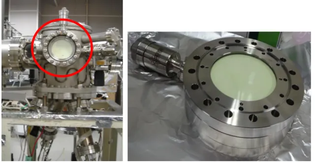
SHG is a well-established surface-specific probe of centrosymmetric media. In the dipole approximation, SHG is forbidden in the bulk of a medium having inversion symmetry, while it is allowed at the surface where inversion symmetry is broken. Because of the symmetry selection property, the SHG method can be one of the ideal methods to measure the contribution of the step structure, while other surface tools such as XPS, TEM, STM, UV-Vis spectroscopy and microscopy technique cannot do because the number of steps are normally lower than the terrace atoms. In this research, stepped bare TiO2 (320) single crystal surface was used as a substrate and Au thin film with the thickness of 2 nm was deposited on the surface in a UHV chamber at a pressure of 2x10⁻⁷ Torr. This Au/TiO2 (320) interface may act as an active sites for showing the catalytic behaviour. As it is well known, surface defects such as steps and kinks play an important role in generating active sites for catalytic reactions, so it is vital to study the structure and the electronic states of such surface defects. Thus, it would be very informative to analyse the SHG response from the interface of TiO2 stepped
symmetry at the interface.
The SHG response from the Au/TiO2 (320) interface and bare TiO2 (320) surface was investigated with the incident photon energies of 1.17 eV and 2.33 eV generated by using a pulsed Nd3+:YAG laser. The isotropic response was found from both samples at the incident photon energy of 1.17 eV. In contrast, we observed the anisotropic response from both Au /TiO2 (320) and bare TiO2 (320) at the incident photon energy of 2.33 eV. From the Au/TiO2 (320) sample, an anisotropic structure was observed to the [ 30] direction for Pin/Pout polarization combination. Here, the Pin denotes the input polarization mode of the incident light and Pout corresponds to the output polarization mode of the SHG light. From the experimental data, I theoretically decomposed the nonlinear susceptibility elements (χijk(2)). Here, i, j and k denote the axis direction of the sample coordinate. I found that there were two groups of the nonlinear susceptibility elements corresponding to step and terrace contribution. More precisely, I have calculated SHG intensity patterns for Au/TiO2 (320) and bare TiO2 (320) based on the terrace and step contributions fitted to the experimental results with photon excitation energies of 2.33 eV. The anisotropic responses were observed due to the contributions of both the step and terrace groups of nonlinear susceptibility elements.
From the calculated results of the step and terrace groups of elements, it was found that the step contribution of the Au/TiO2 (320) is different from that of the bare TiO2 (320) sample for the Pin/Pout polarization combination. In order to discuss the possible reasons for this difference, I considered the four possible mechanism candidates as an origin of the signal enhancement from the Au/TiO2 (320) interface. This four candidates are (a) Enhancement of the incident electric field by surface defects (b) Electronic resonance of Au/TiO2 interface step (c) Surface plasmon effect on SHG enhancement (d) Fresnel factor effect on SHG
I found that Au deposited TiO2 (320) surface contains island structure by the observation of AFM and SEM with EDX and these islands might act as “hot spot” and make the SHG intensity stronger. However, this effect should have an isotropic nature with respect to the rotation of the sample around its normal because these islands are randomly distributed.
The effect would be similar if I consider the enhancement occurring due to the random steps on the surface. This is not the case when I see the SHG pattern for Pin/Pout polarization combination. So, this candidate should be eliminated.
An electronic resonance may occur at the step region of the Au/TiO2 interface and it is the most possible candidate for the enhancement of the SHG signal considering this case.
In this study I observed the enhanced SHG signal correlated with the existence of the Au/TiO2 (320) interface steps. Hence this interface step electronic state is a credibe candidate of the origin of the enhanced signal.
In the case of a thin gold film deposited on pre-patterned TiO2 substrate, local field enhancement may result from the surface plasmon resonance (SPR). I measured the linear optical reflectivity in order to confirm whether there is any influence of surface plasmon resonance and Fresnel factor for the enhancement of the anisotropic SHG signal obtained from the Au/TiO2 (320) interface. From the reflectivity data of the Au/TiO2 (320) interface, it was observed that, the reflectivity for P- and S- polarized light are almost the same at the azimuthal angle, φ = 0ᴼ and 180ᴼ. This result indicates that the linear optical process at frequency ω occurs almost in the same way at φ = 0ᴼ and 180ᴼ and it means that even if the SPR occurred, there is no effect on the enhanced SHG signal due to the different response from the φ = 0ᴼ and 180ᴼ. This discussion is also true for the Fresnel factors.
From the above discussion of four candidates, it seems that electronic resonance at the Au/TiO2 (320) interface step is feasibly responsible for the enhancement of the SHG.
The other three mechanism candidates can be eliminated due to their less feasibility.
Keywords: Second harmonic generation (SHG); Catalyst; Au/TiO2 (320) interface;
Electronic states; Anisotropy; Nonlinear susceptibility elements; Step contribution.
Abstract ………III-VI Table of contents ……….VII-IX Dedication………..X
Chapter 1: Introduction and Research Motivation ………..1-27
1.1 Background ………..………...2-5 1.1.1 Literature of catalytic activities of Au/TiO2 interface………...6-12 1.1.2 Literature on SHG response from Au/TiO2 interface and flat or stepped TiO2
surface……….………..13-22 1.2 Objectives of research ………...23 1.3 Outline of the thesis ………...23-24 References ………...25-27
Chapter 2: Theoretical Background ………28-62
2.1 Maxwell’s equations ………..29-30 2.2 Nonlinear Optics ……….31 2.2.1 Theoretical background of nonlinear optics ………...31-34 2.2.2 Nonlinear polarization induced in noncentrosymmetric media...………35-37 2.3 Second harmonic generation (SHG) ………..37-47 2.4 Nonlinear susceptibility: classical anharmonic oscillator model ………..47-52
2.6 Basic theory of atomic force microscope (AFM) ………..55-57 2.7 Basic theory of surface plasmon resonance (SPR) ………58-60 References ………..61-62
Chapter 3: Experimental Procedure ……….………...63-77
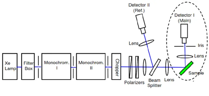
3.1 Sample Preparation ………64 3.1.1 Sample Cleaning by HF etching ……….64 3.1.2 Annealing of TiO2 (320) substrate ………...65-67 3.1.3 Au deposition on TiO2 (320) substrate in a UHV chamber ………...67-71 3.2 Optical Setup ………..72 3.2.1 Linear optical experimental setup ………...72-73 3.2.2 Nonlinear optical system for azimuthal angle dependent SHG experiment…...73-74
3.2.3 Advantage of SHG spectroscopic measurement ………75-76 References……….………….………77
Chapter 4: Results and Discussion ……….………78-109
4.1 Morphological study of Au/TiO2 (320) interface by AFM and SEM with EDX...79-85 4.2 SHG intensity measurement as a function of azimuthal and polarization …………...86 4.2.1 Experimental results ………....86-91 4.2.2 Theoretical investigation of the origin of rotational anisotropic response …....92-107
References.……….…….…...………..109
Chapter 5: General Conclusion ……….………...110-113
Appendix (I) ………..………. 114-116 Appendix (II)………..………..117-133 Appendix (III)………..……….134-140 Appendix (IV)………..……….141-145 Appendix (V)………..………..146-150 Appendix (VI)………..……….151-153
1.1 Background
1.1.1 Literature of catalytic activities of Au/TiO
2interface
1.1.2 Literature on SHG response from Au/TiO
2interface and flat or stepped TiO
2surface 1.2 Objectives of this research
1.3 Outline of the thesis
References
Introduction and research motivation
1.1 Background
Akira Fujishima first discovered that the titanium dioxide has photocatalytic properties in 1967 and its surface exhibits tremendous photocatalytic activity is known as Honda-Fujishima effect [1]. Titanium dioxide can show the catalytic behavior both in thin film and in nanoparticle form [8]. There are mainly three crystal forms of TiO2: rutile, anatase, and brookite. They are polymorph of each other. That means these three forms can be changed to each other at particular temperature although it depends on the morphology and amount of impurity present in the structure [2]. The anatase can be transformed in to rutile at 550-1000ᴼC and brookite can be transformed into rutile form at about 500-600ᴼC [2,3]. The most common form of TiO2 is rutile. It is also very stable and chemically inert.
The UV and visible light can excite rutile [4,5]. However, anatase can excite only by UV light and has ability to transform to rutile form at high temperature. Under the excitation of light both rutile and anatase can act as a photocatalyst through generating the radical species and thus activated the surface [6]. Both of them have crystal structure of tetragonal ditetragonal dipyramidal although the space group lattice is different, whereas brookite has orthorhombic crystal structure and is not excited by UV light irradiation [4,7]. However, it can be converted into rutile or anatase form by heating. The crystal structure and the crystal form are shown by the following Figs. 1.1.1 and 1.1.2.
Figure. 1.1.1. Crystal Structure of rutile, anatase, and brookite titanium dioxide [4,7].
oxygen atoms, respectively.
Figure. 1.1.2. Crystal images of rutile, anatase, and brookite titanium dioxide [8].
The rutile type TiO2 has been used successfully as a potential catalyst to hydrolyze the water molecule into hydrogen and oxygen for the first time in 1970 [5].
The gold is also one of the widely used catalyst although at first it seemed that gold does not have any catalytic activities due to its chemical inertness [9]. It works as a catalyst for oxidative dehydrogenation, isomerization of hydrocarbons and so on [9]. When gold forms alloy with other metals such as palladium and platinum, it also exhibits good catalytic activities. The alloy between gold and other metals acts as a catalyst for the selective oxidation process [9]. Now-a-days the gold nanoparticles are extensively used as a catalyst either in the form of colloids or as a form of junction with supported metal oxides. Indeed, the surface area and the catalytic activity can be greatly enhanced as the size of the gold nanoparticle decreases as shown in Fig. 1.1.3 [10].
Figure. 1.1.3. Relation between the size of gold nanoparticles with the (a) surface area of Au nanoparticles and (b) catalytic activity [10].
reactions. The factors that can have significant influence on catalytic behavior is represented by the following Fig. 1.1.4.
Figure. 1.1.4. The influencing factors on the catalytic activity of bimetallic catalyst in different reactions.
The metal oxide support is one of the important factor for the catalytic performances.
The catalytic activity of AuNPs depends on the supported metal oxide. Miedziak et al.
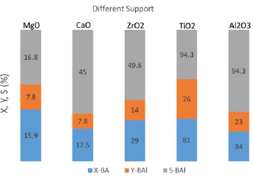
observed that the variation catalytic performances of AuNPs for the oxidation of benzyl alcohol to benzaldehyde depends on different metal oxide supports [11]. They yielded that among the five types of metal supports, the TiO2 supported AuNPs showed the best catalytic performance compared to the others as shown in Fig. 1.1.5. Here, “X” indicates the conversion from benzyle alcohol to benzaldehyde, “Y” denotes the actual yield of the
host chemical that may help to achieve desired amount of main product after the chemical reaction occurred.
Figure 1.1.5. Influence of support on oxidation of benzyl alcohol to benzaldehyde over 1%
Au/M catalysts (M = MgO, CaO, ZrO2, TiO2, Al2O3). Reaction conditions: 30 mL BA, 0.15 g catalyst, 140°C, 5 bar O2, 4 h (X = conversion; Y = yield; S = selectivity).
Not only for this above mentioned chemical reaction but also for many chemical reactions TiO2 supported Au film or nanoparticles exhibit tremendous catalytic performance.
For This reason, I was interested in studying the electronic states of the Au/TiO2 interface to understand the mechanism of the catalytic behavior.
1.1.1.
Literature of catalytic activities of Au/TiO
2interface
The Au/TiO2 interface is a well-known catalyst for many chemical reactions, especially for the oxidation of CO gas at room temperature [12-14]. Gold and titanium dioxide can exhibit catalytic activity when they exist separately. However, the combination of Au and the TiO2 substrate can work as a tremendous catalyst especially for the oxidation of carbon monoxide [14]. Z. Duan et al. studied the electronic structure of Au/TiO2 interface and activation of oxygen atom on the interface through the adsorption and dissociation of oxygen molecule on the interface. This activated oxygen atom plays a vital role for the oxidation of carbon monoxide on the activated interface [13]. The activation sites of oxygen atom on the Au/TiO2 interface for the oxidation reaction of carbon monoxide to carbon dioxide is shown by the following Fig. 1.1.1.1 [13].
Figure. 1.1.1.1. Activation sites of oxygen atom on the Au/TiO2 interface for the oxidation of CO [13].
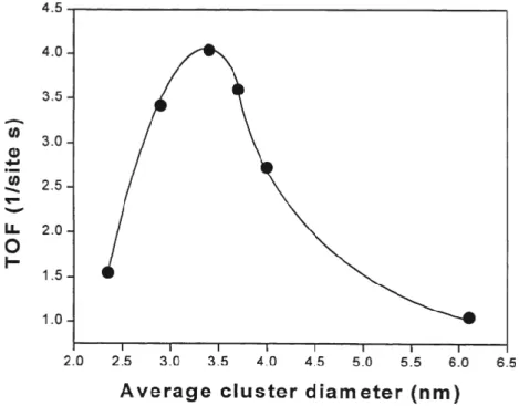
The catalytic activity of Au/TiO2 interface for the oxidation of CO has been observed in terms of particle size of Au [14,15]. M. Valden et al. found that the catalytic performance depends on the size of Au nanoparticle. Around 3.5 nm Au nanoparticle shows
are supported by the TiO2 substrate [15]. Here, turn over frequency (TOF) can be expressed as the reaction rate ((Product molecules) x (surface sites)-1 x s-1) for the conversion of CO to CO2 [15]. Fig. 1.1.1.2 represents the catalytic activity for the oxidation of CO to CO2 as a function of average Au cluster dimeter.
Figure 1.1.1.2. Catalytic activity for the oxidation of CO to CO2 as a function of average Au cluster dimeter.
Haruta et al. found that, when Au/TiO2 has large peripheral part of a jointing interface, it shows high catalytic activity [16]. Tsubota et al. studied the catalytic activity of mechanically mixed Au colloids and TiO2 powder for the oxidation of CO and it shows poor catalytic activity. A tight junction interface was formed between Au colloids and TiO2
powder by calcination at temperature of 673 K and dramatically enhanced the catalytic performance [17]. This result indicates that the effect of the peripheral part of a joining interface exhibit better catalytic activity. X. Z. Li et al. investigated the performance of photocatalytic activity of Au or Au3+ doped TiO2 powder photocatalysts for the
for photodegradation of methyl blue by a number of spectroscopic method through irradiation of visible light [18]. Their results demonstrated that the photooxidation efficiency of methyl blue using Au/Au3+ doped TiO2 powder is much higher than that of conventionally used TiO2
powder [18]. These new types of photocatalyst have the ability to hinder the electron-hole recombination and extend the absorption spectrum to the visible region and also can enlarge the surface area for the photocatalytic activities [18]. I. M. Arabatzis et al. observed the photocatalytic activity of Au/TiO2 composite film for azo-dye methyl orange photodegradation [19]. They improved the photocatalytic activity by the surface deposition of Au particles on TiO2 through the observation of gold valence state, structure and morphological change at the interface of Au and TiO2 by irradiating the UV light [19]. These modified photocatalyst have higher efficiency over conventional TiO2 catalyst for the photodegradation of methyl orange type azo-dye materials [19]. A. S. Castillo et al. observed the photocatalytic activity for the degradation of organic pollutant of anisotropic plasmonic nanoparticles containing hot spots in Au/TiO2 nanostructures [20]. They observed the influence of different Au anisotropic architecture on the performance of catalytic activity due to their different plasmonic excitation by the application of electromagnetic field. Among the three architecture of AuNPs, AuNSTs containing nanocomposite exhibits highest catalytic performance due the creation of intense local field at the spikes of this nanostructures [20].
These local fields create hot spot for the plasmon resonance due to the enhancement of incident electric field and can show tremendous catalytic activity for the organic pollutant degradation [20]. However, the role of Au/TiO2 interface as a catalyst for many chemical reactions and the corresponding electronic phenomena taking place at the active sites are not yet clear enough [21].
oxygen vacancies, step structure and crystal imperfections and electronic states of the Au/TiO2 interface [22]. So far, many researchers studied the electronic states of the Au/TiO2
interface by using various techniques to explore the mechanism of the catalytic properties. X.
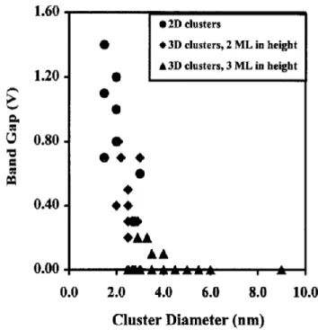
Lai et al. studied the electronic structure of Au/TiO2 interface as a function of different Au particle size by STM method. They found that the bandgap of the Au/TiO2 (110) interface is related to the particle size of Au and the catalytic activity for the oxidation of CO is also closely related to the particle size [23]. Fig. 1.1.1.3 shows that the bandgap is nonzero at lower cluster diameter. When the Au cluster diameter is larger than 4 nm, then the electronic structure is fully metallic due to the lack of bandgap. However, it becomes non-metallic while the cluster diameter is less than 2 nm and the band gap become ≥1V. Therefore, the bandgap is closely related to the Au cluster diameter as well as the electronic states of the Au/TiO2
(110) interface [23].
Figure 1.1.1.3. Relation between the cluster diameter and bandgap of Au/TiO2 (110) interface.
using aberration corrected transmission electron microscope. They showed the perspective view from the [110] direction of the bulk TiO2 (110) structure and defined the IV-site shown in Fig. 1.1.1.4. Here, Ti-only columns contain only Ti-ions, Ti and O are arranged alternately in the Ti-O columns and in case of O-O columns, oxygen ions form a zigzag chain. The defined IV-site is in the center of the oxygen octahedrons where there is a probability to occupy the interstitial Ti ion [24].
Figure1.1.1.4. Perspective view of bulkTiO2 from the [110] direction. Blue and red spheres illustrate Ti and O ions, respectively. Three kinds of atomic columns, viz. “Ti-only”, “Ti–O”, and “O–O” form the bulk TiO2 unit cell. Cross (X) indicates the location of IV-column.
Location of “IV-site” (green) at the center of the oxygen octahedron [24].
They observed the TEM images in Fig. 1.1.1.5 (a) and (b) taken at defocus of +5.5 nm and -5.5 nm, respectively, corresponding to the Fig. 1.1.1.4. All the atomic columns including Ti-only, Ti-O and O-O were bright at the defocus of +5.5 nm but at the defocus of
+5.5 nm and -5.5 nm, respectively. According to the model structure, there were no interstitial Ti-ions at the defined IV-site which in the center of the oxygen octahedrons. So this simulated result agreed with the experimental TEM image result. They concluded that if there is any interstitial Ti-ion, the IV-site should be brighter enough in the TEM image.
Figure 1.1.1.5. (a)–(b) Observed TEM images of defect-free TiO2 viewed along the [110]
direction: Defocus is (a) +5.5 nm (over focus) and (b) −5.5 nm (under focus). (c)–(d) Simulated TEM images of TiO2: Defocus is (c) +5.5 nm and (d) −5.5 nm. Specimen thickness is 3.9 nm. TiO2 specimen and simulated model has no interstitial Ti ions at IV-site [24].
nanoparticle whereas they existed at the perimeter/interface of Au/TiO2 [24]. The following schematic diagram shows the local distribution of interstitial Ti ions in TiO2 substrate and Au/TiO2 perimeter/interface [24].
Figure 1.1.1.6. Schematic model representing a local distribution of interstitial Ti ions in TiO2 substrate and Au/TiO2 perimeter/interface after TEM observation. Green dot denotes the interstitial Ti ions. Light blue region illustrates a TiO2 surface layer. Interstitial Ti ions are deficient in a peripheral region (orange area) of the Au particle, and localized at the Au/TiO2
perimeter/interface. Blue curve indicates the perimeter of the Au/TiO2 interface [24].
Yao et al. measured the broad absorption peak at the visible region from the particle size-dependent Au/TiO2 catalyst by using UV-vis reflectance spectra. They concluded that the observed peak was due to the localized surface plasmon resonance of Au nanoparticle [14]. Strong absorption peak was observed due to the excitation of plasmon of Au nanoparticle from the Au/TiO2 interface reported by other researchers [25-28]. These studies are related to the catalytic activity of Au/TiO2 interface as a function of nanoparticle size distribution. Therefore, the Au/TiO2 (320) interface act as a well-established catalyst for many chemical reactions. So, it emphasized to study the electronic states of this catalyst to understand mechanism of the catalytic behaviour.
stepped TiO
2surface
The precise measurement of atomic scale electronic state from Au/TiO2 interface using specific surface and interface probe such as second harmonic generation (SHG) is desirable to understand the catalytic mechanism properly. SHG is one of the prominent candidate to detect this signal due to its high surface and interface sensitivity. SHG is forbidden in the bulk of medium with inversion symmetry within the electric dipole approximation [29]. It has submonolayer sensitivity to the surface and interface of centrosymmetric media because it is only allowed in a broken symmetric structure [29].
Another attractive feature of SHG is that it is non-invasive and contactless and it can be applied to “in situ” and in “real-time” experiments with a good time resolution [30]. We can deduce information such as electronic structure and molecular orientation from the precise determination of the nonlinear susceptibility tensor [31]. We can extract the electronic level information from the interface of Au/TiO2.
However, the number of studies of the electronic states from the stepped Au/TiO2
interface by SHG is very limited. Only a single report was found regarding the observation of the SHG signal from Au/TiO2 interface. Quelin et al. measured SHG intensity as a function of incident angle and input/output polarization from the thin Au/TiO2 cermet films. Cermet films are the combination of the ceramic and metallic materials. The Au particles are randomly distributed in the TiO2 matrix to form a cermet film. They detected enhanced SHG signal due to the local field enhancement by the surface plasmon excitation on gold clusters [32]. In fact, they studied the electromagnetic mode on Au clusters embedded in TiO2 and were not interested in atomic scale electronic states at the interface between the two materials
beam plot. They observed the highest SHG intensity for the polarization angle of SHG beam θ = 0ᴼ and the intensity is reduced for θ = +45ᴼ and -45ᴼ but it approaches to zero at θ = 90ᴼ [32]. However, they did not analyse the susceptibility tensor to clarify the electronic states between the Au particles and TiO2 matrix interface.
Fig. 1.1.2.1. Dependence of the SH intensity for 37% volume fraction of gold sample versus ω beam polarization (ϕ) for four values of the 2ω beam polarization: o for θ = 0ᴼ, 󠄾 for θ = - 45ᴼ, for θ = +45ᴼ, for θ = 90ᴼ.
For this reason, I intended to observe the atomic level electronic states of the stepped Au/TiO2 interface using SHG method. This research work was one of the great motivation for me to analyse the independent non-vanishing nonlinear susceptibility tensor
interface properly.
In fact, many researchers observed the SHG response from the flat and stepped bare TiO2 surface to understand the electronic states of the surface. Kobayashi et al.
observed SHG response from TiO2 (110) as a function of the polarization and azimuthal angle. They determined surface elements and compared the theoretical patterns with that obtained from the experiment. They concluded that the SHG signal originateed from surface electric dipole [33]. Omote et al. studied the SHG spectra as a function of the photon energy from the TiO2 (110) and (001) surface. In order to study the surface electronic structure, they conducted phenomenological analysis to separate the contribution from various nonlinear susceptibility elements, and the dominant contribution was observed from the surface elements [34]. They noticed that the dominant nonlinear susceptibility element was for both TiO2 surfaces. They detected small contribution of the bulk quadrupoles. They performed an ab initio calculation by using the FLAPW method within the local density approximation and found that Ti-O-Ti-O- chains including the bridging oxygen atoms on the surface act as the main origin of the SHG signal generation from the TiO2 (110) surface [34].
Takahashi et al. detected contribution of steps by comparing the SHG spectra from three kinds of stepped surfaces, TiO2 (15 15 4), (13 9 0) and (671) with that from a flat (110) surface. They observed large SHG intensity from the step surfaces compared to the flat (110) surface that is shown by the following Fig. 1.1.2.2 [22]. In Fig. 1.1.2.2, The SHG intensity patterns of TiO2 (15 15 4) and (671) were very similar for the Pin-Sout polarization combination. It might be due to the same electronic states. The patterns were different for the Sin-Sout polarization combination. More asymmetric shape was observed for the TiO2 (671) compared to the TiO2 (15 15 4) for the Sin-Sout polarization combination. The reason might
intensity of the TiO2 (13 9 0) surface was very different from that of the (15 15 4) and (671) surfaces for both Pin-Sout and Sin-Sout polarization combinations due to the different miscut angle. On the other hand, the SHG intensity of TiO2 (110) was very low for both Pin-Sout and Sin-Sout polarization combinations and it was at the noise level [22]. They could successfully separate the step contribution of surface elements from stepped surfaces of TiO2 (15 15 4), (13 9 0) and (671), respectively, because the symmetry was broken at the periodic steps within the surface plane [22].
Figure 1.1.2.2. SHG intensity patterns as a function of the sample rotation angle for the polarization combinations Sin-Sout and Pin-Sout. Dots represent experimental data, and curves represent phenomenologically fitted curves. Incident angle and photon energy were 2ᴼ and 2.33 eV, respectively. The dashed lines represent the sample rotation angles with SH electric field E(2ω) parallel to the step edges.
In another report [35], they observed strong SHG/SFG responses from the stepped TiO2 (17 18 1) and (15 13 0) surfaces compared to the flat (011) and (110) in order to understand the electronic states of terraces and steps. The resonances in the SHG and SFG
electronic transitions at the steps. More specifically the local density of states at the step of the vicinal surfaces was distributed right above the valence band maximum (VBM) and right below the conduction band minimum (CBM) [35].
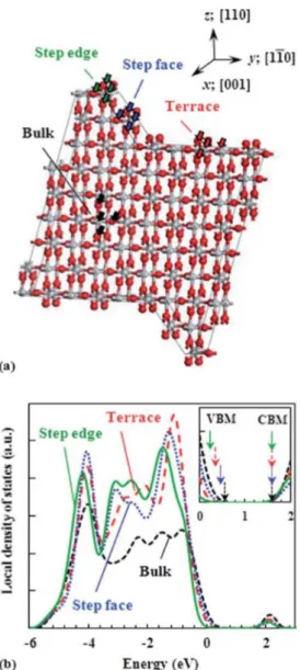
Figure 1.1.2.3. (a) A rutile TiO2 slab model for the density functional theory (DFT) calculation of the local density of states (LDOS). The surface of the slab model consists of a (110) terrace and a double-layer step parallel to [0 0 1] with a (100) step face. (b) Calculated LDOS for bulk, a terrace, a step face, and a step edge. For each structure, the LDOS at the four atoms indicated by the arrows in (a) is summed up to yield the total LDOS at the
arrows in the inset indicate VBM and CBM for each structure. Here, zero corresponds to the energy of the Fermi energy.
Fig. 1.1.2.3 (a) showed the slab model drawn based on the AFM observation for (15 13 0) surface. This surface consists of multilayer step bunches and (110) surface having long terrace with a terrace width of about 20-25 nm towards the [1-10] direction [35].
However, since the size of the model system was limited, they assumed that the surface was composed of a bilayer step with a (100) step face, and a (110) terrace with a terrace width of four unit cells (2.6nm) along [1-10] direction. Thickness of the slab was nine layers. The local density of states (LDOS) was calculated for bulk, a terrace, a step face and a step edge as shown in Fig. 1.1.2.3 (b). According to this figure, it was clear that the LDOS at the VBM varies for the step and terrace significantly but it was very similar around the CBM and had almost the same energy. This energy shift for VBM might be due to the surface relaxation.
However, the red shift for LDOS were significant for the step edge, step face and the terrace [35]. The band-gap energy for the top edges of the step bunches // [001] is larger than that for their hillsides and the (110) terraces. Therefore, it is concluded that the LDOS around the VBM for the top edges of the step bunches // [001] undergoes a red shift from the corresponding DOS for the bulk more significantly than that for their hillside and the (110) terraces does [35].
The red shift of the LDOS around the VBM for the top edges of the step bunches can give high oxidation power to free holes at these sites, as free holes extract electrons from adsorbed molecules more strongly with the increase in their potential energy (redox potential).
However, they could not clearly state that whether these free holes really contribute to
of the high reactivity of the step bunches, especially the ones // [001], as well as that of the (011) face in terms of trap states or LDOS near the band-gap regions. They finally conclude that they should not consider only the static surface electronic properties but also dynamic properties at TiO2 surfaces after UV excitation [35].
S. Nakamura et al. observed electronic states from the anatase TiO2 (101) surface in order to elucidate the mechanism of catalytic reaction on the surface. They found that the optical nonlinearity mainly originated from the surface rather than bulk [36]. H. Sano et al.
theoretically calculated the SHG response on TiO2 (110) face by using self-consistent full potential linearized augmented plane-wave (FLAPW) method [37]. Their theoretical calculation well agreed with the experimental findings. The results exhibited that the strong SHG response originated from the zigzag chains of TiO2 (110) face consisting of bridging oxygen and neighbouring sixfold coordinated titanium atoms at two photon energy of 4eV [37]. H. Takahashi et al. observed the electronic states of stepped TiO2 (15 15 4) by SHG method as a function of photon energy. They successfully separated the step and terrace contribution of the stepped TiO2 (15 15 4) and compared the results with the TiO2 (110) face.
The step and terrace contribution from both the samples were very different indeed due to their different electronic structure [38]. The SHG spectra for the TiO2 (15 15 4) and TiO2
(110) surfaces is shown by the following Fig. 1.1.2.4.
Figure 1.1.2.4. (a) SHG spectra for the TiO2 (110) and (15 15 4) surfaces with the incident angle of 45ᴼ. The polarization combination was Pin-Pout. The incident azimuth was such that k////[0 0 1]. (b) The ratio of SHG intensity for the TiO2 (15 15 4) surface to that for the (110) surface. The curves are guidelines.
Fig. 1.1.2.4 (a) shows the SHG spectra of the TiO2 (110) and (15 15 4) surfaces as a function of photon energy for Pin-Pout polarization combination. The obtained spectra for these two surfaces were different due to their different electronic states. More specifically the spectrum of TiO2 (15 15 4) was different due to its step contribution. In Fig. 1.1.2.4 (b),
surfaces. This intensity ratio was more than unity. Therefore, the deviation of this SHG intensity ratio was due to the contribution of the step surface. The electronic states of the step surface was different from the electronic states of terrace surface. So, the SHG intensity was very strong at photon energy 3.6 eV due to the step contribution.
My motivation came from the above listed research works. Many researches have been conducted on the SHG measurement from the flat or stepped TiO2 surfaces and analyzed their electronic states particularly the electronic states of the steps and terraces.
However, they did not observe the SHG response from the interface between the Au and flat or stepped TiO2 surface. They also did not investigate the electronic states of the Au/TiO2
interface. For this reason, I intended to investigate the SHG signal of the stepped Au/TiO2
(320) interface and study the electronic states of the interface by separating their step and terrace contributions. I choose (320) face because this face contains atomic steps in [1-10]
direction and from the literature it is known that the step structure acts as active sites for the catalytic reactions [35]. So, from the catalytic point of view, the electronic state study of this crystallographic index is important in order to explore the catalytic mechanism. For these reason, I intended to use (320) crystallographic index.
As it is well known, surface defects such as steps and kinks play an important role in generating active sites for catalytic reactions, so it is vital to study the structure and the electronic states of such surface defects in order to understand their catalytic performances.
Thus, it would be very informative if we analyse the SHG response from the interface of TiO2 atomic stepped surface decorated by Au. In this research, a rutile type TiO2 (320) single
can be one of the ideal methods to measure step contribution of this atomic scale step structure by using the symmetry selection property, while other surface tools are not very sensitive because the number of steps are normally lower than the terrace atoms. Because the other tools are not so sensitive like SHG to the symmetry structure, it is difficult to separate the step and terrace contribution precisely. Therefore, only SHG can pick up step contribution for oriented steps and using the advantages of its symmetry selection property. So far as we know, there has been no previous observation of SHG from the stepped Au/TiO2 interface.
In this work, I attempted to detect optical second harmonic generation signal from steps on the TiO2 surface decorated by a Au film of 2 nm thickness and tried to separate the terrace and step contributions. On the TiO2 (320) surface plane there is a structural asymmetry in the [ 30] direction and this should induce SHG. I measured azimuthal angle and polarization dependent SHG intensity patterns at photon energies 1.17 eV and 2.33 eV for Au/TiO2 (320) and TiO2 (320) samples. I calculated the second order nonlinear optical susceptibility elements from the experimental results and separated the step and terrace contributions from both the samples. This analysis provides an important step for the future SHG spectroscopy of the electronic states of the Au/TiO2 (320) interface as a function of the photon energy and the spectrum will be closely related to the catalytic activity of this interface.
The following points summarize the main objectives of my research:
(a) I will fabricate the 2nm Au film on the stepped TiO2 (320) surface and characterized it.
(b) Then I will measure the anisotropic SHG response from the Au/TiO2 (320) interface.
(c) One of the most interesting objective of this research is to separate the step and terrace contribution of the Au/TiO2 (320) interface to understand the electronic states precisely.
1.3 Outline of the thesis
This thesis consists of six chapters and are organized as follows:
Chapter 1 describes the general introduction and research motivation. In this chapter, I list literature of the catalytic properties of TiO2 and Au separately and Au/TiO2 interface. The SHG response on the Au/TiO2 interface and flat and stepped TiO2 are discussed precisely. I also discuss the reason for studying the electronic states of Au/TiO2 (320) interface by using SHG method. This chapter also includes the objectives of this present research and outline of this research work as well.
Chapter 2 explains the theoretical background related to the nonlinear optics, principle of nonlinear optical media. Here I discuss the principle of SHG, brief explanation of nonlinear susceptibility elements, theory of surface plasmon resonance (SPR), theory of scanning electron microscope and atomic force microscope.
including sample cleaning by HF etching, annealing of TiO2 (320) and Au deposition on stepped TiO2 (320) surface in a UHV chamber. I also discuss the optical setup for the SHG and linear experiment and mention the advantages of SHG spectroscopic measurement.
Chapter 4 deals with the main experimental results and discussion of this research work. I discuss the anisotropic SHG response and the origin of the anisotropic behavior. I also elaborately explain the separation of the step and terrace contribution of the nonlinear susceptibility elements originates from step and terrace of Au/TiO2 interface and morphology by using SEM and AFM. I also discuss the linear optical reflectivity and transmission spectra obtained from the Au/TiO2 interface.
Chapter 5 describes the general conclusion based on the obtained main results from this research work.
I also included all the lists of my publications and conference proceedings and the participation in the national and international conferences in the appendix sections. I added some appendixes related to this research work.
[1] Discovery and applications of photocatalysis — Creating a comfortable future by making use of light energy; Japan Nanonet Bulletin Issue 44, 12 May 2005.
[2] Hanaor, Dorian A. H.; Sorrell, Charles C. "Review of the anatase to rutile phase transformation". Journal of Materials Science. 46, 4, 855–874, 2011.
[3] J. G. Li and T. Ishigaki, Acta Materialia, 52, 17, 5143-5150, 2004.
[4] R. Austin and S.-f. Lim, "The Sackler Colloquium on Pormoses and Perils in Nanotechnology for Medicine," PNAS, vol. 105, no. 45, pp. 17217-17221, 2008.
[5] K. Hashimoto, H. Irie and A. Fujishima, "TiO2 Photocatalysis: A Historical Overview and Future Prospects," Japanese Journal of Applied Physics, vol. 44, no. 12, pp. 8269-8285, 2005.
[6] K. Prasad, D. V. Pinjari, A. B. Pandit and S. T. Mhaske, Ultrasonics Sonochemistry, 17, 2, 409-415, 2010.
[7] S. Woodley and C. Catlow, "Structure prediction of titania phases: Implementation of Darwinian versus Lamarckian concepts in an Evolutionary Algorithm," Computational Materials Science, vol. 45, no. 1, pp. 84-95, 2009.
[8] Wikipedia, "Titanium Dioxide," Widipedia, 11 April 2012. [Online]. Available:
http://en.wikipedia.org/wiki/Titanium_dioxide. [Accessed 16 April 2012].
[9] G. C. Bond, The catalytic properties of gold, Gold Bulletin, Vol. 5, Issue 1, pp 11-13, 1972.
[10] A. Alshammahi and V. N. Kalevaru, Supported gold nanoparticles as promising catalysts, http://dx.doi.org/10.5772/64394, 2016.
[11] Hermans LA, Interaction of Nickel ions with silica Geus JW. Stud. Surf. Sci. Catal.
Oxford, UK; 4, 113, 1979.
Knight, S. T. Taylor, C. J. Kiely, G. J. Hutchings, Oxidation of benzyl alcohol using supported gold palladium nanoparticles. Catal. Today. 163(1), 47–54, 2011.
[13] Z. Duan and G. Henkelman, ACS Catal. 5, 1589-1595, (2015).
[14] Q. Yao, C. Wang, H. Wang, H. Yan, and J. Lu, J. Phys. Chem. C 120, 9174-9183, (2016).
[15] M. Valden, S. Pak, X. Lai, and D. W. Goodman, Catal. Lett. 56, 7-10, (1998).
[16] M. Haruta, S. Tsubota, T. Kobayshi, H. Kageyama, M. J. Ganet, and B. Delmon, J. Catal.
144, 175, (1993).
[17] S. Tsubota, T. Nakamura, K. Tanaka, and M. Haruta, Catal. Lett. 56, 131, (1998).
[18] X. Z. Li and F. B. Li, Environ. Sci. Technol., 35, 2381-2387, 2001.
[19] I. M. Arabatzis, T. Stergiopoulos, D. Andreeva, S. Kitova, S. G. Neophytides and P.
Falaras, Journal of Catalysis, 220, 127-135, 2003.
[20] A. S. Castillo, M. C. Hermo, B. R. Gonzalez, M. P. Lorenzo, Z. Wang, X. T. Kong, A. O.
Govorov and M. A. C. Duarte; J. Phys. Chem. C. 120, 11690-11699, 2016.
[21] T. Minato, H. Kato, M. Kawai; Surface Science. Vol. 27, No. 6, 319-325, 2006.
[22] H. Takahashi, R. Watanabe, and G. Mizutani, e-J. Surf. Sci. Nanotech. 8, 84-88, (2010).
[23] X. Lai, T. P. St. Clair, M. Valden, and D. W. Goodman, Surface Science 59, 25-52, (1998).
[24] T. Tanaka, A. Sumiya, H. Sawada, Y. Kondo, and K. Takayanagi, Surface Science 619, 39-43, (2014).
[25] A. Aiboushev, A. Astafiev, O. M. Sarkisov, and V. A. Nadtochenko, Journal of Physics 291, 012040, (2011).
[26] Z. K. Zhou, M. Li, X. R. Su, Y. Y. Zhai, H. Song, J. B. Han, and Z. H. Hao, Phys. Stat.
Sol. (a) 205, 345-349, (2008).
[28] M. Kyoung and M. Lee, Bull. Korean Chem. Soc. 21, 1, (2000).
[29] L. Marrucci, D. Paparo, G. Cerrone, C. de Lisio, E. Santamato, S. Solimeno, S.
Ardizzone, and P. Quagliotto, Optics and Lasers in Engineering 37, 601-610, (2002).
[30] J. J. H. Gielis, P. M. Gevers, I. M. P. Aarts, M. C. M. Van de Sanden, and W. M. M.
Kessels, J. Vac. Technol. A 26, 6, (2008).
[31] S. Cattaneo, E. Vuorimaa, H. Lemmetyinen, and M. Kauranen, J. Chem. Phys. 120, 19, (2004).
[32] X. Quelin, J. Sakars, A. Bourdon, and P. Gadenne, Physica B 279, 102-104, (2000).
[33] E. Kobayashi, T. Wagasugi, G. Mizutani, and S. Ushioda, Surface Science 402-404, 537-541, (1998).
[34] M. Omote, H. Kitaoka, E. Kobayashi, O. Suzuki, K. Aratake, H. Sano, G. Mizutani, W.
Wolf, and R. Podloucky, Journal of Physics: Condensed Matter. 17, 8, S175-S200, (2005).
[35] H. Takahashi, R. Watanabe, Y. Miyauchi and G. Mizutani, J. Chem. Phys. 134, 154704, (2011).
[36] S. Nakamura, K. Matsuda, T. Wakasugi, E. Kobayashi, G. Mizutani, S. Ushioda, T.
Sekiya and S. Kurita, Journal of Luminescence, 87-89, 862-864, 2000.
[37] H. Sano, G. Mizutani, W. Wolf and R. Podloucky, Phys. Rev. B., 70, 125411, 2004.
[38] H. Takahashi, R. Watanabe and G. Mizutani, Surf. Interface Anal. 42, 1659-1662, 2010.
Chapter 2: Theoretical Background 2.1 Maxwell’s equations 2.2 Nonlinear Optics
2.2.1 Theoretical background of nonlinear optics 2.2.2 Nonlinear polarization induced in non- centrosymmetric media
2.3 Second harmonic generation (SHG
2.4 Nonlinear susceptibility: classical anharmonic oscillator model 2.5 Basic theory of scanning electron microscope (SEM)
2.6 Basic theory of atomic force microscope (AFM)
2.7 Basic theory of surface plasmon resonance (SPR)
References
Theoretical background
In this chapter, I will discuss about the theoretical part related to this research and about the nonlinear susceptibility elements along with their symmetric consideration. Further, I will discuss about the brief theory of some microscopic observation.
2.1 Maxwell’s equations
I would like to discuss briefly about the Maxwell’s equations those simply represents the little matter interaction. These equations are very important for describing the electromagnetic theory. The four important Maxwell’s equations are given below,
∇ × 𝐸̃ = − 𝜕𝐵̃
𝜕𝑡 (1) ∇ × 𝐻̃ = 𝜕𝐷̃⁄𝜕𝑡+ 𝐽̃ (2)
∇. 𝐷̃ = 𝜌̃ (3) ∇. 𝐵̃ = 0 (4)
Here, 𝐸̃and 𝐵̃ are the electric field and magnetic induction, respectively. 𝐷̃ and 𝐻̃ are the dielectric vector and magnetic field, respectively. 𝜌̃ and 𝐽̃are the electric charge density and current density, respectively.
The magnetic induction 𝐵̃ and the magnetic field 𝐻̃ is related according to the following equation,
𝐵̃ = 𝜇0𝐻̃ (5) The dielectric vector can be defined as
𝐷̃ = 𝜀0𝐸̃ + 𝑃̃ (6)
Here, 𝜀0 is the electric permittivity of the free space and 𝑃̃ is the polarization induced by the electric field.
The relation between the induced polarization and electric field is almost linear when the value of electric field is very small. The linear polarization equation can be written as follows, 𝑃̃ = 𝜀0𝜒𝐸̃ (7)
Putting the value of 𝑃̃ in to the euation (6)
𝐷̃ = 𝜀0𝐸̃ + 𝑃̃ = 𝜀0𝐸̃ + 𝜀0𝜒𝐸̃ = (1 + 𝜒) 𝜀0𝐸̃ = 𝜀𝜀0𝐸̃ (8) Where, χ is the linear susceptibility and it is related to the index of refraction [1].
𝜒 = 𝑛2− 1
χ acts as a tensor and shows different values with different polarization of light and different symmetry system. In case of large values of electric field, the relation between the polarization and the electric field also changes and it becomes nonlinear. The nonlinear polarization equation can be written as a series of expansion,
𝑃̃ = 𝜀0𝜒(1)𝐸̃ + 𝜀0𝜒(2)𝐸̃𝐸̃ + 𝜀0𝜒(3)𝐸̃𝐸̃𝐸̃ + ⋯ (9)
From the equation (9), it is clear that the polarization have a significant role in the nonlinear optical phenomena and 𝜒(2) is the second order optical nonlinear susceptibility elements.
2.2 Nonlinear Optics
Nonlinear optics (NLO) is the study of nonlinear interaction between the light and matter.
More precisely, the nonlinearity observed due to the nonlinear responses of a material with the interaction of electric field of light and the source of light should be very intense. The only source of intense light is laser [2]. After the discovery of laser by Theodore Harold Maiman in 1960, the revaluation occurred in the field of nonlinear optics by the invention of different types of exciting phenomena namely second harmonic generation (SHG), sum frequency generation (SFG), difference frequency generation (DFG), optical parametric oscillation (OPO), third harmonic generation (THG) [2-3].
2.2.1 Theoretical background of nonlinear optics
In order to understand the interaction between light wave and nonlinear medium properly, it is important to calculate the electromagnetic wave equation for nonlinear medium by using the Maxwell’s equations discussed in the part 2.1.
For the calculation of electromagnetic wave equations, we consider curl on the both side of equation (1) and we have,
∇ × ∇ × 𝑬̃ = ∇ × (− 𝜕𝑩̃
𝜕𝑡 ) ⇒ ∇ × ∇ × 𝑬̃ = − 𝜕
𝜕𝑡 ( ∇ × 𝐵 ̃ ) (10) Substituting the value of equation (5) into equation (10), we have,
∇ × ∇ × 𝑬̃ = −𝜇0 𝜕
𝜕𝑡 ( ∇ × 𝐻 ̃) (11) Now putting the value of equation (2) in to equation (11), We got,
∇ × ∇ × 𝑬̃ = −𝜇0 𝜕
𝜕𝑡 ( 𝜕𝑫̃
⁄𝜕𝑡+ 𝑱̃ ) ⇒ ∇ × ∇ × 𝑬̃ = −𝜇0𝜕2𝑫
𝜕𝑡2 − 𝜇0𝜕𝑱̃
⁄𝜕𝑡 (12) We consider the electric current density is 𝑱̃ = 0, then we have,
∇ × ∇ × 𝑬̃ = −𝜇0𝜕2𝑫̃
𝜕𝑡2
∇ × ∇ × 𝑬̃ + 𝜇0𝜕2𝑫̃
𝜕𝑡2 = 0 (13)
Now from equation (6), replacing the value 𝐷̃ = 𝜀0𝐸̃ + 𝑃̃ into equation (13), we can write,
∇ × ∇ × 𝐸̃ + 𝜇0 𝜕2
𝜕𝑡2(𝜀0𝐸̃ + 𝑃̃) = 0
∇ × ∇ × 𝐸̃ + 1
𝐶2
𝜕2
𝜕𝑡2𝐸̃ = − 1
𝜀0𝐶2
𝜕2𝑃̃
𝜕𝑡2 (14)
In the field of nonlinear optics, equation (14) is generally known as the Maxwell’s source-term equation or nonlinear wave equation. Under the appropriate boundary conditions, it can be written as,
∇ × ∇ × 𝐸̃ = ∇(∇. 𝐸̃) − ∇2𝐸̃ (15)
We can write, ∇. 𝐸 = 0, when ∇. 𝐷 = 0 for the anisotropic media, The equation (15) becomes,
∇ × ∇ × 𝐸̃ = −∇2𝐸̃ (16) Using equation (14) and (16) we have,
−∇2𝐸̃+1
𝐶2
𝜕2
𝜕𝑡2𝐸̃ =− 1
𝜀0𝐶2
𝜕2𝑃̃
𝜕𝑡2 (17)
In equation (17), the 𝑃̃ is denoted by the induced polarization by the electric field application linearly and nonlinearly and the polarization can be divided into linear and nonlinear part according to the equation (18).
𝑃̃ = 𝑃̃(1)+ 𝑃̃𝑁𝐿 (18) The dielectric vector can also be separated into linear and nonlinear parts,
𝐷̃ = 𝐸̃ + 4𝜋𝑃̃(1)+ 4𝜋𝑃̃𝑁𝐿 (19) 𝐷̃ = 𝐷̃(1)+ 𝑃𝑁𝐿 (20) The linear equation of dielectric vector can be written as,
𝐷̃(1) = 𝜀0𝐸̃ + 𝑃̃(1) (21) By using the equation (21) and (14) can be expressed as,
−∇2𝑬̃+ 1
𝜀0𝐶2
𝜕2𝑫̃(𝟏)
𝜕𝑡2 =− 1
𝜀0𝐶2
𝜕2𝑷̃𝑵𝑳
𝜕𝑡2 (22)
Equation (22) is also known as the wave equation for the nondispersive and lossless medium. A new relation among the linear dielectric vector, electric field and linear dielectric tensor for isotropic material can be expressed by the following formula,
𝐷̃(1)= 𝜀0𝜖(1). 𝐸̃ (23)
For the isotropic and nondispersive materials, the dielectric tensor 𝜖(1) is scaler in quantity. So, the equation (22) becomes
−∇2𝑬̃ + 𝜖
(1) 𝐶2
𝜕2
𝜕𝑡2𝑬̃ =− 1
𝜀0𝐶2
𝜕2
𝜕𝑡2𝑷̃𝑁𝐿 (24)
The equation (24) is generally known as inhomogeneous wave equation and the right hand side of this equation can be expressed as nonlinear source term of a medium.
Each frequency components of a particular field can be separated in a dispersive medium.
The sum of the different frequency components can be expressed by the linear dielectric vector, electric field and nonlinear polarization. The equations for the different frequency components can written as,
𝐸̃(𝑟, 𝑡) = ∑ 𝐸̃𝑛 𝑛 (𝑟, 𝑡) 𝑤𝑖𝑡ℎ 𝐸̃(𝑟, 𝑡) = 𝐸𝑛(𝑟)𝑒−𝑖𝜔𝑛𝑡 (25)
𝐷̃(1)(𝑟, 𝑡) = ∑ 𝐷̃𝑛(1) 𝐷𝑛(1)(𝑟, 𝑡)
𝑛
𝑤𝑖𝑡ℎ 𝐷̃𝑛(1) (𝑟, 𝑡) = 𝐷𝑛(1)(𝑟)𝑒−𝑖𝜔𝑛𝑡 =
𝜖(1)(𝜔𝑛). 𝐸̃𝑛 (𝑟, 𝑡) (26)
𝑃̃𝑁𝐿(𝑟, 𝑡) = ∑ 𝑃̃𝑛 𝑛𝑁𝐿(𝑟, 𝑡) 𝑤𝑖𝑡ℎ 𝑃̃𝑛𝑁𝐿(𝑟, 𝑡) = 𝑃𝑛𝑁𝐿(𝑟)𝑒−𝑖𝜔𝑛𝑡 (27)
By introducing equation (25) and (26) into the equation (22), we can deduce a wave equation which is equivalent to the equation (24) and that is valid for each frequency components of the field. Then the equation (24) can be written as,
−∇2𝐸̃𝑛 + 𝜖
(1)(𝜔𝑛) 𝐶2
𝜕2𝐸̃𝑛
𝜕𝑡2 =
−
1𝜀0𝐶2
𝜕2𝑃̃𝑛𝑁𝐿
𝜕𝑡2 (28)
The equation (28) is the key equation in the field of nonlinear optics and helps to determine the origin of the optical nonlinearity.
2.2.2 Nonlinear polarization induced in noncentrosymmetric media
The second-order nonlinear optical process is only allowed from the surface or interface where the symmetry is broken or having no inversion symmetry. On the contrary, it is forbidden in the bulk of medium consisting inversion symmetry [4].However, the third-order nonlinear optical process is allowed in both media having inversion and no inversion symmetry [3]. In order to understand precisely, we will now discuss about the occurrences of optical nonlinearity.
In case of linear medium, the polarization is induced by the applied electric field linearly where the electric field strength is small. The equation can be expressed as,
𝑃̃(𝑡) = 𝜀0𝜒(1)𝐸̃(𝑡) (29)
Here, 𝜒(1) is the linear optical susceptibility and 𝜀0 is known as electric permittivity of vacuum.
In case of nonlinear medium, the polarization is induced by the applied electric field nonlinearly where the electric field strength is significantly large. This relation can be written as the following equation expressed as a power series,
𝑃̃(𝑡) = 𝜖𝑜[𝜒(1)𝐸̃(𝑡) + 𝜒(2)𝐸̃(2)(𝑡) + 𝜒(3)𝐸̃(3)(𝑡) + ⋯ ] (30)
≡ 𝑃̃(1)(𝑡) + 𝑃̃(2)(𝑡) + 𝑃̃(3)(𝑡) + ⋯ (31) 𝑃𝑁𝐿(𝑡) = 𝑃̃(2)(𝑡) + 𝑃̃(3)(𝑡) +…. (32)
Where, 𝜒(2) and 𝜒(3) are denoted as the second and the third order nonlinear optical susceptibilities. In addition 𝑷̃(𝟐)(𝑡) and 𝑷̃(𝟑)(𝑡) can be expressed as the second-order and
third-order nonlinear polarization, respectively. When an electric field is applied to a nonlinear medium, we can write the following expression,
𝑬̃(𝑡) = 𝐸𝑐𝑜𝑠𝜔𝑡 (33)
In case of centrosymmetric medium, the sign of the polarization will be changed obviously along with changing the sign of applied electric field to that medium. So the following equation for second-order nonlinear polarization can be written as,
−𝑷̃(𝟐)(𝑡) = 𝜖0𝜒(2)[−𝑬̃(𝑡)]2 = 𝜖0𝜒(2) 𝑬̃(𝟐)(𝑡) (33)
For centrosymmetric medium, the following expression is valid.
𝑷̃(𝟐)(𝑡) = −𝑷̃(𝟐)(𝑡) (34)
The equation (34) will be satisfied only when there is no nonvanishing nonlinear susceptibility elements and therefore the induced polarization will be equal to zero. So, in case of centrosymmetric medium, the second-order nonlinearity is forbidden according to the dipole approximation rule. Fig. 2.2.2.1 shows a graphical representation for clarifying the linear and nonlinear response with respect to the applied electric field. Fig. 2.2.2.1 (a) exhibits the waveform of electric field, (b) expresses the linear optical response with respect to the applied electric filed as shown in (a). The curves (c) and (d) show the nonlinear optical responses in the centrosymmetric and noncentrosymmetric medium, respectively.
![Figure 2.2.2.1. The graphical presentations for linear and nonlinear polarizations with respect to the applied electric field in various media [3]](https://thumb-ap.123doks.com/thumbv2/123deta/6076525.1073729/50.918.299.639.155.580/figure-graphical-presentations-nonlinear-polarizations-respect-applied-electric.webp)
![Figure 2.5.2. The different types of signal emitted from the different part of the interaction volume [10]](https://thumb-ap.123doks.com/thumbv2/123deta/6076525.1073729/67.918.238.683.413.826/figure-different-types-signal-emitted-different-interaction-volume.webp)
![Figure 2.7.2. Otto configuration for the generation of surface plasmon resonance (SPR) [16]](https://thumb-ap.123doks.com/thumbv2/123deta/6076525.1073729/73.918.297.624.238.438/figure-otto-configuration-generation-surface-plasmon-resonance-spr.webp)