1
Title: Location of the Tibial Tunnel Aperture Affects Extrusion of the Lateral Meniscus Following Reconstruction of the Anterior Cruciate Ligament
Short title: Location of the tibial tunnel
Yuya Kodama, Takayuki Furumatsu*, Shinichi Miyazawa, Masataka Fujii, Takaaki Tanaka, Hiroto Inoue, Toshifumi Ozaki
Department of Orthopaedic Surgery, Okayama University Graduate School of Medicine, Dentistry, and Pharmaceutical Sciences, 2-5-1 Shikatacho, Kitaku, Okayama 700-8558, Japan
* Corresponding author: Takayuki Furumatsu, Department of Orthopaedic Surgery, Okayama University Graduate School of Medicine, Dentistry, and Pharmaceutical Sciences, 2-5-1 Shikatacho, Kitaku, Okayama 700-8558, Japan
Tel: 81-86-235-7273; Fax: 81-86-223-9727; E-mail: matino@md.okayama-u.ac.jp Authors’ Contributions
Yuya Kodama designed the study, prepared the samples, performed experimental work, and wrote the manuscript. Takayuki Furumatsu designed the study and prepared the samples. Shinichi Miyazawa prepared the samples. Masataka Fujii, Takaaki Tanaka, and Hiroto Inoue conducted measurement of experimental samples. Toshifumi Ozaki organized the laboratory works. All authors have read and approved the final version of the manuscript submitted.
2
Abstract
The anterior root of the lateral meniscus provides functional stability to the meniscus. In this study, we evaluated the relationship between the position of the tibial tunnel and extrusion of the lateral meniscus after anterior cruciate ligament reconstruction, where extrusion provides a proxy measure of injury to the anterior root. The relationship between extrusion and tibial tunnel location was retrospectively evaluated from computed tomography and magnetic resonance images of 26 reconstructed knees, contributed by 25 patients aged 17 to 31 years. A measurement grid was used to localize the position of the tibial tunnel based on anatomical landmarks identified from the three- dimensional reconstruction of axial computed tomography images of the tibial plateaus. The reference point-to-tibial tunnel distance (mm) was defined as the distance from the midpoint of the lateral edge of the grid to the posterolateral aspect of the tunnel aperture. The optimal cutoff of this distance to minimize post-operative extrusion was identified using receiver operating curve analysis. Extrusion of the lateral meniscus was positively correlated to the reference point-to-tibial tunnel distance (r² = 0.64; P < 0.001), with a cutoff distance of 5 mm having a sensitivity to extrusion of 83% and specificity of 93%. The mean extrusion for a distance >5 mm was 0.40±0.43 mm, compared to 1.40±0.51 mm for a distance ≤5 mm (P < 0.001). Therefore, a posterolateral location of the tibial tunnel aperture within the footprint of the anterior cruciate ligament decreases the reference tibial-to-tunnel distance and increases extrusion of the lateral meniscus post- reconstruction.
Keywords: Anterior cruciate ligament; lateral meniscus; lateral meniscal extrusion
3
Introduction
During reconstruction of the anterior cruciate ligament (ACL), it is generally accepted that the tibial attachment of the graft should be located in an anteroposterior direction on the tibial plateau, with the anteromedial tunnel localized just anterior to its native anatomical position.1,2 However, although clinical consensus exists regarding the ideal location for the femoral tunnel, no such consensus has been reached for the tibial tunnel. The regional anatomy of the tibial attachment of the ACL is complex, and accurate location of the tibial tunnel is likely to be of clinical importance.
Anatomical studies have identified the tibial attachment of the ACL to be either oval or triangular in shape,3,4 and to be surrounded by several bony landmarks, including the anterior ridge, medial intercondylar ridge and tubercle, intertubercular fossa, lateral intercondylar tubercle, and lateral groove.5 No ligamentous tissue is attached to the lateral intercondylar tubercle and intercondylar fossa.5 In addition, Siebold et al. demonstrated that the tibial ACL insertion is “C”- shaped along the medial tibial spine and the center of the ‘‘C’’ is the bony attachment of the anterior root of the lateral meniscus.6 Cadaveric studies have also described the lateral meniscus (LM) to have an anterior tibial attachment area of 140.7 mm2 and to be inserted deeply beneath the anatomical footprint of the tibial attachment of the ACL, itself having an attachment area of 218.4 mm2.2 Therefore, 63.2% of the LM anterior root and 40.7% of the tibial attachment of the ACL footprint overlap.7 There is also evidence that the LM anterior root dives underneath the tibial attachment of the ACL, while the anterior fibers of the LM blend into the anterolateral part of the ACL. This anatomical coupling between the ACL and the LM is likely to contribute to the extent of LM extrusion after tears of the anterior horn of the meniscus, explaining the significantly higher incidence of cartilage degeneration associated with anterior horn tears, compared to other types of
4
meniscal tears.8,9 A previous histological study of the tibial attachment of the ACL demonstrated that the LM anterior root and the posterior root of the LM form an ACL-LM complex via dense fiber connections located on the tibial surface.9 Using safranin O staining, Furumatsu et al.10 demonstrated that fibers of the ACL-LM complex become gradually more dense from their posterior to anterior points of attachment, with the ACL-LM transition zone being located 7 to 8 mm from the anterior ridge of the tibia. Additionally, in their study of 12 cadaveric knees, LaPrade et al.11 reported a significant reduction in the ultimate failure strength of the attachment of the LM anterior root after reaming of the tibial tunnel during anatomic single-bundle (SB) ACL reconstruction.
Based on this anatomical evidence, we hypothesized that a lateral shift in the location of the aperture of the tibial tunnel within the ACL footprint would increase the likelihood of injury to the attachment of the LM anterior root. Therefore, the aim of our study was to evaluate the relationship between the location of the tibial tunnel aperture and LM extrusion, where LM extrusion is used as a proxy measure of the stability of the LM anterior root after ACL reconstruction in patients with no evidence of meniscal injury at the time of surgery. A secondary aim was to use the measures of the location of the tibial tunnel aperture and LM extrusion to determine the optimal position of the aperture within the tibial attachment of the ACL.
Methods
This study was approved by our Institutional Review Board, and patients provided informed consent prior to participation. The correlation between the location of the tibial tunnel aperture and LM extrusion was evaluated through a retrospective review of three-dimensional reconstructed computed tomography (CT) and magnetic resonance (MR) images of patients who
5
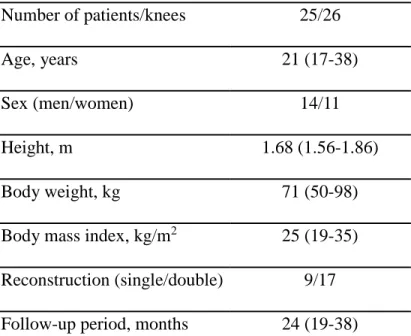
underwent anatomical ACL reconstruction at our institution between January 2011 and November 2014. The medical records for 128 consecutive ACL surgeries were reviewed to exclude patients with associated injury to the medial and/or lateral meniscus. Among these 128 cases, 26 cases (20%) without meniscal injury, contributed by 25 patients, were identified and formed our study group. Relevant characteristics of the study group are reported in Table 1. The average time from injury to pre-operative MR imaging was 2 weeks (range, 1–4 weeks); average time from injury to ACL reconstruction, 3 months (range, 1–10 months); and post-operative MR imaging, 3 months.
For analysis, the location of the tibial tunnel aperture was quantified from the three-dimensional reconstruction of post-operative CT images. LM extrusion was quantified from pre- and post- surgery MR images.
Surgical techniques for ACL reconstructions
Based on patient characteristics, either SB or double-bundle (DB) arthroscopic ACL reconstructions were performed, using semitendinosus tendon (ST) and bone-patellar tendon-bone (BTB) autografts. SB reconstructions were performed for patients with smaller knees by using an ST autograft for recreational athletes and a BTB graft for heavier patients and those playing contact sports. Patients with larger knees underwent DB reconstruction using an ST autograft. All reconstructions were performed by three experienced surgeons.
For DB reconstructions with ST autografts, an outside-in technique was used.12 The harvested tendons were double-looped over an Endobutton fixation device (Smith & Nephew Inc., Andover, MA), with the distal ends anchored using a Krackow suture, thus recreating the anteromedial (AM) and posterolateral (PL) bundles of the ACL. The apertures of the femoral and tibial tunnels were localized at the center of the footprint of each bundle. Femoral fixation was achieved using the Endobutton system. Tibial fixation was performed with the knee flexed at 20°
6
by using double-spike plates (Meira, Aichi, Japan), with an initial tension of 20 N for the PL bundle and 30 N for the AM bundle. The tension in each bundle was independently measured using a tensiometer.13 For SB reconstructions, both ST and BTB autografts were used. The distal end of the graft was again anchored to the tibia using double-spike plates, with the knee in 20° of knee flexion and an initial tension of 30 N.14 The procedures for the BTB grafts were similar, with the exception of using a far anteromedial (FAM) portal to drill the femoral tunnel.15 The initial tension on the graft was set to 30 N.
Post-operative rehabilitation protocols
All patients were maintained in a knee brace for 1 week, to promote initial healing of the graft, fixation points, and affected soft tissues. Knee range of motion exercises and partial weight bearing were initiated at post-operative week 2. Full weight bearing was permitted at 1 month, post-operatively, and running at 5 months, with a return-to-sport permitted at 8 months.
CT image-based measurement
All patients underwent CT imaging at 1 week post-surgery to localize the tunnel aperture, which is difficult to obtain from plain radiographs. CT images were obtained with an Asteion 4 Multislice CT System (Toshiba Medical Systems, Tochigi, Japan) using 120 kVp and 150 mA, and 1-mm slice thickness. CT reconstruction of the tibial condyles in the axial plane was completed using a three-dimensional volume-rendering technique (AZE Virtual Place software; AZE Ltd., Tokyo, Japan). Measurement of the location of the tibial tunnel aperture was obtained using a rectangular measurement grid centered over the tibial plateaus, with the size and position of the grid individually adjusted using the following patient-referenced anatomical landmarks identified from the reconstructed model of the proximal tibia: the medial intercondylar ridge, the anterior ridge, the lateral groove, and the intertubercular fossa. To localize the position of the tibial tunnel
7
aperture, the tibial model was placed in a medial view to identify the posterior corner of the widest part of the medial tibial plateau, and the Amis-Jakob line was drawn, passing through the posterior corner of the tibial plateau and parallel to the medial joint line.16 The model was then rotated to visualize the superior aspect of the proximal tibia, with the internal/external rotation adjusted until the most posterior articular margins of both the medial and lateral tibial condyles were placed on the same horizontal level (Figure 1). The rectangular grid was then drawn over the superior view of the model, with the view adjusted until the anterior ridge line and the inter-tubercular fossa line were parallel to the posterior line. The location of the anterior ridge and inter-tubercular fossa lines was verified on coronal view. The top of the medial intercondylar tubercle was identified in the superior view, and the position of this reference point was confirmed in the coronal view. The medial intercondylar tubercle line was drawn perpendicular to the posterior line. To define the lateral border, a reference point was localized at the midpoint of a line joining the midpoint of the anterior ridge and the inter-tubercular fossa lines on the edge of the lateral groove. The lateral border was defined by a line crossing this reference point and drawn perpendicular to the posterior line. Finally, the reference point-to-tibial tunnel distance (RTD) was defined as the minimum distance (mm) from the midpoint of the lateral edge of the measurement grid and the posterolateral margin of the tunnel aperture (Figure 2). The localization of the posterolateral margin of the tunnel aperture was defined as the posterior and lateral aspect using the measurement grid (Figure 3).
Using these anatomical references, localization of the reference grid was standardized between patients, independent of individual differences in the size of tibial plateaus.
MR image-based measurement of LM extrusion
Change in LM extrusion was measured from pre- and post-surgical MR images, with pre- operative images obtained on average 3 weeks prior to surgery (range, 1–12 weeks) and post-
8
operative images obtained 3 months after surgery. MR images were obtained using an Achieva 1.5 T (Philips, Amsterdam, The Netherlands) or an EXCELART Vantage Powered by Atlas 1.5 T (Toshiba Medical Systems, Tochigi, Japan) coil. The extent of LM extrusion was evaluated independently by two reviewers using a picture archiving and communication system (PACS) workstation (Figure 3). LM extrusion was defined as the distance (mm) from the lateral margin of the meniscus to the lateral margin of the lateral tibial plateau, measured using the digital caliper function of the PACS software. LM extrusion measurements were obtained in the mid-coronal plane by linking the coronal and sagittal image series. Each image sequence was then reviewed to identify the most lateral chondral surface of the tibial plateau.17 The increase in lateral meniscal tissue extending beyond the most lateral border of the tibial chondral surface was measured and recorded. The LM extrusion value was calculated as the difference between pre- and post-operative measurements, with a positive value indicative of a larger extrusion post-operatively, whereas a negative value was indicative of a larger pre-operative extrusion.
Statistical analysis
The mean and associated 95% confidence intervals (95% CI) of the RTD and LM extrusion measurements were calculated. Correlation between RTD and LM extrusion was evaluated using Spearman’s rank correlation analysis. The optimal RTD cut-off associated with a smaller post-operative LM extrusion was determined by constructing the receiver operating (ROC) curve and calculating the Youden index (J). Difference in LM extrusion between patients above or below the identified RTD cut-off was evaluated using the Mann-Whitney U test. Statistical significance was set at P < 0.05, a priori. Measures of aperture localization and LM extrusion were completed by two independent orthopedic surgeons to determine inter-observer reliability using the intra-class correlation coefficient (ICC), and with each observer repeating the measurements
9
after a 4-week interval to determine intra-observer reliability.
Results
Measurement of the RTD was reliable, with ICC values of 0.948 to 0.992 for intra- observer reliability and 0.992 to 0.996 for inter-observer reliability. For the LM extrusion measurement, the ICC for intra-observer reliability ranged between 0.899 and 0.975, and 0.991 and 0.995 for inter-observer reliability. A significant correlation was observed between RTD and LM extrusion (r² = 0.64; P < 0.001; Figure 4), with the linear regression line (y = - 0.34 x + 2.61) confirming an increase in LM extrusion as a function of an increasing posterolateral location of the tibial tunnel aperture within the tibial footprint of the ACL.
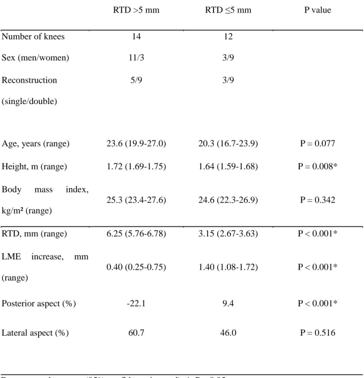
The ROC analysis identified the optimal location of the posterolateral edge of the tibial tunnel aperture to be 5.01 mm from the reference point. This RTD cutoff had a sensitivity of 83%
to LM extrusion and specificity of 93%. Cases were subsequently classified into two groups based on this RTD cut-off, ≤5 mm and >5 mm. The two groups were compared with respect to the extent of LM extrusion. The relevant clinical characteristics of patients in each group are reported in Table 1, with between-group differences in LM extrusion reported in Table 2. A mean LM extrusion value of 1.40 mm (95% CI, 1.08-1.72 mm) was calculated among patients in the RTD ≤5 mm group, compared to a mean LM extrusion of 0.40 mm (95% CI, 0.252-0.748 mm) among patients in the RTD >5 mm group (P < 0.001). The lateral aspect of the RTD ≤5 mm group and the RTD >5 mm group were 60.7% and 46.0%, respectively (P = 0.52). The posterior aspect of the RTD ≤5 mm group and the RTD >5 mm group were -22.1% and 9.4%, respectively (P > 0.001).
Discussion
The most important finding of our study was that a more posterolateral positioning of the
10
tibial tunnel aperture could induce the LM extrusion after ACL reconstruction. An association between tibial tunnel preparation and damage to the LM anterior root has been reported by various other researchers.11,18-22 The primary site of damage is the soft tissue fibers, which couple the deep layers of the ACL to the LM and the LM to the tibial plateau. These fibers specifically act on the LM anterior root attachment itself, explaining the reduction in the ultimate failure strength of the LM anterior root with disruption of these fibers.1,23 Ellman et al.24 reported that the fibers overlying the ACL tibial insertion did not significantly modify the strength or the stiffness of the LM anterior root. Therefore, the LM extrusion identified in our study could have possibly resulted from damage to the fibers of the LM inserting into the subchondral bone. However, as a relatively small reaming area (6-7 mm-diameter tibial tunnel) caused LM extrusion, complete destruction of the insertion of the LM anterior root into the subchondral bone is unlikely in all of our cases. Rather, we propose that the tunnel reaming likely overlapped the area of the LM anterior root attachment, which resulted in LM extrusion. Evidence for this mechanism is confirmed by identification of a wavy appearance of the LM anterior root, due to damage to the anterior insertion of the LM during primary ACL reconstruction, on second-look arthroscopy.25 Our study provides original data on the importance of tunnel location in actual patients, confirming previous reports from cadaveric studies.
Our image-based assessment could improve accuracy in localizing the tibial tunnel during the reconstruction procedure. In current practice, the ‘quadrant technique’ is usually used, a technique that was developed to provide a standardized localization of the tunnel between patients.26-28 However, a significant variation in tibial tunnel placement between surgeons and institutions29 has been reported using the quadrant technique.30-33 Because the center of the ACL insertion was the bony insertion of the LM anterior root,6 this variation could increase the risk for damage to the LM
11
anterior insertion. Our image-based technique uses a rectangular measurement grid to localize the aperture of the tibial tunnel, with positioning of the grid based on patient-specific anatomical references identified from an individualized three-dimensional reconstruction of the tibial plateau.
The reliable identification of the anatomical reference used to place the grid on the tibial plateaus has previously been well described by Tensho et al., including an anterior ridge on the anterior boundary, a lateral groove on the lateral boundary, and an intertubercular fossa on the posterior boundary.5 Therefore, the use of these three bony landmarks is clinically feasible. Currently, the insertion angle of the guide pin dictates the orientation of the tunnel aperture. However, even if the guide angle includes the anterior ridge on the anterior boundary providing a general posterolateral direction for the tibial tunnel, the intra-operative angle of reaming will be influenced by the size and orientation of the tunnel aperture, with a decrease in angle increasing the risk for injury to the LM anterior insertion. Therefore, we measured the distance between the posterolateral aspect of the tibial tunnel aperture and the reference point as the shortest RTD. The lateral groove that we use as a reference includes both the ACL footprint and the LM anterior root. However, due to individual differences in the shape of ACL footprint, classified as oval or triangular in shape,3,5 the safe zone, in which the risk of injury to the LM anterior insertion would be low, will vary depending on the shape of the ACL footprint, as shown in our supplemental figure (Figure S-1). Consequently, we propose that the reference point for tunnel localization should be set outside the lateral groove to lower the risk of injury to the LM anterior insertion, with the lateral wall of the groove being used as a reproducible reference point. Notably, the position of the guide pin aligns with the center of the tunnel aperture. Therefore, even if the insertion position of the guide pin is set outside the lateral groove, injury to the LM anterior root is still possible if a large tibial tunnel is created. As the risk of injury to the LM anterior insertion is influenced by tunnel size, it was difficult to exactly
12
evaluate the relationship between tunnel location and LM extrusion (r2 = 0.10; Figure S-2). By contrast, the distance between the reference point and the posterolateral edge of the tunnel aperture did correlate with LM extrusion (r2 = 0.64; Figure 4).
Use of patient-specific anatomical landmarks provides standardization, eliminating effects of individual variation in the size of the tibial plateaus on aperture localization. Localization of the reference point at the posterior edge of the LM anterior root is not visually identifiable by arthroscopy. Intra-operatively, the reference point can be localized around the intersection provided by the medial margin of the lateral tibial plateau and the posterior edge of the LM anterior root.
Arthroscopic probing can be useful to identify our reference point (Figure 5). Although not directly identifiable by direct vision, the location of this reference point was sufficiently reliable in our study, with a mean (SD) localization at 38.6±5.0% of the anterior-to-posterior depth of the tibial plateau, and 63.3±2.6% of the medial-to-lateral width of the tibial plateau (Figure 6). Based on this reliability for the localization of this reference point, we suggest that the RTD could provide a reliable index to identify optimal aperture location. Our image-based assessment could be used in prospective studies to further characterize the association between a posterolateral location of the tibial tunnel and LM extrusion, as well as to evaluate the clinical outcomes of this relationship on the future development of knee osteoarthritis.34
As an innovation in our study, we also used an ROC analysis (Figure S-3.) to evaluate the relationship between deviation in the location of the tibial tunnel aperture away from an optimal RTD of 5 mm on the extent of LM extrusion. For cases with RTD values >5 mm, the increase in post-surgical LME was 0.4 mm, compared to 1.4 mm for cases with RTD values ≤5 mm (Table 2).
Although a more posterior positioning of the tibial tunnel in the ACL footprint has traditionally
13
been recommended for ACL reconstruction to lower the risk for roof impingement,35,36 current recommendations are for localization of the aperture at the true center of the footprint of the tibial attachment of the ACL, adjacent to the anterior horn of the LM, with the goal of approximating the mechanical response of the reconstructed graft to the native ACL.21,32,37,38 Ziegler et al. and Zantop et al. reported this ‘true’ center to be located 7.5 mm from the center of the LM anterior root.2,21 However, LaPrade et al.7 suggested that the functional importance of the supplemental soft tissue fibers of the LM anterior root should be considered and suggested a location at 5.0 mm from the center of the LM anterior root. We agree with this latter recommendation. A significant difference in height was identified for patients in our two RTD groups: 1.72±0.06 m for the RTD >5 mm group, compared to 1.64±0.06 m for the RTD ≤5 mm group (P = 0.008; Table 2). Height correlated with RTD (r² = 0.18, P = 0.008) but not with LM extrusion (r² = 0.07, P = 0.177; Figure S-4). This relationship between height and RTD did not explain the significant variance in LM extrusion and, therefore, it is unlikely that stature would influence LM extrusion. The relationship between height and RTD may be more appropriately explained by differences in surgical technique, with a significantly lower number of patients having undergone SB reconstruction (n = 8) compared to DB reconstruction (n = 18). In our study group, a SB reconstruction was selected for patients with smaller stature who, therefore, had shorter harvested ST lengths, as well as for recreational athletes.
Interestingly, although the technique is recommended for patients with smaller stature, the SB graft itself is larger than the graft diameter for AM bundles used in DB reconstruction, which could explain the smaller RTD and, therefore, the higher rate of LM extrusion in cases having undergone SB reconstructions. Sex may be another explanatory factor, as women tend to be of smaller stature compared to men and tend to have a narrower area of the tibial attachment of the ACL.1 Consequent to these sex-specific anatomical differences, women would be expected to have a smaller RTD
14
relative to the size of the tibial tunnel and, therefore, to be at higher risk for damage to the LM anterior root and LM extrusion. In our study group, we attempted to obtain an anteromedial location of the tibial tunnel aperture in these cases. Considering our findings, we suggest that, for patients with a narrow tibial attachment of the ACL or of small stature, purposeful selection of a DB reconstruction may be warranted to preserve the structure of the LM anterior root. Future studies are required to provide evidence to guide practice around this issue.
Limitations
The limitations of our study need to be acknowledged. Foremost, the number of cases included in our analysis was limited (n = 26), with variation in relevant demographic features among patients;
this could have influenced outcomes. However, our findings do provide justification for larger, population-based studies. In addition, the specific effect of LM extrusion on short- and long-term surgical outcomes needs to be specifically evaluated. Currently, evidence exists for specific factors being associated with the development of osteoarthritis post-ACL reconstruction, including reduced quadriceps strength, higher BMI, meniscectomy at the time of the ACL reconstruction, loss of knee extension, and increased knee joint laxity.34,38,39 As LM extrusion will affect the mechanics and long-term health of the meniscus, we feel that long-term follow-up observation is needed for patients with identified LM extrusion to ascertain its role in knee health and function.
RTD also varied according to the type of ACL reconstruction, with a smaller RTD for SB reconstruction than for DB reconstruction. Although the correlation between an increased RTD and LM extrusion was high, we were unable to identify a significant difference in risk for LM extrusion between the SB and DB groups (Figure S-5). The fewer cases in the SB group than in the DB group may have been a confounding factor in our statistical analysis. We must also consider that MR
15
images used for the evaluation of LM extrusion were obtained at different time points before surgery and, therefore, factors associated to acute ACL injury, such as swelling, could have resulted in error in the measurement of LM extrusion. However, such error is likely to be relatively small as the intra- and inter-rater reliability in measurement was high. In addition, we did not extend MR imaging beyond the 3-month limit, post-surgery. Therefore, any subsequent change in LM extrusion was not assessed. Finally, the identification of reference points directly under imaging using arthroscopy was difficult for surgeons because the reference point was localized under the LM anterior segment. Intraoperative identification was limited to the approximate location based on a few bony landmarks. No significant deviations should exist between CT and intraoperative images because the reference point was set based on anatomical landmarks. However, a reliability test plan is necessary for the future. We suggest the use of intraoperative CT imaging in the future, because it is the most reliable method to identify a reference point.
Conclusion
A posterolateral location of the tibial tunnel aperture within the ACL footprint decreases the RTD and increases LM extrusion post-ACL reconstruction.
ACKNOWLEDGMENT
We would like to thank Editage (www.editage.jp) for English language editing.
16
REFERENCES
1. Kopf S, Pombo MW, Szczodry M, et al. 2011. Size variability of the human anterior cruciate ligament insertion sites. Am J Sports Med 39: 108–113. doi:10.1177/0363546510377399.
2. Zantop T, Ferretti M, Bell KM, et al. 2008. Effect of tunnel-graft length on the biomechanics of anterior cruciate ligament-reconstructed knees: intra-articular study in a goat model. Am J Sports Med 36: 2158–2166. doi:10.1177/0363546508320572.
3. Ferretti M, Doca D, Ingham SM, et al. 2012. Bony and soft tissue landmarks of the ACL tibial insertion site: An anatomical study. Knee Surg Sports Traumatol Arthrosc 20: 62–68.
doi:10.1007/s00167-011-1592-z.
4. Furumatsu T, Miyazawa S, Tanaka T, et al. 2013. Postoperative change in medial meniscal length in concurrent all-inside meniscus repair with anterior cruciate ligament reconstruction.
Int Orthop 38: 1393–1399. doi:10.1007/s00264-013-2238-1.
5. Tensho K, Shimodaira H, Aoki T, et al. 2014. Bony landmarks of the anterior cruciate
ligament tibial footprint: A detailed analysis comparing 3-dimensional computed tomography images to visual and histological evaluations. Am J Sports Med 42: 1433–1440.
doi:10.1177/0363546514528789.
6. Siebold R, Schuhmacher P, Fernandez F, et al. 2014. Flat midsubstance of the anterior cruciate ligament with tibial “C”-shaped insertion site. Knee Surg Sports Traumatol Arthrosc 23(11):3136–3142.
7. LaPrade CM, Ellman MB, Rasmussen MT, et al. 2014. Anatomy of the anterior root
attachments of the medial and lateral menisci: a quantitative analysis. Am J Sports Med 242:
2386–2392. doi:10.1177/0363546514544678.
8. Hatayama K, Terauchi M, Saito K, et al. 2013. The importance of tibial tunnel placement in
17
anatomic double-bundle anterior cruciate ligament reconstruction. Arthroscopy 29: 1072–
1078. doi:10.1016/j.arthro.2013.02.003.
9. Siebold R, Peter S, Francis F, et al. 2015. Flat midsubstance of the anterior cruciate ligament with tibial “C”-shaped insertion site. Knee Surg Sports Traumatol Arthrosc 23: 3136–3142.
doi:10.1007/s00167-014-3058-6.
10. Furumatsu T, Kodama Y, Maehara A, et al. 2016. The anterior cruciate ligament-lateral meniscus complex: A histological study. Connect Tissue Res 57: 91–98.
doi:10.3109/03008207.2015.1081899.
11. LaPrade CM, Smith SD, Rasmussen MT, et al. 2014. Consequences of tibial tunnel reaming on the meniscal roots during cruciate ligament reconstruction in a cadaveric model, Part 1:
The anterior cruciate ligament. Am J Sports Med 43: 200–206.
doi:10.1177/0363546514554769.
12. Lubowitz JH, Konicek J. 2010. Anterior cruciate ligament femoral tunnel length: Cadaveric analysis comparing anteromedial portal versus outside-in technique. Arthroscopy 26: 1357–
1362. doi:10.1016/j.arthro.2010.02.014.
13. Hamada M, Shino K, Horibe S, et al. 2005. Changes in cross-sectional area of hamstring anterior cruciate ligament grafts as a function of time following transplantation. Arthroscopy 21: 917–922. doi:10.1016/j.arthro.2005.05.006.
14. Anderson CJ, Westerhaus BD, Pietrini SD, et al. 2010. Kinematic impact of anteromedial and posterolateral bundle graft fixation angles on double-bundle anterior cruciate ligament reconstructions. Am J Sports Med 38: 1575–1583. doi: 10.1177/0363546510364841.
15. H Shino K, Suzuki T, Iwahashi T, et al. 2010. The resident’s ridge as an arthroscopic landmark for anatomical femoral tunnel drilling in ACL reconstruction. Knee Surg Sports
18
Traumatol Arthrosc 18: 1164–1168. doi:10.1007/s00167-009-0979-6.
16. Amis AA, Jakob RP. 1998. Anterior cruciate ligament graft positioning, tensioning and twisting. Knee Surg Sports Traumatol Arthrosc 6: S2-S12. doi:10.1007/s001670050215.
17. Furumatsu T, Miyazawa S, Tanaka T, et al. 2013. Postoperative change in medial meniscal length in concurrent all-inside meniscus repair with anterior cruciate ligament reconstruction.
Int Orthop 38: 1393–1399. doi:10.1007/s00264-013-2238-1.
18. Goldsmith MT, Jansson KS, Smith SD, et al. 2013. Biomechanical comparison of anatomic single- and double-bundle anterior cruciate ligament reconstructions: an in vitro study. Am J Sports Med 41: 1595–1604. doi:10.1177/0363546513487065.
19. Middleton KK, Hamilton T, Irrgang JJ, et al. 2014. Anatomic anterior cruciate ligament (ACL) reconstruction: a global perspective. Part 1. Knee Surg Sports Traumatol Arthrosc 22:
1467–1482. doi:10.1007/s00167-014-2846-3.
20. Monaco E, Maestri B, Conteduca F, et al. 2014. Extra-articular ACL reconstruction and pivot shift: In vivo dynamic evaluation with navigation. Am J Sports Med 42: 1669–1674.
doi:10.1177/0363546514532336.
21. Ziegler CG, Pietrini SD, Westerhaus BD, et al. 2011. Arthroscopically pertinent landmarks for tunnel positioning in single-bundle and double-bundle anterior cruciate ligament reconstructions. Am J Sports Med 39: 743–752. doi:10.1177/0363546510387511.
22. Watson JN, Wilson KJ, LaPrade CM, et al. Iatrogenic injury of the anterior meniscal root attachments following anterior cruciate ligament reconstruction tunnel reaming. Am J Sports Med. 2014; 23(8): 2360–2366.
23. Hauch KN, Villegas DF, Haut Donahue TL. 2010. Geometry, time- dependent and failure properties of human meniscal attachments. J Biomech 43: 463–468.
19
doi:10.1016/j.jbiomech.2009.09.043.
24. Ellman MB, Laprade CM, Smith SD, et al. 2014. Structural properties of the meniscal roots.
Am J Sports Med 42: 1881–1887. doi:10.1177/0363546514531730.
25. Furumatsu T, Ozaki T. 2016. Iatrogenic injury of the lateral meniscus anterior insertion following anterior cruciate ligament reconstruction: a case report. J Orthop Sci. [in press]
26. Ahn JH, Jeong HJ, Ko CS, et al. 2013. Three-dimensional reconstruction computed tomography evaluation of tunnel location during single-bundle anterior cruciate ligament reconstruction: A comparison of transtibial and 2-incision tibial tunnel-independent techniques. Clin Orthop Surg 5: 26–35. doi:10.4055/cios.2013.5.1.26.
27. Lertwanich P, Martins CA, Asai S, et al. 2011. Anterior cruciate ligament tunnel position measurement reliability on 3-dimensional reconstructed computed tomography. Arthroscopy 27: 391–398. doi:10.1016/j.arthro.2010.08.018.
28. Sadoghi P, Kröpfl A, Jansson V, et al. 2011. Impact of tibial and femoral tunnel position on clinical results after anterior cruciate ligament reconstruction. Arthroscopy 27: 355–364.
doi:10.1016/j.arthro.2010.08.015.
29. Wolf BR, Ramme AJ, Wright RW, et al. 2013. Variability in ACL tunnel placement:
Observational clinical study of surgeon ACL tunnel variability. Am J Sports Med 41: 1265–
1273. doi:10.1177/0363546513483271.
30. Forsythe B, Kopf S, Wong AK, et al. 2010. The location of femoral and tibial tunnels in anatomic double-bundle anterior cruciate ligament reconstruction analyzed by three- dimensional computed tomography models. J Bone Joint Surg Am 92: 1418–1426.
doi:10.2106/JBJS.I.00654.
31. Lorenz S, Elser F, Mitterer M, et al. 2009. Radiologic evaluation of the insertion sites of the
20
2 functional bundles of the anterior cruciate ligament using 3-dimensional computed tomography. Am J Sports Med 37: 2368–2376. doi:10.1177/0363546509341577.
32. Parkinson B, Gogna R, Robb C, et al. 2015. Anatomic ACL reconstruction: the normal central tibial footprint position and a standardised technique for measuring tibial tunnel location on 3D CT. Knee Surg Sports Traumatol Arthrosc July 1 [Epub ahead of print]
doi:10.1007/s00167-015-3683-8.
33. Tsukada H, Ishibashi Y, Tsuda E, et al. 2008. Anatomical analysis of the anterior cruciate ligament femoral and tibial footprints. J Orthop Sci 13: 122–129. doi:10.1007/s00776-007- 1203-5.
34. Kessler MA, Behrend H, Henz S, et al. 2008. Function, osteoarthritis and activity after ACL- rupture: 11 Years follow-up results of conservative versus reconstructive treatment. Knee Surg Sports Traumatol Arthrosc 16: 442–448. doi:10.1007/s00167-008-0498-x.
35. Howell SM, Taylor MA. 1993. Failure of reconstruction of the anterior cruciate ligament due to impingement by the intercondylar roof. J Bone Joint Surg Am 75: 1044–1055.
http://www.ncbi.nlm.nih.gov/pubmed/8335664.
36. Marchant BG, Noyes FR, Barber-Westin SD, Fleckenstein C. 2010. Prevalence of
nonanatomical graft placement in a series of failed anterior cruciate ligament reconstructions.
Am J Sports Med 38: 1987–1996. doi:10.1177/0363546510372797.
37. Bedi A, Maak T, Musahl V, et al. 2011. Effect of tibial tunnel position on stability of the knee after anterior cruciate ligament reconstruction: is the tibial tunnel position most important?
Am J Sports Med 39: 366–373. doi:10.1177/0363546510388157.
38. Pinczewski LA, Lyman J, Salmon LJ, et al. 2007. A 10-year comparison of anterior cruciate ligament reconstructions with hamstring tendon and patellar tendon autograft: a controlled,
21
prospective trial. Am J Sports Med 35: 564–574. doi:10.1177/0363546506296042.
39. Salmon LJ, Russell VJ, Refshauge K, et al. 2006. Long-term outcome of endoscopic anterior cruciate ligament reconstruction with patellar tendon autograft: minimum 13-year review.
Am J Sports Med 34: 721–732. doi:10.1177/0363546505282626.
22
Tables
Table 1. Demographic and clinical characteristics.
Variables Number or mean (range) Number of patients/knees 25/26
Age, years 21 (17-38)
Sex (men/women) 14/11
Height, m 1.68 (1.56-1.86)
Body weight, kg 71 (50-98)
Body mass index, kg/m2 25 (19-35) Reconstruction (single/double) 9/17 Follow-up period, months 24 (19-38)
23
Table 2. Between-group comparisons of the extent of extrusion of the lateral meniscus among patients with a reference point-to-tibial tunnel distance (RTD) ≤5 mm and among those with a RTD >5 mm.
RTD >5 mm RTD ≤5 mm P value
Number of knees Sex (men/women)
14 11/3
12 3/9 Reconstruction
(single/double)
Age, years (range)
5/9
23.6 (19.9-27.0)
3/9
20.3 (16.7-23.9) P = 0.077 Height, m (range) 1.72 (1.69-1.75) 1.64 (1.59-1.68) P = 0.008*
Body mass index, kg/m² (range)
25.3 (23.4-27.6) 24.6 (22.3-26.9) P = 0.342
RTD, mm (range) 6.25 (5.76-6.78) 3.15 (2.67-3.63) P < 0.001*
LME increase, mm (range)
0.40 (0.25-0.75) 1.40 (1.08-1.72) P < 0.001*
Posterior aspect (%) -22.1 9.4 P < 0.001*
Lateral aspect (%) 60.7 46.0 P = 0.516
Data reported as mean (95% confidence interval); *, P < 0.05.
24
FIGURE LEGENDS
Figure 1. Images of the reconstructed proximal tibia, showing: (a) a medial view of the tibia after ACL reconstruction, with the Amis-Jakob line (white solid line) drawn parallel to medial tibial plateau (white dotted line) and a line drawn perpendicular to the Amis-Jakob line (blue dotted line);
(b) the proximal tibia; and (c) a top view of the tibial articular surface, with the rotation aspect adjusted until both articular surfaces were placed on the same horizontal level. Bar, 10 mm.
Figure 2. Determination of the reference point and RTD. Top view of the tibial surface (a), showing the anterior ridge (yellow line and dot) and intertubercular fossa (orange line and dot) set parallel to the posterior line (blue). The anterior ridge line is identified by the intersection of intercondylar ridge (black arrow) and anterior ridge (yellow arrow) to the index (b, c). The intertubercular fossa is identified on superior view (d).The medial intercondylar tubercle line (a and e) is shown as a green line and dot. The lateral border line is moved parallel to the lateral groove so as to be parallel to the medial intercondylar tubercle line (f). The reference point (red circle) is localized at the midpoint between the anterior ridge line and the intertubercular fossa line on the edge of the lateral groove. The lateral border line crosses the reference point (a). The smallest distance between the reference point and the tibial tunnel aperture defines the RTD (a, red dotted double-headed arrow). The measurement grid scale is placed in this box. One scale indicates 1.5 mm (a). Bar, 10 mm.
Fig. 3. Posterior and lateral aspect using the measurement grid. The posterior and lateral aspect was defined using the measurement grid. The posterolateral margin of the tunnel aperture: purple dot; the reference point: black dot. (a) The reference point was defined as 0%, with anterior being a minus value (0 to -50%) and posterior being a plus value (0 to 50%). The reference point to the medial edge of the grid was defined as 0 to 100%. The plot of the aspect point of the
25
posterolateral margin of the tunnel aperture and the tunnel area is shown. The less LM extrusion group (RTD > 5 mm) is shown as a blue circle (the posterolateral margin of the tunnel aperture) and blue area (tunnel aperture). The larger LM extrusion group (RTD ≤5 mm) is shown as red circle and red area (b).
Figure 4. Representative MR images of pre- and post-reconstruction positions of the LM for a 23-year-old woman who underwent single-bundle reconstruction, showing: (a) the pre-operative lateral meniscus position, with the lateral border of the lateral tibial plateau identified by a white- dotted line; (b) the post-operative extrusion of the lateral meniscus, with the lateral margin of the extruded lateral meniscus identified by the red line. The white arrowhead shows the absolute extrusion of the lateral meniscus. Bar, 10 mm.
Figure 5. Correlation analysis between the reference point-to-tibial tunnel distance (RTD) and extrusion of the lateral meniscus.
Figure 6. Three-dimensional images showing a superior view with the measurement grid and arthroscopic findings identified. (a) lateral intercondylar tubercle; (b) reference point; (c) reference point-to-tibial tunnel distance (RTD). The reference point can be localized along the lateral intercondylar tubercle on the outside wall of the lateral groove by gradually shifting the probe in an anterolateral direction until a ‘flat point’ under the lateral meniscus was identified, and defined as our reference point. AM; anteromedial tunnel, LFC; lateral femoral condyle.
Figure 7. Top view of the tibial plateaus (a), with an overlay of the rectangular grid (b), showing the relative distribution of the reference points (blue dots). The representative reference point was located at a mean anteroposterior depth of 38.6% and at a mean width of 63.3% (red dot).
Figure S-1. Top view of the tibial plateaus, showing the central portion of the plateaus after ACL reconstruction, with the lateral groove identified by a yellow area, for: (a) an oval ACL footprint
26
type, with a narrow distance to the lateral groove, and (b) a triangle ACL footprint type, with a large distance to the lateral groove. Bar, 10 mm.
Figure S-2. Correlation analysis between the reference point and the center of the tibial tunnel and extrusion of the lateral meniscus (r² = 0.10).
Figure S-3. The receiver operating characteristic (ROC) curve calculated from the data relating the reference point-to-tibial tunnel distance (RTD) and extrusion of the lateral meniscus; AUC, area under the curve.
Figure S-4A. Correlation analysis between reference point-to-tibial tunnel distance (RTD) and patient height (r² = 0.18).
4B. Correlation analysis between extrusion of the lateral meniscus and patient height (r² = 0.07).
Figure S-5. Correlation analysis between the reference point-to-tibial tunnel distance (RTD) and extrusion of the lateral meniscus for (a) single bundle reconstruction (r² = 0.86) and (b) double bundle reconstruction (r2=0.48).