─研究ノート─
Scientific Note
Percoll 密度勾配を用いた遠心分離によるイシクラゲの
細胞外多糖(EPS)除去細胞の単離法
小杉真貴子1*・菓子野康浩2・工藤 栄1, 3・伊村 智1, 3
Establishment of an isolation method of Nostoc commune cells free from extracellular polysaccharides (EPS) using Percoll centrifugation
Makiko Kosugi
1*, Yasuhiro Kashino
2, Sakae Kudoh
1, 3and Satoshi Imura
1, 3(2012年
7
月3
日受付;2012年8
月17
日受理)Abstract: The terrestrial cyanobacterium Nostoc commune Vaucher ex Bornet et
Flahault occurs worldwide, including in Japan and Antarctica. N. commune has a large amount of extracellular polysaccharides (EPS) that hold moisture and protect the cells and at the same time accumulate light-blocking substances which is believed to play an important part in adaptation to a severe environment. To evaluate the photoadaptation processes in N. commune and clarify the role(s) of EPS under ambient environmental condition at Antarctica, separation of cells from EPS is necessary. High yield is a prereq- uisite for the use of only small amount of natural N. commune from Antarctica. For this purpose, we developed a separation method by improving the Percoll density gradient centrifugation method using an EPS-coated field-grown Nostoc population. We established the most suitable condition to separate naked cells from EPS at high yield retaining high photosynthetic activity. The method is composed of centrifugation of cell homogenated N. commune in 10% (v/v) Percoll to separate cells efficiently from EPS followed by fractionating centrifugation to remove impurities using the gradient of Percoll (80% and 50%, v/v).
要旨: 陸生ラン色細菌のイシクラゲ(Nostoc commune)は南極や日本にも生育 する汎存種である.イシクラゲの細胞の周りに存在する細胞外多糖(EPS)は,
水分保持や細胞の保護に役立つと共に遮光性物質を蓄積しており,これが苛酷な 環境への適応に重要な役割を果たしていると考えられている.南極産の貴重なイ シクラゲを材料にしてこの仮説を検証するためには,EPSを除去した細胞を高収 率で回収する必要がある.そこで,これまでに報告のある
EPS
除去方法を改良し,Percoll
を用いた遠心分離で細胞の回収率向上を図った. その結果 , イシクラゲ破
砕懸濁液に
10%
濃度のPercoll
を混合して遠心することでEPS
を効率良く除去で1 情報 ・ システム研究機構国立極地研究所.National Institute of Polar Research, Research Organization of
Information and Systems, Midori-cho 10
⊖3, Tachikawa, Tokyo 190-8518.
2 兵庫県立大学大学院生命理学研究科.Graduate School of Life Science, University of Hyogo, 3⊖
2
⊖1 Koto, Kamigori-cho, Ako-gun, Hyogo 678-1297.
3 総合研究大学院大学複合科学研究科極域科学専攻.
Department of Polar Science, School of Multidisciplinary Sciences, The Graduate University for Advanced Studies (SOKENDAI), Midori-cho 10
⊖3, Tachikawa, Tokyo 190-8518.
*
Corresponding author. E-mail: [email protected]
南極資料,Vol. 56,No. 3,285-293,2012
Nankyoku Shiryo
^(Antarctic Record), Vol. 56, No. 3, 285
-293, 2012
Ⓒ 2012 National Institute of Polar Research
きることが分かった.更に細胞画分を
80%
と50%
のPercoll
に上層して遠心する ことで他の不純物の除去が達成され,少量のイシクラゲを生理学実験に用いるの に適した細胞単離法が確立された.1. は じ め に
昭和基地周辺の露岩域に大きな群落を作って生育する光合成生物のひとつとして,ラン色 細菌のイシクラゲ(Nostoc commune Vaucher ex Bornet et Flahault)が知られている(Ohtani,
1986).イシクラゲの細胞の周りに存在する細胞外多糖(EPS: extracellular polysaccharides)は
乾燥重量の60%
以上を占め(Hillet al., 1994, 1997),水分の保持や乾燥状態での細胞の保護
に重要な役割を果たし,さらにそこに強光から身を守るための遮光性物質を蓄積する(Proteauet al., 1993; Tamaru et al., 2005).南極のイシクラゲと日本のものでは,吸光スペクトルに大
きな差が見られ,南極に生育するイシクラゲは遮光性物質,特に紫外線遮光物質を多量に蓄 積している(Kosugi et al., 2010).これらの物質はイシクラゲが南極の陸域において繰り返 される凍結・乾燥環境に耐え,日射と紫外線の強い夏季の環境で生育できるような適応にも 重要な役割を果たしていると考えられる.我々は,南極の陸上生態系において光合成生物として繁殖し,生態系の一翼を担う重要な 構成メンバーであるイシクラゲの光合成特性と
EPS
の重要性を,生理学的実験手法により 明らかにしていくことを目指して研究を進めている.EPS
に含まれる遮光性物質は,内部の 細胞に到達する光の強度や波長特性を調節する役割を果たしており,生育環境によって蓄積 量が変化するため,その多寡が見た目の光合成速度に反映されると考えられる.イシクラゲ の細胞本来の光合成特性を知るためには,EPSの除去が望ましい.EPSは粘性が高く数珠状 に連なる細胞群落を取り囲むように存在しており,その量は細胞の体積に比べて非常に多い.EPS
を取り除く手法としては,これまでTamaru et al.
(2005)によって適度に磨細した試料を フィルター(キムワイプ)と遠心分離により分別する方法が報告されている.しかし,この 方法で細胞を分離しても,多くの細胞がフィルター上に細胞外多糖と共に残ってしまうため か,高収率での細胞の分離が不可能であった.そのため,採集試料として確保できる量が限 られる希少な南極イシクラゲから生理学的実験に必要な量の細胞を得るためには,高収率の 細胞単離法を確立する必要があった.我々はイシクラゲから高収率で細胞を単離するため,Percoll密度勾配遠心分離法を検討 した.Percollを用いた密度勾配遠心分離法は細胞小器官を分離する際によく使われており
(Gutierrez
et al., 1979; Mills and Joy, 1980; Schumacher et al., 1978),物質の密度差を利用した
分離法である.したがって,大きさや重量が一定でない同種の細胞を,密度差のある他の物 質から分離するのにも適した方法である(Sharpe et al., 1988).EPSとイシクラゲ細胞の大き な密度差に着目し,細胞を単離する過程で光合成活性を失活させずに高収率でEPS
と細胞 を分離する条件を詳細に検討した.2. 材料と方法 2.1. 材料
単離方法の検討には,南極のものと同種とされている日本産のイシクラゲを用いた.イシ クラゲは
2012
年2
月に兵庫県立大学播磨学園都市キャンパスで採集し,純水で洗浄した後に
25℃,60%
の湿度に保たれた暗所で乾燥させ,実験まで室温で保存した.そして,実験には暗所で
24
時間以上吸水させたものを使用した.2.2. EPS
の除去と細胞の単離方法図
1
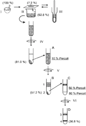
に操作の一連の流れを図解で示す.50 mM HEPES-NaOH(pH 7.5)の緩衝液(Buffer A)図 1 EPS分離操作手順の図解
I
:すり潰し,II:フィルター濾過,III:ガラスホモジナイザーでのすり潰し,IV:7000×g,4℃で 10
分間遠心分離,V:7000×g, 4℃で7
分間遠心分離,VI:12000×g, 4℃で15
分 間遠心分離,( )内はクロロフィルa
を指標とした細胞の回収率を示している.試験 管横のアルファベットと数字は図2
と対応している.Fig. 1. Procedure of the separation of extracellular polysaccharaides.
I: grinding the N. commune colony, II: separation of the suspended cells from the debris of N. commune
colony, III: homogenization of the debris of N. commune colony by a glass homogenizer, IV: centrifugation
at 7000×g, 4℃ for 10 min, V: centrifugation at 7000×g, 4℃ for 7 min, VI: centrifugation at 12000×g,
4℃ for 15 min. The numbers in the parentheses are the yield of cells based on the amount of Chl a. The
alphabets and the numbers on the side of test-tubes correspond to those in Fig. 2.
に浸したイシクラゲを氷上で乳鉢と乳棒を使ってすり潰し(図
1-I),150
ミクロンのナイロ ンメッシュフィルターで濾過した(図1-II).フィルターの上に残ったイシクラゲはガラス
ホモジナイザーを用いてさらにすり潰した後,再度上記フィルターで濾過した(図1-III).
濾液を
4℃,7000×g
で10
分間遠心し,上澄みを除いた後Buffer A
に沈殿を再懸濁させて遠心を行う操作を
5
回繰り返し,水溶性の夾雑物を取り除いた(図1-IV).次に,沈殿を再度 Buffer A
に懸濁し,そこへPercoll(GE Healthcare)を終濃度 10%
(v/v)となるように加え,アングルロータを使って,4℃,7000×gで
7
分間遠心した(図1-V).この沈殿を少量の Buffer A
で懸濁し,80% Percollと50% Percoll
を1:3
程度の分量比で重層させた溶液上に上 層し,12000×gで15
分遠心することで細胞外多糖やその他の不純物を除去した(図1-VI).
2.3. クロロフィル濃度測定
イシクラゲのクロロフィル濃度測定は,100%メタノールにより色素抽出して
Porra et al.
(1989)の計算式を用いて行った.吸光度の測定は
UV-2500PC(島津)を用いた.A
665は抽
出液の波長665 nm
の吸光度である.Chl a
(μg/ml)=12.53×A665.
⑴2.4. 酸素発生活性の測定
クラーク型酸素電極を用いて
25℃で行った.反応液には Buffer A(50 mM HEPES-NaOH
(pH7.5))に,5 mM NaHCO3
を加えたものを用い,5 μg Chl a/ml
になるよう細胞を懸濁した.光量は
1500 mol photons/m
2・sとした.2.5. アルシアンブルーによる EPS
の染色最終標品のイシクラゲ細胞に
EPS
が付着しているかを調べるため,アルシアンブルーに よるEPS
の染色と観察を行った.アルシアンブルーは,pH 2.5で酸性ムコ多糖の硫酸基と カルボキシル基を特異的に染色する試薬である.野生のイシクラゲをこの条件で処理するとEPS
が青く染まる(Hill et al., 1994).本研究ではTamaru et al.
(2005)の方法を参考に,最終 標品のイシクラゲ細胞を純水で洗浄した後,0.33%のアルシアンブルー 8 GX(和光),3%酢 酸溶液で30
分間処理し,その後純水ですすいだものを光学顕微鏡で観察した.2.6. ウロン酸の定量
カルバゾールによる呈色反応法を用いて
EPS
の主成分であるウロン酸の定量を行った(Bowness, 1956; Dische, 1947; Tamaru et al., 2005).1回の反応に
1 mg
の単離細胞と0.5 mg
の イシクラゲを用いてそれぞれ3
サンプルずつ定量を行った.イシクラゲでは1 mg
処理する と吸光度A
530が 2
を超えたため,単離細胞よりも少量で反応を行った.イシクラゲは自然乾燥した状態でセラミックお茶ミル(CM-45GT, Kyocera)によりパウダー状にすり潰し,
1 mg/ml
となるようBuffer A
を加え,単離細胞は凍結乾燥したものを2 mg/ml
となるようBuffer A
を加 えてそれぞれマイクロチューブ用ペッスルで均一になる程度にすり潰した.懸濁液を0.5 ml
ずつ共栓付き硬化ガラス試験管に移し,氷冷しながら濃硫酸(純度96%
)を3 ml
加えてボ ルテックスで混合し,沸騰したお湯に20
分間浸けてEPS
を加水分解処理した.反応液を室 温に戻した後,分光光度計でA
530を測定し,100 μl
のカルバゾール溶液(0.1%(wt/wt)カル バゾール,95%エタノール)を加え室温で2
時間静置してA
530を測定し,吸光度の増加量を
算出した.ウロン酸の定量は,10⊖100 μg/ml
の濃度のD-
(+)-グルクロノラクトンで検量線
を作成して行った.この定量法でのグルクロノラクトンの検出限界は5 μg/ml
であった.3 . 結果と考察
吸水したイシクラゲを乳鉢ですり潰す際は力を入れすぎず,丁寧にすり潰してできるだけ 未破砕の断片が残らないようにした(図
1: I).フィルターで濾過できなかった残渣をガラス
ホモジナイザーでさらにすり潰すことで収率の向上を図った(図1: III).最終的にフィルター
上に残った画分は乾燥重量比(w/w)で野生株の7.2%
(±1.4%)であった.濾液を4℃, 7000×g
で10
分間遠心すると,細胞とEPS
は共に沈殿したように見えた(図1: IV).沈殿を Buffer A
に筆を用いて再懸濁し,7000
×g
で10
分間遠心する操作を5
回繰り返して水溶性の夾雑物 を取り除いた.その後,Buffer Aに懸濁した沈殿画分にPercoll
を10%
(v/v)となるように混 合した(図1: A).Percoll
を用いてEPS
を除去する際のポイントは,細胞懸濁液を十分に希 釈し,EPSの粘性を抑えて細胞を移動させやすくすることである.通常の方法として用いら れているようにPercoll
に試料を上層して遠心すると,Percollに対する試料の体積が少量に なるためEPS
に絡んだまま分離されない細胞が増える.ここで,Percollの遠心による密度 勾配自己形成能が有用である.Percollをイシクラゲ懸濁液に最初から均一に混ぜて遠心す ることで,Percollの濃度勾配形成に伴って低密度のEPS
は上方へ,高密度の細胞は下方へ と分離されるようにした(図2: A,B).遠心前の細胞の希釈割合は 0.8 μg Chl a/ml程度とした.
この操作だけでは
EPS
以外の不純物が除去しきれなかったため(図3: B-2),さらに 80%
と
50%
濃度のPercoll
に上層して遠心分離することで(図1: VI),不純物の非常に少ないイ
シクラゲ細胞を回収することができた(図
2: C, D).図 2
のA
とB
は10% Percoll
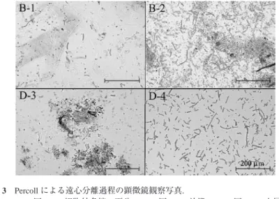
による遠心 分離前後の遠沈管の写真である.このときの上層画分と沈殿を顕微鏡で観察したものが図3
のB-1
とB-2
で,上層画分にはイシクラゲ細胞はほとんど含まれていなかったが,沈殿には イシクラゲの数珠状細胞のほかに茶色いスキトネミンと思われる色素をもったEPS
破片や,イシクラゲ群落に混在していたと思われる石や他の生物などが観察された.この沈殿を
80%
と50% Percoll
によってさらに精製することで,これら不純物をほぼ除去することができた(図3: D-4).また,最終標品であるイシクラゲ細胞の周りに EPS
の付着がないことをアルシア図 2 イシクラゲからの細胞単離の過程
(A)イシクラゲのすり潰し懸濁液に
Percoll
を終濃度10%
になるように混合したもの(B)Aを
7000×g
で5
分間遠心後の結果.1の部分は細胞外多糖,2の部分はイシクラゲ細胞の画分.
(C)Percoll濃度
80%
と50%
の2
段階ステップにB
の沈殿画分(画分2)を上層した状況
(D)Cを
12000×g
で15
分遠心後の結果.3の部分はイシクラゲ細胞をほとんど含まない画分,4の部分がイシクラゲ細胞の画分.
Fig. 2. Each step of Percoll centrifugation to purify cells from N. commune colony.
(A) N. commune homogenate in the 10% Percoll solution. (B) Fractions after centrifugation at 7000
×g for 5 min. The cell fraction (band 2; Fig. 3-B-2) was separated from the EPS fraction (band 1;
Fig. 3-B-1). (C) Cell fraction from B-2 on the top of two phase Percoll (80% and 50%). (D) Purified cell fraction (band 4) separated from impurity (band 3) after centrifugation at 12,000×g for 15 min.
図 3 Percollによる遠心分離過程の顕微鏡観察写真.
B-1
:図2B
の細胞外多糖の画分,B-2:図2B
の沈殿,D-3:図2D
の上層画分,D-4
:図2D
の下層画分.Fig. 3. Microscopic observation of fractions obtained at each step in the Percoll centrifugation process. B-1 and B-2 correspond to the fractions of Fig. 2-B-1 and -2, respectively.
D-3 and D-4 correspond to Fig. 2-D-3 and -4 fractions, respectively.
ンブルー染色法によって確認した.EPSの除去を定量的に評価するため,カルバゾール呈色 反応法により
EPS
の主成分であるウロン酸の定量を行った(Bowness, 1956; Dische, 1947;Tamaru et al., 2005).硫酸で加水分解反応後,両サンプルとも薄い茶色を呈した.検量線作成
に用いたウロン酸のグルクロノラクトン単体ではこの呈色は起こらなかったため,夾雑して いる糖類やアミノ酸が反応したものと考えられる.検量線を用いてグルクロノラクトンに換 算したウロン酸の濃度は,イシクラゲでは216±16 μg/mg(dry wt),単離細胞では 22±2.0 μg/
mg(dry wt)となり, 90%
近くのEPS
が除去されていることが示唆された.なお,カルバゾールによる呈色はウロン酸だけでなく還元糖などでも若干起こり,例としてグルコースとガラ クトースはウロン酸の
10
分の1
程度のモル吸光係数を示す(Bowness, 1956).EPSの加水 分解でこうした還元糖が生成すること(Helm et al., 2000),細胞内にも還元糖が存在すると 考えられることから,ウロン酸の定量としては今回の方法は不十分であるが,EPSの除去の 程度を確認するには十分な目安になると考えられる.乾燥重量
0.5 g
のイシクラゲ群体からの単離操作を独立して3
回行い,クロロフィルa
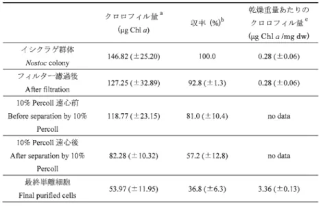
の 量で換算することにより,この手法による細胞の収率を求めた(表1).すり潰し前の野生
のイシクラゲ群体は採集した場所によって細胞密度が異なり,また,一つの群体内において もEPS
中に存在する細胞の密度が一様でなく,クロロフィル含有量に大きな差が認められる のが普通であるため,収率測定のための均一な試料を得ることが難しかった.そこで,すり 潰し後にフィルター濾過した濾液のクロロフィル量と濾過残渣の乾重量から,すり潰し前の クロロフィル量を計算した.イシクラゲ群体をすり潰してフィルターで濾過した懸濁液中の クロロフィル量を100%
として計算すると,収量は39.7
(±7.2)%
であった.フィルター上に残 存したイシクラゲ群体は乾重量にして,すり潰し前の群落の7.2
(±1.4)%
(w/w)であった.す り潰し前のイシクラゲ群体のクロロフィル量を100%
として計算すると,収率は36.8
(±6.3)%
であった.乾燥重量当たりのクロロフィル量は,イシクラゲ群体で0.28
(±0.06)μg Chl a/mg,
最終単離細胞標品では3.36
(±0.13)μg Chl a/mg
であった.イシクラゲ群落では乾燥重量に占 めるEPS
の割合が大きいためクロロフィル濃度は低いが,EPSの除去により最終単離細胞 のクロロフィル濃度が高くなったものである.すり潰し前のイシクラゲ群体と最終標品の光 合成活性を酸素電極によって測定したところ,イシクラゲ群体は154.4
(±25.7)μmoles O
2/mg Chl a/h, 単離後は 133.4(±23.1) μmoles O
2/mg Chl a/h
で,単離前の86%
程度の活性が保持さ れていた.Tamaru et al.
(2005)の方法では単離後の酸素発生活性は単離前の73%
であるので,単離操作前後の活性変化の程度はほぼ同等であった.
今回用いたイシクラゲの乾燥重量当たりのクロロフィル量は,Tamaru et al.(2005)の値よ り低いものであった.野生の土壌藻類として生育するシアノバクテリアは採集された季節や 環境条件の違いによって,細胞当たりのクロロフィル
a
の量が変化することが報告されてい る(Bowker et al., 2002, Lakatos et al., 2001).Bowker et al.(2002)は,シアノバクテリア群集の細胞当たりのクロロフィル
a
の含有量が春から秋にかけて2
倍ほどになり,その変化割合 は生育環境によっても変動することを示している.したがって,本研究と前出研究のクロロ フィル量の差は採集した生育環境の違いに由来するものであり,精製後の乾燥重量当たりの クロロフィルの割合が小さな値となったのは,単離手法の違いによるものではないと推察さ れる.今後この単離法を活用し,南極に生育するイシクラゲの光合成特性と極域環境で
EPS
が 果たしている役割の重要性を明らかにしていく予定である.謝 辞
本研究の一部は国立極地研究所プロジェクト研究「環境変動に対する極域生物の生態的応 答プロセスの研究」,および日本学術振興会科学研究費助成事業(研究課題番号:24770030)
により行われた.
a
100%
メタノールでクロロフィルの抽出を行い,665 nmでの吸光度によりクロロフィル量を求めた.
b
出発材料を 100%
として,各単離過程のクロロフィル量から計算した.c
乾燥後の重量でクロロフィル量を割った値.フィルター濾過後の乾燥重量はすり潰
し前の値から濾過残渣の乾燥重量を引いて求めた.
a
Chlorophyll contents: Chlorophyll a was extracted using 100% methanol, and the Chl a amounts were calculated using the absorbance at 665 nm.
b
Yields (%): These were estimated based on the amount of Chl a at each purification step.
c
The chlorophyll contents per dry weight: The dry weight of filtrate was estimated as: (dry weight of Nostoc colony before grind)-(dry weight of filtration residue).
表 1 乾燥重量
0.5 g
のイシクラゲからEPS
を除去する過程におけるクロロフィル量の 変化および収率Table 1. The yields of Chl a during the process of cell purification from 0.5 g of Nostoc commune colony.
文 献