Acta Med. Nagasaki 44 : 37-41
Surgical Resection Following Induction Chemoradiotherapy for Locally Advanced Lung Cancer
Masatoshi MORI 1), Tomonori MORITAKA 2)
1) Departments of Chest Surgery 2) Internal Medicine,
Ehime Prefectural Central Hospital, 83 Kasuga-machi, Matsuyama, Japan
Fourteen patients with primary bronchogenic carcinoma, in- cluding 6 stage IIIA patients and 7 IIIB disease patients, under- went a pulmonary resection following cisplatin-based chemo- therapy concurrent with 40 Gy of irradiation. The operations included 10 lobectomies, a bilobectomy, 2 pneumonectomies and a sleeve lobectomy. Dissection of the branches of the pulmonary artery was difficult, and an angioplastic proce- dure was employed in 2 patients. The average operating time was 306 minutes, and average blood loss was 338g. All pa- tients were managed routinely, without any special measures.
No major postoperative complication occurred, and patients were discharged from 11 to 35 days following surgery. Opera- tive mortality rate was 0%. The median survival time was 36 months, and the 2-year survival rate was 68%.
There is usually a tremendous amount of fibrosis in the mediastinum and hilum following induction therapy, and therefore, dissection of the adhesion is difficult. However, with meticulous dissection, it is possible to perform a suc- cessful operation without fatal complications.
Key Words: lung cancer, resection, induction chemoradiotherapy
Introduction
The prognosis of primary bronchogenic carcinoma remains poor despite recent advances in operation tech- niques, chemotherapy and radiotherapy. Surgery can provide a cure for approximately 70% of the patients
with stage I lung cancer. However, long-term survival rates following resection for stages IIIA and IIIB dis- ease is unsatisfactory.
In an effort to improve upon the poor results given
Address Correspondence: Masatoshi Mori, M.D.
Department of Chest Surgery, Ehime Prefectural Central Hospital, 83 Kasuga-machi, Matsuyama, Ehime 790-0024, Japan TEL: +81-89-947-1111 FAX: +81-89-943-4136
by conventional therapy, induction chemotherapy and concurrent chemoradiotherapy followed by surgical re- section have been becoming more promising treatments for locally advanced lung cancer. These multimodality therapies may contribute to an improved outlook on treatment of this deadly illness, however, surgeons must be aware of the higher risk of injuring vital organs in the thorax compared to conventional pulmonary resec- tion.
This retrospective study summarizes the results of sur- gical resection following concurrent chemoradiotherapy for treatment of lung cancer, and discusses the intra- and post-operative morbidity and mortality.
Materials and Methods
Patient Population
During a period from July 1992 to January 1998, 14 patients with primary bronchogenic carcinoma con- secutively underwent thoracotomy after concurrent chemoradiotherapy at the Ehime Prefectural Central Hospital. The age of the patients at the time of opera- tion ranged from 48 to 70 years, with the average being 59. There were 13 men and one woman in the group. Four tumors were on the right, and 10 on the left. A histological diagnosis of squamous cell carci- noma was made in 7 patients, of adenocarcinoma in 4 and of large cell carcinoma in 2. In one patient, post- operative pathologic examination revealed no viable cells as a result of the induction therapy. Before begin- ning the induction therapy, 6 patients were judged to have had stage IIIA tumors and 7 to have had stage IIIB disease (Table 1).
Combined Treatment Modality
The chemotherapy regimen consisted of cisplatin (20
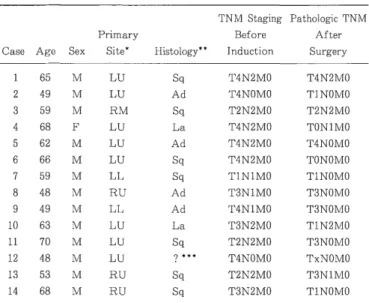
Table 1. Data of patients undergoing resection of lung with pri- mary bronchogenic carcinoma after concurrent chemoradiotherapy
TNM Staging Pathologic TNM
Primary Before After
Case Age Sex Site* Histology** Induction Surgery
1 65 M LU Sq T4N2M0 T4N2M0
2 49 M LU Ad T4NOMO TINOM0
3 59 M RM Sq T2N2MO T2N2M0
4 68 F LU La T4N2M0 T0N1M0
5 62 M LU Ad T4N2M0 T4NOM0
6 66 M LU Sq T4N2M0 TONOM0
7 59 M LL Sq T1N1M0 T1NOM0
8 48 M RU Ad T3N1M0 T3NOM0
9 49 M LL Ad T4N1M0 T3NOM0
10 63 M LU La T3N2M0 T1N2M0
11 70 M LU Sq T2N2M0 T3NOM0
12 48 M LU ? * * * T4NOM0 TxNOM0
13 53 M RU Sq T2N2M0 T3N1M0
14 68 M RU Sq T3N2M0 T1NOM0
* LU=left upper lobe; RM=right middle lobe; LL=left lower lobe;
RU=right upper lobe.
** Sq=squamous cell carcinoma; Ad=adenocarcinoma; La=large cell carcinoma.
* * * No viable tumor cell was seen .
Mg/M2 on days 1 through 5) and etoposide (40 mg/m2 on days 1 through 5) infusion, given every third week. In 3 patients, ifosfamide, vindesine or 5-fluorouracil was given as an option to etoposide. Total doses ranged from 250 to 450 mg for cisplatin, and from 500 to 700 mg for etoposide, administered over 5 weeks. A Corticosteroid (125 mg of methylprednisolone or 60 mg of prednisolone ) was given from the start of chemotherapy with the dose tapering off from 15 to 112 days.
External radiation therapy to the primary tumor and adjacent mediastinum, a dose of 40 Gy (2 courses of 20 Gy in 10 fractions), was given concurrently with chemotherapy over a period of 6 to 7 weeks in 13 pa- tients. One 48-year-old man underwent 70 Gy of radia- tion in 56 fractions on days 1 through 38.
Two to 34 days, with an average 10 days, following the end of irradiation regimen, patients were evaluated for clinical response and resectability of their tumors by means of fiberoptic bronchoscopy, computed tomography (CT) or magnetic resonance imaging (MRI) of the chest.
Twenty-four to 76 days, with an average 40 days, following the completion of induction treatment, a thoracotomy was carried out. The patients underwent a lobectomy, bilobectomy or pneumonectomy with hilar and mediastinal lymph node dissection. All operations were performed under general anesthesia in combina- tion with epidural anesthesia, and with one-lung venti- lation by means of a double lumen endotracheal tube.
In general, dissection of pleural adhesions, mobilization
of the lung, and exposure and dissection of the branches, of a pulmonary artery was not easy. Not infrequently, it took an hour or more to identify and divide a branch of the pulmonary artery. A cord tape tourniquet for proximal control of the main pulmonary artery was placed for ready use, as a contingency, should the pul- monary artery become injured. The bronchial stump was closed with 3-0 nylon interrupted sutures accord- ing to Sweet's method. The closed stump was not cov- ered with pleura or a fat pad. In one case of a right upper sleeve lobectomy, the anastomosis was carried out with 4-0 monofilament polyglyconate sutures and wrapped with a pedicled anterior mediastinal fat pad.
Three patients underwent either a combined wedge resection of another lobe, a resection of partial pleura adjacent to the tumor, or a resection of the adventitia of the thoracic aorta. Two patients underwent a left upper lobectomy with partial resection of the left main pulmonary artery. A right upper sleeve lobectomy was carried out on one patient.
All patients except one who underwent a left pneu- monectomy was extubated immediately following com- pletion of the operation. Postoperative management at the intensive care unit (ICU) was discontinued the next day in all patients. In the postoperative period, corticosteroid was not administered to any patient. All patients were managed routinely, and no special meas- ures were undertaken. Only one patients underwent post-operative adjuvant chemotherapy or radiotherapy.
Histological typing and TNM staging were classified and the effect of the induction therapy (Ef) was graded according to the General Rule for Clinical and Patho- logical Record of Lung Cancer').
Result
Thirteen of the fourteen patients underwent radical resection with histologically negative resection margins.
One patient who underwent a right upper sleeve lobectomy had a microscopically positive margin on the proximal end of the bronchial sleeve and was treated with post- operative radiotherapy (30 Gy in 10 fractions) combined with chemotherapy (10 mg of cisplatin for 10 days).
The operating time ranged from 210 to 440 minutes, with an average of 306 minutes. The amount of bleeding during the operations ranged from 115 to 668 g, with an average of 338 g, and 3 patients received transfu- sion of 1 to 3 units of packed red blood cells.
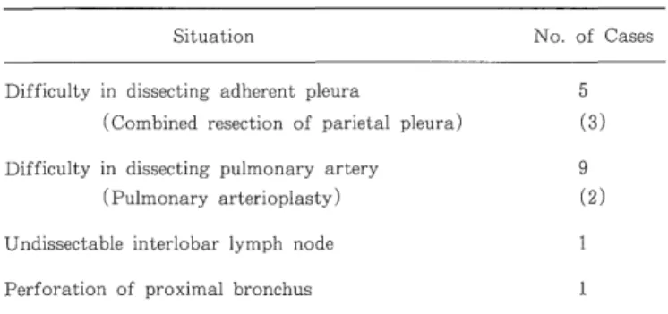
Trouble with performing a resection of the lung were
encountered in 12 of the 14 cases, and included diffi-
culty in dissecting dense pleural adhesion in 5, difficulty
in dissecting the pulmonary artery in 9, an undissectable
interlobar lymph node in 1 and perforation of the bron- chus in 1 patient (Table 2).
Pleural adhesion was awkward, and combined resec- tion of the parietal pleura was carried out in 3 cases.
A part of the adventitia of the descending aorta was resected in one patient.
Table 2. Intraoperative Difficulties
Situation No. of Cases
Difficulty in dissecting adherent pleura 5
(Combined resection of parietal pleura) (3) Difficulty in dissecting pulmonary artery 9
(Pulmonary arterioplasty) (2)
Undissectable interlobar lymph node 1
Perforation of proximal bronchus 1
In 2 of the 9 patients, perivascular fibrosis caused difficulty in dividing the branches of the pulmonary artery, so techniques of arterioplasty were employed.
A portion of the side wall of the left pulmonary artery was excised and the defect in the artery was oversewn using 5-0 polypropylene suture. In the remaining 7 cases, meticulous dissection of the vascular sheath re- sulted in the successful dividing of the branches of the pulmonary artery.
An enlarged lymph node suggesting metastasis was fixed onto the side wall of the left pulmonary artery in a 70-year-old patient, who underwent a pneumonectomy.
In one patient perforation of the intermediate trunk of the right bronchus occurred while dissecting the bron- chus with adhesion of the interlobar lymph node, but the site of the injury was distal to the ultimate line of trans- ection of the bronchus, so that a successful bilobectomy was carried out without a bronchoplastic procedure.
Post-surgical pathological examination revealed that the effects of induction chemoradiotherapy on the tumors were classified as Ef.c (no viable cancer cell) in 4 pa- tients, Ef.2 (viable cancer cells seen in less than one- third of the tumor) in 6 patients, and Ef.1 (viable can- cer cells seen in one-third or more of the tumor) in 4 patients.
Post-operative course was uneventful in 10 of the 14 patients. There was no operative death, and the hospi- tal mortality rate was 0%. Three patients had an al- veolar air leak requiring chest tube drainage for 8 days or longer. Major complications such as atelectasis, pneumonia, respiratory failure, bronchopleural fistula, hemothorax and empyema did not occur in any pa- tient. Hospitalization after the surgery ranged from 11 to 35 days, with an average of 20 days (Table 3).
Table 3. Post-operative Course
Event No. of Patients
Duration of chest tube drainage
3- 5 days 7
9-11 days 3
21-22 days 2
Hospital stay following Surgery
11-14 days 4
15-21 days 6
22-35 days 4
Post-operative complications
Prolonged air leak 3
Pleural effusion 1
Estimates of survival for the 14 patients, with follow- up of 5 months to 6 years, showed a median survival time of 36 months, and the 2-year survival rate for these patients was 68% (Fig. 1). Six patients died of cancer 5 to 36 months after the surgery, and there were documented disease recurrences in 5 patients, includ- ing brain metastasis in 2, pulmonary metastasis in 1, relapse in the bronchial stump in 1 and carcinomatous pleuropericarditis in 1 patient. Two patients who sur- vived for 37 and 45 months died of perforation of a gastric ulcer and infarction of the brain stem, respec- tively (Table 4).
Fig. 1 Overall probability of survival (death from any cause) of 14 patients undergoing pulmonary resection following in- duction chemoradiotherapy for primary bronchogenic carci- noma. Zero time on abscissa represents the date of pulmo- nary resection.
Discussion
The prognosis of primary lung cancer is unsatisfac-
tory, although surgery may provide a long-term cure
for selected patients. Long-term chance of survival fol-
lowing the resection of stage III lung cancer is disap-
pointing, and the treatment of advanced disease remains
Table 4. Response and Outcome of Pulmonary Resection Following Induction Chemoradiotherapy
Effect of
Induction
Patient Age Surgery Therapy Follow-up
1 65 Lobectomy Ef. 2 5 mo., died
2 49 Lobectomy Ef. 2 37 mo., died
3 59 Bilobectomy Ef. 1 16 mo., died
4 68 Lobectomy Ef. 2 36 mo., died
5 62 Lobectomy Ef. C 9 mo., died
6 66 Lobectomy Ef. C 31 mo., died
7 59 Lobectomy Ef. C 45 mo., died
8 48 Lobectomy Ef. 2 7 mo., died
9 49 Pneumonectomy Ef. 2 29 mo., alive
10 63 Lobectomy Ef. 2 27 mo., alive
11 70 Pneumonectomy Ef. 1 26 mo., alive
12 48 Lobectomy Ef. C 17 mo., alive
13 53 Sleeve lobectomy Ef. 1 10 mo., alive
14 68 Lobectomy Ef. 1 5 mo., alive
controversial2 3 4>. Preoperative chemoradiotherapy in pa- tients with lung cancer is a theoretically attractive con- cept, in which the tumor size can be reduced, facilitating a complete resection and treatment of micrometastases, but these theoretical advantages have yet to be proven to yield clinical benefit or improve chance of survival.
Since the advent of cisplatin, the results of chemo- therapy for advanced lung cancer have been improv- ing, and employing irradiation in combination with this tumorcidal drug may contribute to survival benefits".
Improved survival rates for patients with stage III lung cancer treated with combination chemotherapy and irra- diation preoperatively has been reported 6'''8'9,10,">. However, physicians should be aware of the risk of perioperative mortality and morbidity in this very aggressive treat- ment"). Actually, a review of the literature revealed that the operative mortality for thoracotomy following in- duction chemoradiotherapy ranged from 5% to 23%, although no case of death on the operating table has ever been reported678910)
.