Abstract. Although overt hepatitis B virus (HBV) infec-
tion promotes the onset of hepatocellular carcinoma (HCC) in hepatitis C virus (HCV)-infected patients, the effect of occult HBV infection remains unclear. The aim of this study was to investigate the effect of occult HBV infection on the early-onset of HCC in HCV-infected patients. A total of 173 HCC patients with HCV infection were enrolled and classified into 2 groups according to the median age of HCC onset: the early-onset group (n=91; 61.1±5.6 years) and the late-onset group (n=82; 73.8±3.7 years). Independent factors associated with the early-onset of HCC were assessed by multivariate analysis. In the overall analysis, independent risk factors for the early-onset of HCC were the white blood cell count and
alanine aminotransferase level, but not the presence of HBV DNA. In a stratification analysis according to albumin levels of ≥3.5 g/dl, the presence of HBV DNA was a significant independent risk factor for the early-onset of HCC (OR 145.18, 95% CI 1.38-15296.61, P=0.036), whereas the presence of antibodies against hepatitis B core antigen was not found to be a risk factor. The presence of HBV DNA was not a risk factor for the early-onset of HCC in the overall analysis.
However, its presence was an independent factor for the early-onset of HCC in HCV-infected patients with an albumin level of ≥3.5 g/dl. Thus, occult HBV infection may accelerate hepatocarcino genesis in HCV-infected patients with relatively low carcinogenic potential.
Introduction
Hepatocellular carcinoma (HCC) is one of the most common malignancies worldwide. It ranks third in men and fifth in women as the cause of death from malignancies in Japan (1).
Chronic hepatitis C virus (HCV) infection is the major cause of HCC and accounts for ~60-70% of HCC cases in Japan (2). In addition to hepatic inflammation and subsequent fibrosis, various other factors including aging, obesity and diabetes mellitus are involved in the hepatocarcinogenesis in HCV-infected patients (3-5).
Co-infection of HCV with hepatitis B virus (HBV) is thought to synergistically increase the development of HCC (6). The status of HBV infection is evaluated by the pres- ence of hepatitis B surface antigen (HBsAg), antibodies against hepatitis B core antigen (HBcAb), and HBV DNA. In some cases, HBV DNA can be detected in the serum or liver tissue of patients who are negative for HBsAg, a condition referred to as ‘occult HBV infection’ (7,8). In Japan, the prevalence of occult HBV infection in HCV-infected patients is reported
Effect of occult hepatitis B virus infection on the early-onset of hepatocellular carcinoma in patients with
hepatitis C virus infection
MASAHITO NAKANO
1, TAKUMI KAWAGUCHI
1,2, SHINGO NAKAMOTO
3,
ATSUSHI KAWAGUCHI
4, TATSUO KANDA
3, FUMIO IMAZEKI
3,5, RYOKO KUROMATSU
1, SHUJI SUMIE
1, MANABU SATANI
1, SHINGO YAMADA
1, TAKUJI TORIMURA
1,6,
TATSUYUKI KAKUMA
7, OSAMU YOKOSUKA
3and MICHIO SATA
1,21
Division of Gastroenterology, Department of Medicine, Kurume University School of Medicine;
2
Department of Digestive Disease Information & Research, Kurume University School of Medicine, Kurume;
3
Department of Gastroenterology and Nephrology, Graduate School of Medicine, Chiba University, Chiba;
4
Biostatistics Center, Kurume University, Kurume;
5Safety and Health Organization, Chiba University, Chiba;
6
Liver Cancer Research Division, Research Center for Innovative Cancer Therapy, Kurume University;
7
Department of Biostatistics, Kurume University Graduate School of Medicine, Kurume, Japan Received June 22, 2013; Accepted August 2, 2013
DOI: 10.3892/or.2013.2700
Correspondence to:
Dr Takumi Kawaguchi, Division of Gastro- enterology, Department of Medicine, Kurume University School of Medicine, 67 Asahi-machi, Kurume 830-0011, JapanE-mail: [email protected]
Abbreviations:
HCC, hepatocellular carcinoma; HCV, hepatitis C virus; HBV, hepatitis B virus; HBsAg, hepatitis B surface antigen; BMI, body mass index; WBC, white blood cell; HbA1c, hemoglobin A1c; AST, aspartate aminotransferase; ALT; alanine aminotransferase; AFP, α-fetoprotein; DCP, des-γ-carboxy prothrombin; HOMA, homeostasis model assessment; APRI, AST to platelet ratio index; AUROC, area under the receiver operating characteristic curve analysis; MAPK, mitogen activated protein kinaseKey words:
latent HBV infection, hepatoma, liver cancer, oncogenesis, white blood cellto be between 37.7% and 90% (9-11). Occult HBV infection is associated with a poor response to interferon therapy for chronic hepatitis C (12,13) and is also known to accelerate the progression of liver fibrosis, resulting in cirrhosis in patients with HCV infection (9,14,15). Several previous studies have examined the impact of occult HBV infection on the development of HCC in HCV-infected patients, but no clear conclusions have emerged (14,16,17). Moreover, the effects of occult HBV infection on the early-onset of HCC have not been investigated in HCV-infected patients.
Albumin is produced by hepatocytes, and the level of serum albumin is used to evaluate hepatic function (18). Albumin plays a significant role in maintaining colloid osmotic pressure and transports drugs and endogenous substances including bilirubin and unesterified free fatty acids (19). In addition, albumin exerts antioxidative properties (19), and hypoalbuminemia has been shown to be an independent risk factor for mortality among residents of a hyperendemic area of HCV infection in Japan (20).
A serum albumin level of ≥3.5 g/dl is an independent predictor of survival in HCC patients (21,22) and in cirrhotic patients with a serum albumin levels of <3.5 g/dl, branched-chain amino acids increase serum albumin levels and subsequently suppress hepatocarcinogenesis (23,24). Thus, the serum albumin level is an important factor in hepatocarcinogenesis.
The aim of this study is to investigate the impact of occult HBV infection on the early-onset of HCC in HCV-infected patients. We also performed a stratification analysis according to the serum albumin level.
Subjects and methods
Subjects. We conducted a retrospective study to investigate the effect of the presence of HBV DNA on the early-onset of HCC in HCV-infected patients. Between 1995 and 2011, 325 patients underwent hepatic resection at the Kurume University Hospital. The inclusion criteria were histologically proven HCC, a positive result for serum anti-HCV, and a negative result for serum HBsAg. Exclusion criteria were the presence of autoimmune hepatitis, primary biliary cirrhosis, and hemochromatosis, no test results for serum HBV DNA, and a histological diagnosis of combined hepatocellular and
cholangiocellular carcinoma. Although 214 patients met the inclusion criteria, 41 patients had to be excluded because of one or more of these reasons. The remaining 173 HCC patients with HCV infection were therefore enrolled in this study and classified into 2 groups according to the median age of HCC onset: the early-onset group (n=91; 61.1±5.6 years) and the late-onset group (n=82; 73.8±3.7 years).
The study protocol was approved by the institutional review board, and informed consent for participation in the study was obtained from each subject. None of the subjects were institutionalized.
Data collection. Demographic data were collected at the time of hepatic resection including age, gender, and alcohol intake.
Body mass index (BMI) was calculated as body weight in kilograms divided by the square of height in meters (kg/m
2).
Venous blood samples were taken in the morning after a 12-h overnight fast. The presence of serum anti-HCV, HBsAg, and HBcAb was tested using standard clinical methods (Department of Clinical Laboratory, Kurume University Hospital). Blood platelet count, white blood cell (WBC) count, prothrombin time %, plasma glucose levels; hemoglobin A1c (HbA1c) levels, and serum levels of aspartate aminotrans- ferase (AST), alanine aminotransferase (ALT), albumin, total bilirubin, insulin,
α-fetoprotein (AFP), and des-
γ-carboxy prothrombin (DCP) were also measured using standard clin- ical methods. Insulin resistance was evaluated on the basis of fasting levels of plasma glucose and insulin, according to the homeostasis model assessment for insulin resistance (HOMA- IR), as previously described (25).
The stage of hepatic fibrosis was assessed using the AST-to-platelet ratio index (APRI), which is calculated as the serum AST level (U/l)/upper limit of normal AST (U/l) x100/platelet count (x10
4/ml). Patients with APRI values of ≤1.5 were diagnosed as having chronic hepatitis, and patients with APRI values >1.5 were diagnosed as having liver cirrhosis, as previously described (26). The degree of liver cirrhosis was categorized according to the Child-Pugh classification (27). Diabetes mellitus was diagnosed on the basis of fasting blood glucose levels >126 mg/dl or HbA1c levels >6.5%, in accordance with the Diagnostic Criteria for Table I. Nucleotide positions and sequences of TaqMan PCR primers and probes.
Primer/Probe Sequence Position
S-sense TGTACAAAACCTTCGGACGGAAA 442-464
S-antisense TGCGAAAGCCCAGGATGATG 485-504
S-probe CTGCACTTGTATTCCC 465-480
C-sense ACTGTGGTTTCACATTTCCTGTCTT 2072-2096
C-antisense GGCATTTGGTGGTCTGTAAGC 2163-2183
C-probe CCACACTCCAAAAGAC 2132-2147
X-sense CTACTGTTCAAGCCTCCAAGCT 1729-1750 X-antisense GCTCCAAATTCTTTATACGGGTCAATG 1778-1804
X-probe AAGCCACCCAAGGCAC 1751-1766
Nucleotide positions are based on the sequence of hepatitis B virus subtype adr4 (GenBank accession no. X01587) (29).
Diabetes Mellitus of the Japan Diabetes Society (28), or the use of antidiabetic agents.
Nucleic acid extraction from serum. Total nucleic acid was extracted from 300 µl of plasma using a commercially avail- able kit (High Pure Viral Nucleic Acid kit; Roche Diagnostics, Tokyo, Japan) according to the manufacturer's instructions.
The extracted nucleic acid was eluted in 25 µl of elution buffer.
PCR for HBV DNA. Serum HBV DNA was analyzed for the presence of HBs, HBc, and HBx (S, C and X) regions using TaqMan real-time PCR according to the manufac- turer's instructions (TaqMan Fast Universal PCR Master mix; Applied Biosystems, Tokyo, Japan). The oligonucleotide primers and probes that were optimized for the HBV subtype adr4 (29) were specific for the S, X and C region sequences are listed in Table I. The full-length HBV DNA (GenBank accession no. X01587) (29) was used as an internal standard in the quantitative real-time detection PCR. We used 8 µl of nucleic acid-containing serum in our study for better sensi- tivity. The limit of sensitivity of our TaqMan Real-time PCR methods was 4.5 copies/well, and the detection limit of our tests was 45 copies/ml (1.7 log copies/ml). A real-time PCR assay (COBAS TaqMan HBV Auto; Roche Diagnostics) was
also performed to detect the core region of HBV DNA (limit of quantification, 1.8 log copies/ml). The presence of HBV DNA was defined as any positivity of S, X or C region.
Statistical analysis. Data are expressed as the absolute value or the mean ± SD. Differences between the early-onset and late-onset groups were analyzed using the Mann-Whitney U test. A logistic regression model with the Firth's correction 30 was used for multivariate stepwise analysis to identify independent variables associated with the early-onset of HCC, as previously described (31,32). All P-values were 2-tailed, and a level of <0.05 was considered statistically significant.
All statistical analyses were conducted using SAS version 9.2 (SAS Institute, Cary, NC, USA) or R packages version 2.15.2 (URL http://www.R-project.org/).
Results
Univariate analysis between the early-onset and late-onset groups. AFP levels, DCP levels, and maximal HCC size did not differ between the early-onset and late-onset groups (Table II).
Furthermore, although BMI, WBC count, and serum insulin levels were significantly higher in the early-onset group than in the late-onset group, there were no significant differences Table II. Differences in the clinical characteristics between the early-onset and late-onset groups.
Variable Reference value Early-onset Late-onset P
Number of patients 91 82
Age (years) 61.1±5.6 73.8±3.7 <0.001
AFP (ng/ml) <8.7 1876±12163 769±3246 0.588
DCP (mAU/ml) <40 1083±4120 1071±3845 0.378
Maximal HCC size (mm) N/A 30.4±19.6 33.2±16.2 0.055
Gender (female/male) N/A 23/68 20/62 0.893
BMI (kg/m
2) 18.5-22.0 23.6±3.6 22.4±3.2 0.045
Daily alcohol intake (none/0-60 g/>60 g) N/A 21/42/14 23/36/10 0.676
Platelet count (x10
4/mm
3) 13-36 13.8±5.4 13.5±4.6 0.988
WBC count (/mm
3) 4000-9000 5009±1526 4420±1210 0.012
AST (U/l) 13-33 56.2±29.5 52.8±27.2 0.412
ALT (U/l) 6-30 62.2±40.9 51.7±31.7 0.104
Albumin (g/dl) 4.0-5.0 3.87±0.45 3.85±0.38 0.520
Prothrombin time (%) 70-130 90.0±11.2 91.7±12.2 0.272
Total bilirubin (mg/dl) 0.3-1.2 0.84±0.35 0.79±0.29 0.346
Chronic hepatitis/Child-Pugh class A/Child-Pugh class B N/A 40/49/2 36/44/2 0.994
Complication of diabetes mellitus (yes/no) N/A 30/61 20/62 0.214
Fasting blood glucose (mg/dl) 80-109 119±39 107±31 0.060
Insulin (µU/ml) 5-20 13.1±10.4 9.8±8.0 0.014
HOMA-IR <2.5 3.05±2.47 2.11±1.03 0.622
HbA1c (%) 4.6-6.2 5.77±0.88 5.50±0.78 0.053
HBcAb positive/negative N/A 49/42 50/32 0.344
HBV DNA positive/negative N/A 6/85 3/79 0.385
Values are expressed as the mean ± SE. AFP, α-fetoprotein; DCP, des-γ-carboxy prothrombin; HCC, hepatocellular carcinoma; BMI, body mass index; WBC, white blood cell; AST, aspartate aminotransferase; ALT, alanine aminotransferase; HOMA-IR, homeostasis model assessment for insulin resistance; HbA1c, hemoglobin A1c; HBcAb, antibody for hepatitis B core antigen; HBV, hepatitis B virus; N/A, not applicable.
in the daily alcohol intake, platelet count, prothrombin time, Child-Pugh classification, presence of diabetes mellitus as a comorbidity, fasting blood glucose level, HOMA-IR value, HbA1c levels, and the serum levels of AST, ALT, albumin, and
total bilirubin (Table II). The presence of HBcAb and HBV DNA did not differ either between the early-onset and late- onset groups (Table II).
Multivariate stepwise analysis for early-onset of HCC.
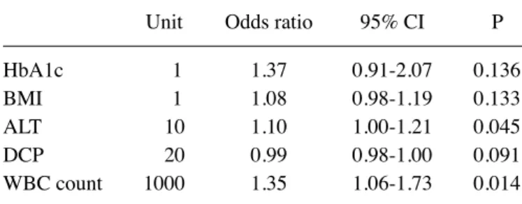
Multivariate stepwise analysis showed that the serum ALT level and WBC count were independent risk factors for the early-onset of HCC (OR 1.10; 95% CI 1.00-1.21; P=0.045 and OR 1.35; 95% CI 1.06-1.73; P=0.014, respectively; Table III), but not the presence of HBcAb or HBV DNA.
Stratification analysis according to serum albumin level.
Differences in the clinical characteristics between patients with the albumin level of ≥3.5 g/dl and <3.5 g/dl were summa- rized in Table IV. There were no significant differences in AFP levels, DCP levels, and maximal HCC size between the albumin level of ≥3.5 g/dl and <3.5 g/dl groups (Table IV). In the albumin level of ≥3.5 g/dl group, a significant elevation was seen in platelet count, prothrombin time and the number of patients with chronic hepatitis and a significant depletion was seen in AST level than in the albumin level of <3.5 g/
dl group. However, other biochemical parameters and the Table III. Multivariate stepwise analysis for factors associated
with the early-onset of hepatocellular carcinoma.
Unit Odds ratio 95% CI P HbA1c 1 1.37 0.91-2.07 0.136
BMI 1 1.08 0.98-1.19 0.133
ALT 10 1.10 1.00-1.21 0.045
DCP 20 0.99 0.98-1.00 0.091
WBC count 1000 1.35 1.06-1.73 0.014
All P-values were 2-tailed, and a level of <0.05 was considered statis- tically significant. HbA1c, hemoglobin A1c; BMI, body mass index;ALT, alanine aminotransferase; DCP, des-γ-carboxy prothrombin;
WBC, white blood cell.
Table IV. Differences in the clinical characteristics between patients with the albumin level of ≥3.5 g/dl and <3.5 g/dl.
Albumin level of ---
Variable Reference value ≥3.5 g/dl <3.5 g/dl P
Number of patients 138 35
Age (years) 67.8±8.1 67.9±6.7 0.895
AFP (ng/ml) <8.7 786±3219 3262±18195 0.248
DCP (mAU/ml) <40 854±3269 1961±5977 0.306
Maximal HCC size (mm) N/A 30.6±15.9 36.8±23.8 0.171
Gender (female/male) N/A 35/103 8/27 0.759
BMI (kg/m
2) 18.5-22.0 23.0±3.5 23.0±3.4 0.918
Daily alcohol intake (none/0-60 g/>60 g) N/A 21/58/38 3/20/6 0.172
Platelet count (x10
4/mm
3) 13-36 14.3±4.9 11.3±4.8 0.001
WBC count (/mm
3) 4000-9000 4798±1395 4291±1331 0.052
AST (U/l) 13-33 51.2±26.2 67.1±32.6 0.001
ALT (U/l) 6-30 54.9±37.4 63.4±32.7 0.057
Albumin (g/dl) 4.0-5.0 4.02±0.28 3.23±0.20 <0.001
Prothrombin time (%) 70-130 91.6±12.0 88.0±10.0 0.038
Total bilirubin (mg/dl) 0.3-1.2 0.82±0.34 0.80±0.28 0.822
Chronic hepatitis/Child-Pugh class A/Child-Pugh class B N/A 69/69/0 7/24/4 <0.001
Complication of diabetes mellitus (yes/no) N/A 38/100 12/23 0.431
Fasting blood glucose (mg/dl) 80-109 112±37 121±49 0.694
Insulin (µU/ml) 5-20 10.1±6.4 17.6±16.3 0.063
HOMA-IR <2.5 3.12±3.87 4.23±2.23 0.315
HbA1c (%) 4.6-6.2 5.61±0.79 5.68±1.04 0.905
HBcAb positive/negative N/A 75/63 24/11 0.129
HBV DNA positive/negative N/A 6/132 3/32 0.315
Values are expressed as the mean ± SE. AFP, α-fetoprotein; DCP, des-γ-carboxy prothrombin; HCC, hepatocellular carcinoma; BMI, body mass index; WBC, white blood cell; AST, aspartate aminotransferase; ALT, alanine aminotransferase; HOMA-IR, homeostasis model assessment for insulin resistance; HbA1c, hemoglobin A1c; HBcAb, antibody for hepatitis B core antigen; HBV, hepatitis B virus; N/A, not applicable.
presence of HBcAb and HBV DNA did not differ between the albumin level of ≥3.5 g/dl and <3.5 g/dl groups (Table IV).
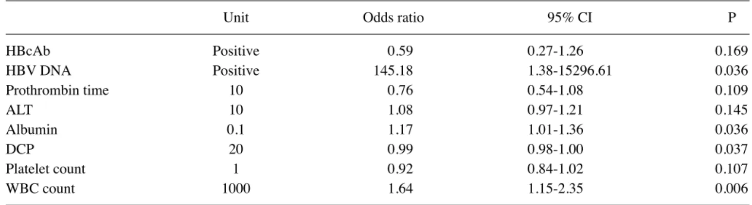
In patients with a serum albumin level of ≥3.5 g/dl, the WBC count and serum levels of albumin and DCP were iden- tified as independent factors associated with the early-onset of HCC (OR 1.64; 95% CI 1.15-2.35; P=0.006, OR 1.17; 95% CI 1.01-1.36; P=0.036, and OR 0.99; 95% CI 0.98-1.00; P=0.037, respectively; Table V). Although the presence of HBcAb was not found to be a significant risk factor for the early-onset of HCC, the presence of HBV DNA was identified as a significant independent risk factor associated with the early-onset of HCC (OR 145.18; 95% CI 1.38-15296.61; P=0.036; Table VI).
In patients with a serum albumin level of <3.5 g/dl, the serum AFP level was the only significant risk factor found to be associated with the early-onset of HCC (Table V). The presence of HBcAb and HBV DNA was not found to be a significant risk factor for the early-onset of HCC.
Discussion
In the overall analysis, the presence of HBV DNA in serum was not identified as a risk factor for the early-onset of HCC in HCV-infected patients. However, a stratification analysis according to a serum albumin level of ≥3.5 g/dl revealed that the presence of HBV DNA was an independent factor for the
early-onset of HCC. These findings suggest that occult HBV infection may accelerate hepatocarcinogenesis in HCV-infected patients with a relatively low carcinogenic potential.
Although co-infection of HCV and HBV is thought to synergistically increase the risk of HCC (6), the overall analysis in this study showed that occult HBV infection was not significantly associated with the early-onset of HCC in HCV-infected patients. Similarly, several studies conducted in Asia have also failed to show any significant effect of occult HBV infection in these patients (33-35). Recently, Lok et al (36) performed a nested case-control study using a large number of patients enrolled in the HALT-C cohort and reported no significant difference in the prevalence of occult HBV infection between HCC and non-HCC patients with HCV infection. Taken together, these results suggest that occult HBV infection may not be an intensive promoter of HCC development in the presence of a potent carcinogenic factor such as HCV infection.
In contrast with these previous studies and with our own findings for all patients, a stratification analysis according to a serum albumin level of ≥3.5 g/dl showed that occult HBV infec- tion was an independent risk factor for the early-onset of HCC.
In patients with occult HBV infection, it is unclear whether a presence of HBV DNA is due to full-length HBV DNA repli- cated from covalently closed circular DNA in hepatocytes or fragmented HBV DNA integrated into the hepatocyte genome.
However, the HBx gene is frequently integrated into cellular genes in HCC (37). The HBx protein upregulates the expres- sion of proto-oncogenes including c-jun, c-fos and c-myc, all of which can promote hepatocarcinogenesis (38,39). In addition, albumin plays a crucial role in the development of various diseases, as it is a major antioxidant (19). In cirrhotic patients with a serum albumin level of <3.5 g/dl, branched-chain amino acids increase serum albumin levels, and this subsequently suppresses hepatocarcinogenesis (23,24). In this study, we found a significant association between occult HBV infection and the early-onset of HCC in patients with a serum albumin level of ≥3.5 g/dl, but not in patients with a serum albumin level of <3.5 g/dl. Taken together, these findings suggest that HBV DNA may promote hepatocarcinogenesis in HCV-infected patients with relatively low carcinogenic potential.
Table V. Multivariate stepwise analysis for factors associated with the early-onset of hepatocellular carcinoma in patients with a serum albumin level of ≥3.5 g/dl.
Unit Odds ratio 95% CI P
HBcAb Positive 0.59 0.27-1.26 0.169
HBV DNA Positive 145.18 1.38-15296.61 0.036
Prothrombin time 10 0.76 0.54-1.08 0.109
ALT 10 1.08 0.97-1.21 0.145
Albumin 0.1 1.17 1.01-1.36 0.036
DCP 20 0.99 0.98-1.00 0.037
Platelet count 1 0.92 0.84-1.02 0.107
WBC count 1000 1.64 1.15-2.35 0.006
All P-values were 2-tailed, and a level of <0.05 was considered statistically significant. HBcAb, antibody for hepatitis B core antigen; HBV, hepatitis B virus; ALT, alanine aminotransferase; DCP, des-γ-carboxy prothrombin; WBC, white blood cell.
Table VI. Multivariate stepwise analysis for factors associated with the early-onset of hepatocellular carcinoma in patients with a serum albumin level of <3.5 g/dl.
Unit Odds ratio 95% CI P
HbA1c 1 1.83 0.75-4.47 0.183
HBV DNA Positive 0.00 0.00-2.96 0.093
AFP 20 1.39 1.01-1.93 0.045
All P-values were 2-tailed, and a level of <0.05 was considered statis- tically significant. HbA1c, hemoglobin A1c; HBV, hepatitis B virus;
AFP, α-fetoprotein.
Although we designed this study to investigate the effect of HBV DNA on the early-onset of HCC in HCV-infected patients, we found instead that an elevated WBC count is an indepen- dent risk factor for the early-onset of HCC in HCV-infected patients. An elevated WBC count may reflect the conse- quences or underlying pathogenesis of the early-onset of HCC.
One possible explanation is aging, because the WBC count declines in old age (40). Alternatively, an elevated WBC count still within the reference range is known to be associated with the development of various malignancies including gastric, colorectal, endometrial and lung cancers (41,42). The WBC count is a well-validated biomarker of inflammation. Chronic inflammation is a possible risk factor for hepatocarcinogenesis as it leads to the activation of receptors for chemokine and advanced glycation-end products (43,44). Another inflamma- tion marker, C-reactive protein, is reported to be a diagnostic and prognostic marker of HCC (45,46). Taken together, these findings suggest that inflammation may promote the early- onset of HCC in HCV-infected patients.
A limitation of this study is that there were only a small number of HBV DNA-positive patients. Previous studies regarding occult HBV infection had a similar limita- tion (33,47,48). Since occult HBV infection is not frequently seen in HCV-infected patients with HCC, a multicenter study is needed to confirm our findings.
In conclusion, the presence of HBV DNA in serum was not a risk factor for the early-onset of HCC in HCV-infected patients. However, a stratification analysis based on a serum albumin level of ≥3.5 g/dl revealed that presence of HBV DNA in serum was an independent risk factor for the early-onset of HCC. These findings suggest that occult HBV infection may accelerate hepatocarcinogenesis in HCV-infected patients with relatively low carcinogenic potential.
Acknowledgements
This study was supported, in part, by Health and Labour Sciences Research Grants for Research on Hepatitis from the Ministry of Health, Labour and Welfare of Japan.
References
1. Kiyosawa K, Umemura T, Ichijo T, et al: Hepatocellular carcinoma: recent trends in Japan. Gastroenterology 127:
S17-S26, 2004.
2. Taura N, Fukushima N, Yastuhashi H, et al: The incidence of hepatocellular carcinoma associated with hepatitis C infection decreased in Kyushu area. Med Sci Monit 17: PH7-PH11, 2011.
3. Koike K: Hepatitis C as a metabolic disease: implication for the pathogenesis of NASH. Hepatol Res 33: 145-150, 2005.
4. Kawaguchi T and Sata M: Importance of hepatitis C virus- associated insulin resistance: therapeutic strategies for insulin sensitization. World J Gastroenterol 16: 1943-52, 2010.
5. Sumie S, Kawaguchi T, Kuromatsu R, et al: Total and high molecular weight adiponectin and hepatocellular carcinoma with HCV infection. PLoS One 6: e26840, 2011.
6. Wu Q and Liu Q: Do hepatitis B virus and hepatitis C virus co-infections increase hepatocellular carcinoma occurrence through synergistically modulating lipogenic gene expression?
Hepatol Res 42: 733-740, 2012.
7. Torbenson M and Thomas DL: Occult hepatitis B. Lancet Infect Dis 2: 479-486, 2002.
8. Blackard JT, Martin CM, Sengupta S and Forrester J: Limited infection with occult hepatitis B virus in drug users in the USA.
Hepatol Res 43: 413-417, 2013.
9. Koike K, Kobayashi M, Gondo M, Hayashi I, Osuga T and Takada S: Hepatitis B virus DNA is frequently found in liver biopsy samples from hepatitis C virus-infected chronic hepatitis patients. J Med Virol 54: 249-255, 1998.
10. Fukuda R, Ishimura N, Niigaki M, et al: Serologically silent hepatitis B virus coinfection in patients with hepatitis C virus-associated chronic liver disease: clinical and virological significance. J Med Virol 58: 201-207, 1999.
11. Nirei K, Kaneko M, Moriyama M and Arakawa Y: The clinical features of chronic hepatitis C are not affected by the coexistence of hepatitis B virus DNA in patients negative for hepatitis B surface antigen. Intervirology 43: 95-101, 2000.
12. Mrani S, Chemin I, Menouar K, et al: Occult HBV infection may represent a major risk factor of non-response to antiviral therapy of chronic hepatitis C. J Med Virol 79: 1075-1081, 2007.
13. Berberova M, Mendizova A, Popchristova E, Krastev N and Genov J: Disease and treatment outcome in chronic active hepatitis C with occult HBV infection. Hepatogastroenterology 50: 2009-2012, 2003.
14. Squadrito G, Pollicino T, Cacciola I, et al: Occult hepatitis B virus infection is associated with the development of hepato- cellular carcinoma in chronic hepatitis C patients. Cancer 106:
1326-1330, 2006.
15. Cacciola I, Pollicino T, Squadrito G, Cerenzia G, Orlando ME and Raimondo G: Occult hepatitis B virus infection in patients with chronic hepatitis C liver disease. N Engl J Med 341: 22-26, 1999.
16. Hasegawa I, Orito E, Tanaka Y, et al: Impact of occult hepatitis B virus infection on efficacy and prognosis of interferon-alpha therapy for patients with chronic hepatitis C. Liver Int 25:
247-253, 2005.
17. Ikeda K, Marusawa H, Osaki Y, et al: Antibody to hepatitis B core antigen and risk for hepatitis C-related hepatocellular carcinoma:
a prospective study. Ann Intern Med 146: 649-656, 2007.
18. Kawaguchi T, Izumi N, Charlton MR and Sata M: Branched-chain amino acids as pharmacological nutrients in chronic liver disease.
Hepatology 54: 1063-1070, 2011.
19. Sakata M, Kawaguchi T, Taniguchi E, Abe M, Koga H and Sata M: Quick and simple method for increasing the reduced albumin fraction in human serum albumin preparations by using stronger neo-minophagen C. Hepatol Res 41: 1120-1125, 2011.
20. Nagao Y and Sata M: Serum albumin and mortality risk in a hyperendemic area of HCV infection in Japan. Virol J 7: 375, 2010.
21. Pacella CM, Francica G, Di Lascio FM, et al: Long-term outcome of cirrhotic patients with early hepatocellular carcinoma treated with ultrasound-guided percutaneous laser ablation: a retro- spective analysis. J Clin Oncol 27: 2615-2621, 2009.
22. Nishikawa H, Osaki Y, Iguchi E, et al: Radiofrequency ablation for hepatocellular carcinoma: the relationship between a new grading system for the ablative margin and clinical outcomes. J Gastroenterol 48: 951-965, 2013.
23. Muto Y, Sato S, Watanabe A, et al: Effects of oral branched- chain amino acid granules on event-free survival in patients with liver cirrhosis. Clin Gastroenterol Hepatol 3: 705-713, 2005.
24. Muto Y, Sato S, Watanabe A, et al: Overweight and obesity increase the risk for liver cancer in patients with liver cirrhosis and long-term oral supplementation with branched-chain amino acid granules inhibits liver carcinogenesis in heavier patients with liver cirrhosis. Hepatol Res 35: 204-214, 2006.
25. Kawaguchi T, Yoshida T, Harada M, et al: Hepatitis C virus down-regulates insulin receptor substrates 1 and 2 through up-regulation of suppressor of cytokine signaling 3. Am J Pathol 165: 1499-1508, 2004.
26. Itou M, Kawaguchi T, Taniguchi E, et al: Altered expression of glucagon-like peptide-1 and dipeptidyl peptidase IV in patients with HCV-related glucose intolerance. J Gastroenterol Hepatol 23: 244-251, 2008.
27. Pascal JP and Cales P: Propranolol in the prevention of first upper gastrointestinal tract hemorrhage in patients with cirrhosis of the liver and esophageal varices. N Engl J Med 317: 856-861, 1987.
28. The Committee of Japan Diabetes Society on the diagnostic criteria of diabetes mellitus: Report of the committee on the classification and diagnostic criteria of diabetes mellitus. J Japan Diab Soc 53: 450-467, 2010 (In Japanese).
29. Fujiyama A, Miyanohara A, Nozaki C, Yoneyama T, Ohtomo N and Matsubara K: Cloning and structural analyses of hepatitis B virus DNAs, subtype adr. Nucleic Acids Res 11: 4601-4610, 1983.
30. Firth D: Bias reduction of maximum likelihood estimates.
Biometrika 80: 27-38, 1993.
31. Otsuka M, Uchida Y, Kawaguchi T, et al: Fish to meat intake ratio and cooking oils are associated with hepatitis C virus carriers with persistently normal alanine aminotransferase levels. Hepatol Res 42: 982-989, 2012.
32. Taniguchi E, Kawaguchi T, Sakata M, Itou M, Oriishi T and Sata M: Lipid profile is associated with the incidence of cognitive dysfunction in viral cirrhotic patients: a data-mining analysis.
Hepatol Res 43: 418-424, 2013.
33. Adachi S, Shibuya A, Miura Y, Takeuchi A, Nakazawa T and Saigenji K: Impact of occult hepatitis B virus infection and prior hepatitis B virus infection on development of hepatocellular carcinoma in patients with liver cirrhosis due to hepatitis C virus.
Scand J Gastroenterol 43: 849-856, 2008.
34. Kao JH, Chen PJ, Lai MY and Chen DS: Occult hepatitis B virus infection and clinical outcomes of patients with chronic hepatitis C. J Clin Microbiol 40: 4068-4071, 2002.
35. Shintani Y, Yotsuyanagi H, Moriya K, et al: The significance of hepatitis B virus DNA detected in hepatocellular carcinoma of patients with hepatitis C. Cancer 88: 2478-2486, 2000.
36. Lok AS, Everhart JE, Di Bisceglie AM, Kim HY, Hussain M and Morgan TR: Occult and previous hepatitis B virus infection are not associated with hepatocellular carcinoma in United States patients with chronic hepatitis C. Hepatology 54: 434-442, 2011.
37. Tamori A, Nishiguchi S, Kubo S, et al: Possible contribution to hepatocarcinogenesis of X transcript of hepatitis B virus in Japanese patients with hepatitis C virus. Hepatology 29:
1429-1434, 1999.
38. Kim CM, Koike K, Saito I, Miyamura T and Jay G: HBx gene of hepatitis B virus induces liver cancer in transgenic mice. Nature 351: 317-320, 1991.
39. Koike K, Moriya K, Iino S, et al: High-level expression of hepatitis B virus HBx gene and hepatocarcinogenesis in transgenic mice. Hepatology 19: 810-819, 1994.
40. MacKinney AA Jr: Effect of aging on the peripheral blood lymphocyte count. J Gerontol 33: 213-216, 1978.
41. Margolis KL, Rodabough RJ, Thomson CA, Lopez AM and McTiernan A: Prospective study of leukocyte count as a predictor of incident breast, colorectal, endometrial, and lung cancer and mortality in postmenopausal women. Arch Intern Med 167:
1837-1844, 2007.
42. Iida M, Ikeda F, Ninomiya T, et al: White blood cell count and risk of gastric cancer incidence in a general Japanese population:
the Hisayama study. Am J Epidemiol 175: 504-510, 2012.
43. Barashi N, Weiss ID, Wald O, et al: Inflammation induced hepatocellular carcinoma is dependent on CCR5. Hepatology:
Mar 21, 2013 (Epub ahead of print). doi: 10.1002/hep.26403.
44. Pusterla T, Nèmeth J, Stein I, et al: Receptor for advanced glycation endproducts (RAGE) is a key regulator of oval cell activation and inflammation-associated liver carcinogenesis in mice. Hepatology 8: 363-373 2013.
45. Lee FY, Lee SD, Tsai YT, Wu JC, Lai KH and Lo KJ: Serum C-reactive protein as a serum marker for the diagnosis of hepato- cellular carcinoma. Cancer 63: 1567-1571, 1989.
46. Dufour JF: C-reactive protein, a prognostic marker in HCC.
Hepatology 57: 2103-2105, 2013.
47. Assar S, Arababadi MK, Ahmadabadi BN, Salehi M and Kennedy D: Occult hepatitis B virus (HBV) infection: a global challenge for medicine. Clin Lab 58: 1225-1230, 2012.
48. Matsuoka S, Nirei K, Tamura A, et al: Influence of occult hepatitis B virus coinfection on the incidence of fibrosis and hepatocellular carcinoma in chronic hepatitis C. Intervirology 51: 352-361, 2008.