Clinicopathological and Molecular Features of Laterally Spreading Tumors
Kenichi K ONDA
1), Kazuo K ONISHI
1), Atsushi K ATAGIRI
1), Hisako N OZAWA
1), Yutaro K UBOTA
1), Takashi M URAMOTO
1),
Yuichiro Y ANO
1), Toshihiro K IHARA
1), Masayuki T OJO
1), Kensuke S HINMURA
1), Teppei T AGAWA
1), Fumito Y ANAGISAWA
1),
Reiko M AKINO
2)and Hitoshi Y OSHIDA
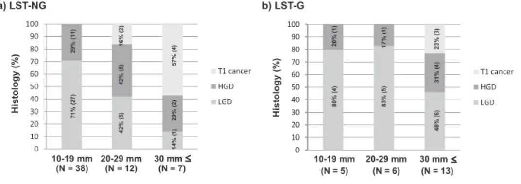
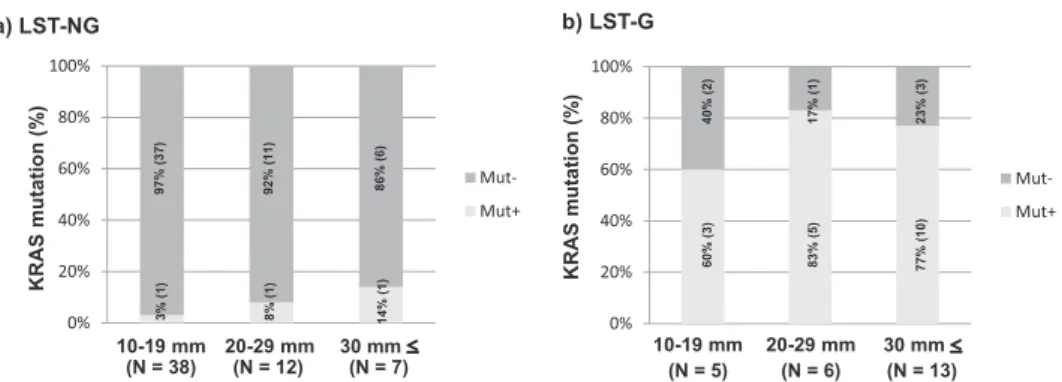
1)Abstract : Colorectal flat-elevated neoplasms can be classified into small-flat adenoma and laterally spreading tumors (LSTs) , which can then be sub-categorized into granular-type (LST-G) and nongranular-type (LST-NG) LSTs with possible biological differences between them. We evaluated clinicopathological features and KRAS / BRAF mutations in 24 LST-Gs and 57 LST-NGs. PCR-based pyro- sequencing assays were used to determine the presence of activating mutations in codons 12 and 13 of KRAS and in codon 600 of BRAF. Significant differences between LST-Gs and LST-NGs were observed in tumor size (30 mm vs. 15 mm, P <0.0001) and the frequency of KRAS mutations (75%, 18 / 24 vs. 5%, 3 / 57, P <0.0001) . For LST-NGs, the histological grade was increased with an increase in the tumor size. The frequency of submucosal cancer (SM-ca) was also higher in tumors of at least 20 mm than in tumors smaller than 20 mm (P< 0.05) . In contrast, there was no indication of a size-dependent increase in the histological grade. No significant difference in the frequency of KRAS mutation in LST-Gs and LST-NGs was related to tumor size. Two subtypes of LSTs were observed to have different clinicopathological and molecular characteristics. These findings sug- gest that different molecular mechanisms could exist in these subtypes of colorectal flat-type neoplasms.
Key words : colorectal adenoma, colorectal carcinoma, laterally spreading tumor, KRAS mutation, flat adenoma
Introduction
Colorectal cancer (CRC) can develop via various molecular pathways. Most CRCs develop over a long period of time by a multistep process called the adenoma-carcinoma sequence
1). Approximately two-thirds of sporadic CRCs arise from conventional adenomas and usually show a protruding (polypoid) macroscopic appearance. The process of colorectal carcinogenesis often begins with inactivation of the adenomatous polyposis coli (APC) gene / β -catenin signaling pathway (the Vogelstein mode
l), followed by KRAS and TP53 mutations
2).
Original
1)
Department of Medicine, Division of Gastroenterology, Showa University School of Medicine, 1—5-8 Hatanodai, Shinagawa-ku, Tokyo 142-8666, Japan.
2)