Title Studies on the Control of Avian Influenza by Enhancement of Bio-security( 本文(Fulltext) )
Author(s) THAMMAKARN Chanathip
Report No.(Doctoral Degree) 博士(獣医学) 甲第445号 Issue Date 2015-09-24 Type 博士論文 Version ETD URL http://hdl.handle.net/20.500.12099/53640 ※この資料の著作権は、各資料の著者・学協会・出版社等に帰属します。
Studies on the Control of Avian Influenza
by Enhancement of Bio-security
(バイオセキュリティ強化による
鳥インフルエンザの制御方法に関する研究)
2015
The United Graduate School of Veterinary Sciences,
Gifu University
(Tokyo University of Agriculture and Technology)
Studies on the Control of Avian Influenza
by Enhancement of Bio-security
(バイオセキュリティ強化による
鳥インフルエンザの制御方法に関する研究)
CONTENTS
Chapter 1 General Introduction.………1
I-1. Current poultry production 2
I-2. Poultry diseases and their global effects on the economy 3 I-3. Transmission and spreading of important poultry diseases 5 I-4. Disease control for a modern poultry industry 9
I-5. Biosecurity in poultry farming 10
I-6. Enhancement of biosecurity systems 10
I-7. Ceramic powders 11
I-8. Objective of thesis dissertation 13
Chapter 2 Preliminary Materials Selection………...………15
II-1. Background 16
II-2. Materials and methods 17
II-3. Results 20
II-4. Discussion 21
Chapter 3 Efficacy of Scallop Shell Powder………..28
III-1. Introduction 29
III-2. Materials and methods 30
III-3. Results 34
Chapter 4 Efficacy of Bioceramic Derived from Chicken Feces……….46
IV-1. Background 47
IV-2. Efficacy of BCX on poultry viruses and its stability in environment 47
IV-2-1. Materials and methods 47
IV-2-2. Results 50
IV-2-3. Discussion 51
IV-3. Fecal-oral transmission inhibition of virus using BCX 53
IV-3-1. Materials and methods 53
IV-3-2. Results 59
IV-3-3. Discussion 63
IV-4. BCX application in rehabilitation aviary 67
IV-4-1. Materials and methods 67
IV-4-2. Results 70
IV-4-3. Discussion 73
Chapter 5 General Conclusion and Discussion……….94
V-1. General conclusion 95
V-1-1. Conclusion for efficacy of SSP on poultry diseases control 96 V-1-2. Conclusion for efficacy of BCX on poultry diseases control 97
V-2. General discussion 99
ACKNOWLEDGEMENTS………..………102 REFERENCES……….…...104
ABBREVIATION
ADG Average Daily Gain
AI Avian Influenza
AIV Avian Influenza Virus
ANOVA Analysis of Variance
B: BW Bursa Index
BCX Bioceramic Derived From Chicken Feces
BF Bursa of Fabricius
bp Base Pair
C Celsius
CEF Chicken Embryo Fibroblasts
CFU Colony Forming Unit
CPE Cytopathic Effect
CRBCs Chicken Red Blood Cells
DHL Desoxycholate Hydrogen Sulfide Lactose
DNA Deoxyribonucleic Acid
dpi Days Post-Inoculation
dW2 Redistilled Water
E. coli Escherichia coli
EMEM Eagle’s Minimum Essential Medium
FAO Food and Agriculture Organization of the United Nations
FBS Fetal Bovine Serum
FCR Feed Conversion Ratio
g Gram
GM Growth Medium
GPV Goose Parvovirus
h Hour
HA Hemagglutination Assay
HACCP Hazard Analysis and Critical Control Points
HEPA High Efficiency Particular Air
HPAI Highly Pathogenic Avian Influenza
HPAIV Highly Pathogenic Avian Influenza Virus
HSD Honest Significant Difference
IBD Infectious Bursal Disease
IBDV Infectious Bursal Disease Virus
LB Lysogeny Broth
LBM Live Bird Markets
LPAI Low Pathogenic Avian Influenza
LPAIV Low Pathogenic Avian Influenza Virus
M Molar
MAFF Ministry of Agriculture, Forestry and Fisheries
MD Marek’s Disease
MDCK Madin-Darby Canine Kidney
MDEF Muscovy Duck Embryo Fibroblasts
MHLW Ministry of Health, Labor and Welfare
min Minute
ml Milliliter
mM Millimolar
mm Millimater
NBRC National Institute of Technology and Evaluation Biological Resource Center
ND Newcastle Disease
NDV Newcastle Disease Virus
NT Not Tested
nt Nucleotide
OIE World Organization for Animal Health
PBS Phosphate Buffered Saline
PFU Plaque Forming Units
PVC Bioceramic Derived From Polyvinyl Chloride
RF Reduction Factor
RNA Ribonucleic Acid
RT-PCR Reverse Transcription Polymerase Chain Reaction
SB Bioceramic Derived From Soybean
SD Standard Deviation
sec Second
SI Salmonella Infantis
SL Slaked Lime
SPF Specific Pathogens Free
SSP Scallop Shell Powder
ta Converted Titer into Log10 of the Recovered Virus of Treated Samples
Tpc Converted Titer into Log10 of the Recovered Virus of Untreated Samples µg Microgram µl Microliter µm Micrometer VN Virus Neutralization VP2 Viral Protein 2 w/v Weight by Volume w/w Weight by Weight 2 Chi-square
Chapter 1
2
I-1. Current poultry production
Today, products from poultry species are the major meat sources for mankind. The cost of the protein production per unit in the poultry industry is relatively cheap compared with meat sources produced from other animals. This is due to genetic selection of the poultry and as a result of the high efficiency of the feed conversion ratio (FCR). The protein from poultry sources can be obtained from various species such as chicken, duck, goose, quail and ostrich etc. Poultry production across the world has increased according to increase of the human population which elevates the protein consumption.
This situation has boosted the growth of the poultry industry and the related businesses that supply the industry. For example industries that supply poultry feed, additives, drugs, vaccines, instruments and transportation etc, have all grown alongside the poultry industry, which helps drive the world economy (Fig. 1-1).
Fig. 1-1. Number of chickens (a), carcass weight (b) and egg production per animal (c) from
1961 to 2008, data from FAO (2010) [105].
b
c
a
3
The poultry production system over the past two decades has been transformed to be an industry of integrated farming, for mass production supplies to the human food chain. To meet the consumption demand, large scale production has helped to keep the prices of poultry products relatively low. Many investments have been made in this area and the related businesses. However, the supply to the global market is still insufficient. Hence, small scale poultry production systems by smallholders and backyard farming still play important role for supplying the protein sources in a family and within small communities (Fig. 1-2).
Fig. 1-2. Global distributions of chickens (a) and ducks (b), data from Livestock Geo-Wiki
(http://www.livestock.geo-wiki.org) [84].
I-2. Poultry diseases and their global effects on the economy
Success in optimal production yield is affected by breed, feed and management, which also includes disease control. Control of diseases is one of the crucial factors which directly effects poultry population and production yields. The diseases can be infectious and
non-4
infectious. The non-infectious diseases are usually caused by inappropriate management, which can usually be resolved by management correction. On the other hand, the infectious diseases cause big damages to production systems. Causative agents may circulate in the farm or surrounding areas and possibly reappear under certain conditions. Some diseases are highly contagious and harmful, often causing outbreaks in many areas around the world. Examples of these are avian influenza (AI), Newcastle disease (ND) and infectious bursal disease (IBD). The infectious diseases can be transmitted through various routes. However, the main routes attributed to many impacting pathogens usually occur by ingestion and airborne transmission [31, 38, 89, 122].
Current trend in consumption, and the need for clean and high-quality food at a relatively low price, food borne pathogens and chemical residues are seriously concerned. As a result, global trading barriers have been established using these reasons as criteria. Some poultry diseases, especially AI and ND, are highly focused, because of their huge impacts on the poultry production system, as well as human health concerns. If these diseases re-emerge in countries around the world, there would be serious implications and restrictions regarding the import and export of poultry products.
So far, disease prevention has been implemented using various tools such as vaccines, disinfectants and bio-security systems etc. However, highly pathogenic avian influenza (HPAI) virus (HPAIV) H5N1 subtype emerged in many countries especially in Southeast Asia since late 2003 [55, 91]. HPAIV H5N1 has very harmful effects to poultry health and becomes endemic in many regions (Fig. 1-3). Similar to other contagious livestock diseases, HPAI affects poultry production yield via morbidity and mortality. This disease pushed government for interventions
5
aimed at disease control including mass culling of infected flocks, movement restrictions of birds and surveillance of the disease.
I-3. Transmission and spreading of important poultry diseases
The emergence or re-emergence of some infectious diseases have enormous impacts on the poultry industries as mentioned. Spreading of AI viruses (AIVs) was found to be relating to weather, a temperature drop shortly before these outbreaks in birds in each of the Eurasian regions stricken in 2005 and 2006 [56]. Moreover, a wealth of evidence discloses that wild aquatic birds are reservoirs for influenza viruses [24, 25, 33, 36, 39, 41, 43, 53, 63, 75, 76]. AIV is therefore spread each other in the resting or breeding areas via migratory birds. The transmission of AIV may occur among migrating birds and domestic poultry through the local avian species or direct contact with birds in the poultry farms.
HAIV H5N1 affects global health and the economic loss of poultry production, as well as human health concerns due to the human fatalities caused by the virus transmission through live birds and live bird markets (LBM). The LBM plays a crucial role in the maintenance, amplification and dissemination of AIVs as potential zoonotic transmission of influenza viruses to humans [69, 95]. HPAIV can also persist in chicken meat for several weeks under frozen storage [20, 71], as well as in feathers or feces [13, 115].
6
Fig. 1-3. Disease distribution maps of avian influenza virus during January-June 2015. Low
pathogenic avian influenza (a) and highly pathogenic avian influenza (b), data from OIE (2015) [113].
a
7
AIVs can be spread in a variety of ways during migration of wild aquatic birds. Poor hygiene practices are especially problematic in some Asian countries containing large populations of backyard poultry, as well as domestic ducks raised as adjunct or in the rice fields [65].
Similar to HPAI, ND has a huge impact on poultry industries. It is one of the global trading barriers, because of its high mortality and morbidity. ND virus (NDV) has also been found in wild or migratory birds [14, 37, 57, 85, 94]. Along with strict hygiene management, vaccination programs with inactivated and live attenuated viruses have been used for the prevention and control of ND. However, this disease still occurs occasionally in some countries (Fig. 1-4) [14, 48].
Fig. 1-4. Disease distribution maps of Newcastle disease during July-December 2014, data from
8
IBD, also known as Gumboro disease, is one of the most highly contagious diseases that effects chicken production worldwide (Fig. 1-5). This virus resists heat inactivation [3] as well as chemical treatments [77]. It can survive in various environments, even during composting procedures [30]. The most efficient way to prevent this disease is vaccination, as chickens are susceptible to IBDV in their first week of life. It’s possible that some vaccines may not give full protection against very virulent IBD virus strain, depending on its characteristics or vaccination procedures. Early vaccination with live vaccines, however, may be interfered by maternally derived specific antibodies against IBDV [68].
Fig. 1-5. Disease distribution maps of infectious bursal disease during July-December 2014, data
from OIE (2015) [113].
Although chickens are highly susceptible to IBD, other poultry species such as turkeys and ducks show minimal or no clinical signs under natural conditions [49], However, IBDV was isolated from clinically healthy ducks which were negative for IBD antibody [49]. These results
9
suggest that turkeys and ducks can be reservoirs and pass this virus to domestic chickens, especially in non-vaccinated chickens, which increase the risk of IBDV as compared to vaccinated birds [119].
Transmission risks of infectious diseases in backyard poultry was affected by making contact with wild birds, neighboring backyard waterfowls and local LBM [110], The disease pathogens can also be introduced into a farm area by human, mechanical or biological vectors.
I-4. Disease control for a modern poultry industry
According to the different types of poultry farming, the disease control measures vary depending upon the management. Generally, the industrial farming systems have much higher intensive management systems than smallholders or backyard farming. The production systems in the industry are usually operated by management of the companies. The production systems are quite complex. The need to supply products continuously may result in companies performing sub-standard disease control measures. The consequences of the sub-standard disease control measures may contribute to the invasion of pathogens and the spread of diseases in farms.
Intensive poultry farming provides ideal conditions for the mutation and transmission of pathogens, as huge numbers of birds can be crowded in narrow spaces. A warm dusty environment can facilitate the transmission of disease pathogens. In addition to this, genetic selections of birds to generate new breeds for their faster growth rates and the higher meat or egg yields have made immune systems of birds less able to cope with infections.
Since HPAI became the most important disease of interest, some countries have decided to use vaccines as the strategy to stop the disease from spreading [21, 54, 78, 81, 98, 121]. However, the live-attenuated vaccine could cause the circulation of the virus in the area, bringing
10
genetic evolution under vaccination pressure [22] and vaccine using was found to be unsuccessful in eradicating the virus from the countries [98]. Thus, many bio-security measures have been the topic of interest. They are important for reducing the risk of infection and protecting the poultry from diseases such as HPAI. There are many materials tested for inactivating the virus, and several methods for application have been developed which are the essential tools for reducing AIV from spreading [34, 74, 98, 103, 104, 106, 108]. Until now, many chemicals have been used as virucide in the biosecurity system. But the efficacy of them is usually not prolonged and can be affected by many factors when used under field conditions. Hence, studies on how to control AI by enhancement of biosecurity should be carried out for finding the way to prevent and control this disease.
I-5. Biosecurity in poultry farming
Biosecurity is a term originally described efforts to prevent infectious disease in crops and livestock, particularly poultry [72]. Good biosecurity should be practiced at all times, not only during a disease outbreak, to protect birds and poultry industries from infectious diseases. However, occasionally biosecurity measures can fail, enabling outbreaks to occur on intensive production farms. The rise of emerging and re-emerging poultry diseases has forced the operators to establish tools and measures to prevent the diseases from entering their farms.
I-6. Enhancement of biosecurity systems
Intensive biosecurity systems are the topic of interest in terms of disease prevention and control. Nowadays, vaccination and disinfection are the core measures for disease prevention. Disinfectants play a major role for farm cleaning and disinfection. But most of the disinfectants
11
usually lose their efficacy under farm conditions [58], especially in the presence of organic materials [18, 26]. Their efficacy may not last long enough after being applied [108]. Thus, the restriction of disinfectant applications brings about vulnerability of biosecurity systems. Therefore, searching for new candidate materials that can be applied in livestock farming is essential for the enhancement of biosecurity systems serving farming.
I-7. Ceramic powders
Ceramic powders are materials derived by a process of sintering or burning. The ceramics can be prepared by various sources, i.e. chicken feces, scallop shell, cow feces, Japanese cypress, soybean (byproduct from tofu production), polyvinyl chloride etc. The ceramic powders derived from organic matter, prepared by sintering process with the plant developed by NMG Environmental Development Co., Ltd. (Tokyo), were tentatively named bioceramics (Fig. 1-6).
12
The bioceramic derived from chicken feces at pH 10.6 that was first reported as a virucidal material by Takehara et al. had virucidal effects on AIV and avian adenovirus [103]. The ceramic powder could maintain the virucidal activity even after washing with 1M Tris-HCl (pH 8.0) and the virucidal activity was not affected by presence of organic material [103]. This material is safe for applying in chick feed as feed additive, even adding up to 5% in feed. There was no difference in body weight between normal feeding and the ceramic-mixture nurturing. The mode of action of the ceramic powder remains unknown, but it possibly works by its alkalinity and by adsorbing the virus [103].
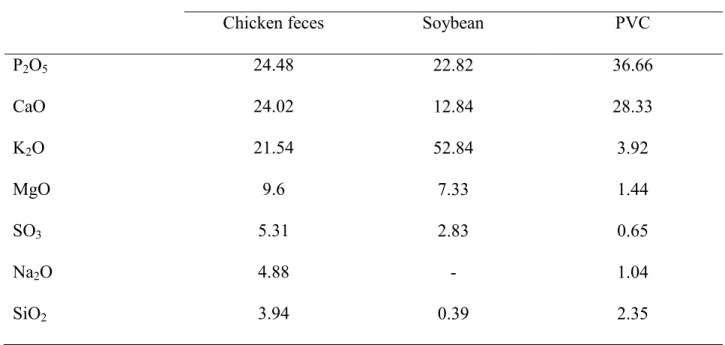
Table 1-1. Composition of ceramics derived from some materials.
Components Amount in ceramics from various sources (%)
Chicken feces Soybean PVC
P2O5 24.48 22.82 36.66 CaO 24.02 12.84 28.33 K2O 21.54 52.84 3.92 MgO 9.6 7.33 1.44 SO3 5.31 2.83 0.65 Na2O 4.88 - 1.04 SiO2 3.94 0.39 2.35 PVC: Polyvinyl chloride
13
Table 1-1 shows composition of ceramics derived from chicken feces, soybean and polyvinyl chloride that were evaluated by fluorescent x-ray analysis method (data provided by NMG Environmental Development Co. Ltd.). There are 3 major components, namely P2O5, CaO and K2O. These 3 components were preliminary supposed to be the main active components of each ceramics. However, Tsujimura et al. [106] reported that ceramic powder made from scallop shell by burning process, which contains 99.9% of CaO illustrated the excellent efficacy to inactivate low pathogenic AIV and goose parvovirus, even in a short contact time as 3 min. It could maintain virucidal efficacy for several weeks when kept in a natural environment. This report displayed the potency of CaO to inactivate the virus, which was the evidence that CaO could possibly be the main active component for viral inactivation.
I-8. Objective of thesis dissertation
According to the information described previously, the recent situation regarding poultry diseases around the world is still unstable. Bio-security systems still play a major role in poultry disease control at farm level. However, the intensive measurement for diseases prevention is still limited to industrial scale farms. Bio-security systems require big investment, which small farm owners cannot obtain. The farms with low bio-security systems tend to be portal entry sites of infectious agents through wild animals. This is one of the reasons for the continuous outbreaks of infectious diseases.
This thesis dissertation aims to find materials or methods for enhancing bio-security systems at farms. Ceramic powders are interesting for evaluating and optimizing methods of application in poultry farms, either at industrial scale, small holder or backyard level. Therefore,
14
various sources of ceramics, their efficacies should be evaluated to find the best way to apply them at the farms for enhancement bio-security systems in the poultry industry.
Chapter 2
16
II-1. Background
Since 2003, outbreaks of highly pathogenic avian influenza (HPAI) by avian influenza virus (AIV) subtype H5N1 in Asia have been causing serious problems to poultry production and spread widely in Asia, Europe, and Africa [55, 91]. It affects global health and the world economy, with a decrease of poultry productivity, and has become one of the major factors regarding trade barriers.
For HPAI control, the stamping out policy is a key point and the strategy of vaccination only is not preferred as a general concept [15]. Even with vaccination, to control many diseases, especially Marek’s disease (MD) or infectious bursal disease (IBD), general hygiene management including cleaning and disinfection are very important. Prevention of invasion and the elimination of pathogens at farms are very critical. Hence, biosecurity measures have become the topic of interest and it is very important to search for good materials that can inactivate pathogens and are stable in the environment.
The products produced from biological materials such as animal feces, food residue, and food processing byproducts by sintering process with the plant developed by NMG Environmental Development Co., Ltd. were tentatively named bioceramics in this thesis. Efficacy of a bioceramic powder derived from chicken feces (BCX) at pH 10.6 to inactivate AIV was reported by Takehara et al. in 2009 [103]. In the present experiment, bioceramic powders from various sources had been preliminary evaluated for the efficacy to inactivate AIV. The possibility of bioceramics application in the environment was also evaluated.
17
II-2. Materials and methods
II-2-1. Ceramic Powders
The ceramics used in the present experiment were derived from various sources, including chicken feces (BCX), soybean (byproduct from tofu production; SB), and polyvinyl chloride (PVC), and these ceramics were provided by NMG Environmental Development Co., Ltd (Fig. 2-1). The other ceramic powder produced from scallop shell (scallop shell powder: SSP) was supplied by C&C Co., Ltd. (Tokyo, Japan) (Fig. 2-1). As already described in I-7, SSP had been tested in our lab and showed good efficacy to inactivate viruses [106]. All the ceramics were examined for the pH after conducted 20 % w/w suspension with double distilled water (dW2). The pH strips (PEHANON, pH 9.5-12.0 and pH 12.0-14.0, Macherey Nagel GmbH & Co. KG, Germany) were applied to check the pH of the supernatants after centrifugation of the suspension by 340 g for 5 min.
II-2-2. Viruses
In this experiment, two viruses, including low pathogenic AIV (LPAIV) H7N1 (A/duck/Aomori/395/04) [43] and goose parvovirus (GPV) strain IHC [100] were applied for the evaluation of the efficacy of the bioceramics to inactivate the viruses. AIV was propagated in 10-day-old embryonated chicken eggs and GPV in Muscovy duck embryo fibroblasts (MDEF). The harvested allantoic fluid and the supernatant of MDEF culturing medium which contained the viruses were stored at -80 oC until further testing.
18
II-2-3. Cell cultures
Two kinds of cells were used for culturing the viruses during the evaluation procedure in the experiment. One was Madin-Darby canine kidney (MDCK) cells. This cell line was used for AIV culturing. The other one was MDEF, the primary cells prepared from 14-day-old embryonated Muscovy duck eggs provided from Gin-no Kamo, Agricultural union (Aomori, Japan). The MDEF had been subjected for culturing the GPV strain IHC, a MDEF adapted virus [100]. Both kinds of the cells were cultured in growth medium (GM), consisting of Eagle’s minimum essential medium (EMEM; Nissui Pharmaceutical Co., Ltd., Tokyo, Japan), supplemented with 0.3% of tryptose phosphate broth, 5% fetal bovine serum (FBS), L-glutamine (0.3 mg/ml), NaHCO3 (1.4 mg/ml), and an antibiotic-fungicide cocktail (penicillin 100 IU/ml, Streptomycin 0.1 mg/ml and amphotericin B 0.5 µg/ml).
II-2-4. Experimental designs
II-2-4-1. Virucidal efficacy of ceramics on AIV and GPV
For inactivation efficacy testing, the ceramics of 200 mg were mixed with 100 µl of viruses, and then kept at room temperature (25oC) for 20 h., except SSP which was kept for 3 min. After that, viruses were recovered by adding 900 µl of maintenance medium (MM) (GM as mentioned above, without FBS), vortexed vigorously and centrifuged by 17,400 g for 3 min. The resulted supernatant was then the subject for making 10 fold dilutions serially with MM and titrated on MDCK cells for AIV H7N1 with 1 ug/ml trypsin. The cytopathic effect (CPE) was observed and determined at 3 days post-inoculation (dpi), which was confirmed by hemagglutination assay (HA) for measuring the virus growth.
19
The GPV inactivation was also performed as described above. But the virus titration was performed using MDEF without adding trypsin and CPE was determined at 7 dpi, which was confirmed by 0.5% crystal violet (w/v) staining.
II-2-4-2. Stability in environment testing
All the ceramics were assessed for their stability efficacy to inactivate AIV after being applied into the environment. The ceramics with the amount of 3 g were poured into 90 mm petri dishes (3 g/57cm2 = 526 g/m2) (Fig. 2-1). Then all the dishes were kept under the sunlight from morning to evening for 7 weeks.
Another set of dishes containing each ceramic was also prepared and kept separately, aiming to measure the efficacy after being kept under wet and dry conditions. These ceramics in dishes were suspended with chlorine free tap water, 10 ml per dish. This water had been stored in an open container at room temperature at least one day before using for ensuring the chlorine vapored out. Then these dishes were kept with the lids open under the sunlight until the ceramics became dried up. Each dish was re-suspended with the same volume of water followed the evaporation. This procedure was repeated 7 times.
The portions of the ceramics in the dishes were collected for 7 weeks under the sunlight, as well as for 7 times after being dried. All the collected samples were stored at room temperature for further virucidal efficacy testing. The ceramics of 100 mg in micro-tubes were incubated with 50 µl of AIV. The virus was recovered with 450 µl of MM, made serial 10 fold dilution, then inoculated into MDCK cells.
20
Virus titers were calculated as 50% tissue culture infectious dose (TCID50)/ml, according to Behrens-Kärber’s method [64]. Reduction factor (RF) was calculated using the following equation:
RF = tpc – ta
The tpc is converted titer into log10 of untreated samples, while ta is converted titer of the recovered virus of treated samples. Inactivation was considered to be satisfactory when RF≥3 [5, 34, 58, 103, 106].
II-3. Results
The results obtained from the pH measurement of supernatant mixed with ceramic revealed that all the ceramics had alkalinity properties. The lowest value was illustrated by PVC, of which the pH was 9.5-10. The highest values were for BCX and SSP, up to pH 13 (Table 2-1).
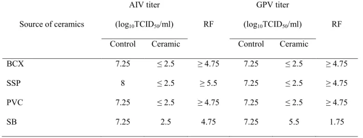
After 20 h of the incubation period, all the ceramics showed the sufficient efficacy to inactivate AIV, the titer of which could be reduced more than 10,000 times (RF>4) when compared with positive control, namely the untreated virus which was kept for the same period as the incubated virus (Table 2-2). At the same incubation period as mentioned for AIV, BCX, SSP and PVC showed the satisfactory efficacy to reduce GPV titer, with more than 10,000 times (RF>4) of reduction, compared with the virus control. Solely, SB had the ability to inactivate GPV with less than 100 times reduction (Table 2-2).
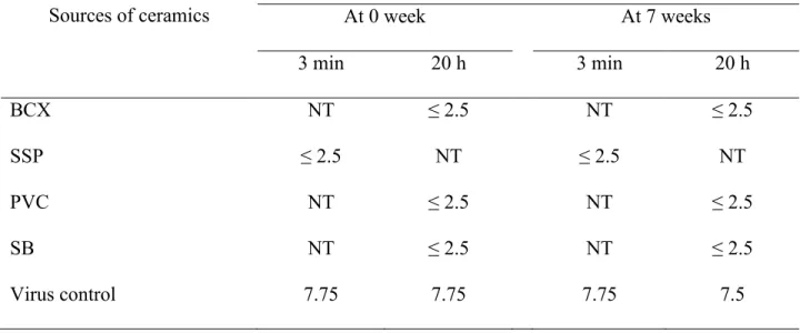
After keeping the ceramics derived from various sources under the sunlight for 7 weeks, the ceramics treated viral titers were as shown in Table 2-3. The ceramics were incubated with AIV for 20 h except SSP, which was incubated for 3 min. The complete inactivation was found with all the collected samples with 20 h incubation. All the samples reduced the viral titer to the
21
one lower than the detectable level (≤2.5 log10TCID50/ml) at the beginning, and even after 7 weeks under the sunlight. The SSP sample collected after 7 weeks also showed virucidal efficacy (RF≥5.75) with 3 min incubation.
Under the wet and dry conditions, namely being washed with water and dried under the sunlight 7 times, the collected samples could still inactivate the virus to the undetectable level (≤ 2.5 log10TCID50/ml) if they were incubated with AIV for 20 h (Table 2–4). The SSP sample collected after 7 times washing lost its virucidal efficacy with 3 minute incubation periods, however, if the sample was incubated with the virus for 20 h, it still showed the sufficient virucidal efficacy (RF=3.75) (Table 2-4).
II-4. Discussion
Influenza virus is an enveloped RNA virus [97], stable at a slightly basic pH (7.4–8.2) [11]. In the present study, the high basic pH of ceramics was found sufficient to inactivate AIV when 20 h of incubation periods was desired for testing. GPV was applied in the experiment with the aim to compare the inactivation efficacy of ceramics between AIV and GPV, which is an RNA virus, and a physico-chemically resistant DNA virus. Although AIV was inactivated by all ceramics in the present experiments, SB could not illustrate the satisfactory efficacy to inactivate GPV.
Tsujimura et al. [106] reported that SSP showed the excellent efficacy to inactivate AIV and GPV within 3 min. However, in their field test, SSP was placed in a planter with 10 kg/m2 and the amount of SSP was 10-20 times more than SSP in the normal field application (0.5-1.0 kg/m2) [106]. In the present experiment, ceramics of 526 g/m2 was used to mimic application at poultry farms.
22
The main component of SSP is CaO, and it accounts for 99.9% of all the components of SSP [106]. As shown in Table 1-1, CaO in BCX and PVC accounts more than 24%, but only 12.8% in SB. As shown in Table 2-2, BCX, PVC and SSP could inactivate AIV and GPV to under detectable lever, however, SB showed good virucidal activity against AIV but not against GPV. These results suggest that the account of CaO contents relates virucidal activity. Bioceramic derived from chicken feces at pH 10.6 required at least 4 h incubation period to inactivate AIV (RF>4.1), but 12 h for avian adenovirus (RF>5.9) [103]. In the present experiment, therefore, incubation of 3 min for SSP and 20 h for other ceramics were chosen. The present experiment demonstrated the inactivating efficacy of BCX, SSP, PVC and SB to LPAIV H7N1. All ceramics disclosed the good efficacy to inactivate LPAIV, in which the titer can be reduced more than 10,000 times (RF>4) when compared with positive control, the untreated virus which was kept at the same incubation period.
The measurement to see if it’s possible to use these ceramics in the environment was designed by using the simulated conditions (0.5 kg/m2 in the field) under the sunlight and the wet-dry conditions. The results in the present study indicated that all ceramics still had satisfactory efficacy even after kept under the sunlight for up to 7 weeks. Previously SSP was placed in 10 kg/m2 [106], but in the present study the ceramics were placed in the small amount (0.5 kg/m2) to mimic the field conditions and the results were satisfactory.
Under the wet and dry conditions, all ceramics were examined by 20 h of incubation period. They illustrated good efficacy after being re-suspended, 7 times. There are some differences among the obtained titers. However, they are all satisfying due to the high level of viral reduction which was demonstrated, as more than 1,000 times (RF>3) (Table-2-4).
23
As shown here, both simulation methods can be referred to, the harsh condition in the field application especially at the high temperature, long exposure time under the sunlight, and wet conditions after being exposed to rain. The results indicated that all ceramics can be used in the field due to their efficacy to inactivate LPAIV and remain stably under harsh conditions. These ceramics may be able to apply in bio-security and HACCP at farms for controlling AIV due to their long lasting efficacy to inactivate AIV.
However, when their efficacy on GPV is considered, BCX, SSP and PCV illustrated satisfactory virucidal efficacy on both viruses (AIV and GPV). Among these three materials, PVC is derived from synthetic resin, not bio-materials (not bioceramic for definition in this thesis dissertation), and caking usually occurred during storage. Hence it’s possible to ignore PVC for further evaluation.
Table 2-1. The pH of the supernatants of 20% ceramics suspentions derived from various sources.
Source of Ceramics pH
Chicken feces: BCX 13
Scallop shell powder: SSP 13
Polyvinyl chloride: PVC ≤ 10
Soybean: SB 11
All the ceramics were examined for the pH after conducted 20 % w/w suspension with dW2.
24
Table 2-2. Viral titers of AIV and GPV after treated with ceramics.
Source of ceramics
AIV titer
(log10TCID50/ml) RF
GPV titer
(log10TCID50/ml) RF
Control Ceramic Control Ceramic
BCX 7.25 ≤ 2.5 ≥ 4.75 7.25 ≤ 2.5 ≥ 4.75
SSP 8 ≤ 2.5 ≥ 5.5 7.25 ≤ 2.5 ≥ 4.75
PVC 7.25 ≤ 2.5 ≥ 4.75 7.25 ≤ 2.5 ≥ 4.75
SB 7.25 2.5 4.75 7.25 5.5 1.75
BCX: bioceramic derived from chicken feces, SSP: scallop shell powder, PVC: ceramic derived from polyvinyl chloride, SB: bioceramic derived from soybean.
Ceramics were incubated with viruses for 20 h, except SSP for 3 min. RF: Reduction factor
25
Table 2-3. Virucidal efficacies of ceramics on AIV after kept under the sunlight.
Sources of ceramics
Viral titer (log10TCID50/ml)
At 0 week At 7 weeks 3 min 20 h 3 min 20 h BCX NT ≤ 2.5 NT ≤ 2.5 SSP ≤ 2.5 NT ≤ 2.5 NT PVC NT ≤ 2.5 NT ≤ 2.5 SB NT ≤ 2.5 NT ≤ 2.5 Virus control 7.75 7.75 7.75 7.5
BCX: bioceramic derived from chicken feces, SSP: scallop shell powder, PVC: ceramic derived from polyvinyl chloride, SB: bioceramic derived from soybean.
NT: Not tested
These ceramics were kept under the sunlight for 7 weeks. The virucidal efficacies on viruses were evaluated by incubation with viruses for 20 h. SSP was also evaluated by incubation for 3 min.
26
Table 2-4. Virucidal efficacies of ceramics on AIV after kept under the wet and dry conditions.
Sources of ceramics
Viral titer (log10TCID50/ml)
At 0 times At 7 times 3 min 20 h 3 min 20 h BCX NT ≤ 2.5 NT 2.5 SSP ≤ 2.5 NT 6 3 PVC NT ≤ 2.5 NT ≤ 2.5 SB NT ≤ 2.5 NT 2.5 Virus control 8 7.75 7.75 6.75
BCX: bioceramic derived from chicken feces, SSP: scallop shell powder, PVC: ceramic derived from polyvinyl chloride, SB: bioceramic derived from soybean.
NT: Not tested
These ceramics were kept under the wet and dry conditions for 7 times. The virucidal efficasies on viruses were evaluated by incubation with viruses for 20 h. SSP was also evaluated by incubation for 3 min.
27
Fig. 2-1. Ceramic powders derived from chicken feces (BCX), soybean (SB), polyvinyl chloride
(PVC) and scallop shell powder (SSP) pouring in 90 mm dishes. The color of each ceramic is different according to the sources. BCX illustrates brown color, SSP shows white color, while grey color is found from others.
Chapter 3
29
III-1. Introduction
Regarding preliminary materials selection described in Chapter 2, three materials which had been shown to have the excellent efficacy to inactivate avian influenza virus (AIV) and goose parvovirus (GPV) are ceramics derived from chicken feces, scallop shell and polyvinyl chloride (PVC). Bioceramic derived from chicken feces (BCX) is authorized as feed for domestic animals by Ministry of Agriculture, Forestry and Fisheries (MAFF), Japan (Feed No. 538, Livestock hygiene service center). Scallop shell powder (SSP) is one of the calcinated calcium that is recognized as food additives by Ministry of Health, Labour and Welfare (MHLW), Japan. PVC is not derived from bio-materials but a synthetic resin, and usually used as insulating materials. Hence PVC was ignored for further evaluation.
In this Chapter, SSP was investigated to inactivate AIV, Newcastle disease virus (NDV) and GPV. SSP is the materials prepared by burning process. Calcinated powder made from scallop shells with an average 20 μm diameter of the powder particles has been shown to possess virus-inactivating ability even in the presence of organic materials [106]. These powders can be used as “trapping” disinfection materials instead of slaked lime, because of their long lasting virus inactivating ability. Slaked lime became hardened in the shape of a board, but SSP remained as soft powder until the end of the experiment (8 months post-scattering) [106]. SSP had been evaluated in field test with large amount (10 kg/m2) [106], then it was evaluated with amount for normal field application (0.5 kg/m2) as described in Chapter 2, Here in chapter 3, SSP was further evaluated in suspensions or modified for liquid application.
30
III-2. Materials and methods
III3-2-1. Viruses
Low pathogenic AIV (LPAIV) H7N1 (A/duck/Aomori/395/04) [43], NDV strain Sato [99] and GPV strain IHC [100] were applied in the evaluation system. The viruses were propagated in 10-day-old embryonated chicken eggs for AIV and NDV or Muscovy duck embryo fibroblasts (MDEF) prepared from 14-day-old Muscovy duck embryos for GPV. After aliquot, these viruses were kept at −80°C until used.
III-2-2. Tested powder
SSP used in the present experiment was kindly supplied by C&C Co., Ltd. (Tokyo, Japan). SSP further ground into nano sized particles (the average size of diameter is 500 nm – thereafter called CaO-Nano) and 20% suspension (weight per volume, w/v) of CaO-Nano in redistilled water (dW2) were obtained from Takara Yojo Co., Ltd. (Kawasaki, Japan). Suspensions of 3%, 6% and 10% (w/v) of SSP and 2% suspension of CaO-Nano were prepared in dW2. Just before use, these suspensions were centrifuged at 12,000 × g for 3 min, and the resulted supernatants were used as SSP solutions or CaO-Nano solution. Commercially available slaked lime (SL) was purchased.
III-2-3. Cell cultures
AIV was titrated in Madin-Darby canine kidney (MDCK) cells as described [42], NDV in chicken embryo fibroblasts (CEF) [99, 100], and MDEF for GPV. All cells were cultured in growth medium (GM), consisting of Eagle’s minimum essential medium (MEM, Nissui Pharmaceutical Co., Ltd., Tokyo, Japan) supplemented with 0.3% of tryptose phosphate broth,
31
5% fetal bovine serum (FBS), L-glutamine (0.3 mg/ml), NaHCO3 (1.4 mg/ml), and an antibiotic-fungicide cocktail (penicillin 100 units/ml, streptomycin 100 μg/ml, amphotericin B 0.5 μg/ml and 4 mM L-glutamine).
III-2-4. Viral inactivation efficacy testing
The efficacy evaluation system for SSP was conducted by incubating the powder 100 mg with 50 µl of AIV, and then keeping it at room temperature for 3 min or 20 h. After incubation, the virus was recovered by adding 450 µl of maintenance medium (MM; GM as mentioned above, without FBS), followed by vigorous vortex, and centrifuged by 17,400 g for 3 min. Finally, supernatant was subjected for performing 10 fold dilutions serially and titrated on MDCK with MM containing 1 µg/ml of trypsin at final concentration.
The efficacies of SSP solution and CaO-nano-size solution were evaluated by 450 µl of the solution mixed with 100 μl of virus in a microtube, incubated at indicated time (5, 15, 30 and 60 sec) and then neutralized with 450 μl of 1M Tris-HCl (pH 7.2). The neutralized samples were titrated immediately for remained viruses in each sensitive cell. To evaluate virus-inactivating activity of the solutions with organic materials, 200 μl of FBS was added to 100 μl of viruses, mixed with 450 μl of solutions in a microtube and then neutralized with 250 μl of 1M Tris-HCl (pH 7.2). To confirm the effect of 1 M Tris-HCl (pH 7.2) for neutralizing the tested solutions, 1 M Tris-HCl (pH 7.2) was added to each solution before adding viruses (treatment for zero second). All the virus-inactivating activity tests were repeated at least 3 times and conducted at room temperature, namely 25°C±1. Hundred μl of each dilution were inoculated onto susceptible cells seeded in 96 well cell-culture plates (4 wells per dilution, 200 μl final volume in each well). For AIV, 1 μg/ml (final concentration) trypsin was added to MM.
32
Plates were incubated at 37°C in the presence of 5% CO2 and observed for virus-induced cytopathic effect (CPE). At 3 days-post inoculation (dpi) for AIV and at 5 dpi for NDV, hemagglutinin (HA) activity of the culture supernatant was checked with 0.5% chicken red blood cells (CRBCs) [99]. For GPV, at 7 dpi, endpoint cell viability was assayed by crystal-violet staining [93].
Virus titers were calculated as 50% tissue culture infectious dose (TCID50) /ml, according to Behrens-Kärber’s method [64]. Reduction factor (RF) was calculated using the following equation:
RF = tpc – ta
The tpc is converted titer into log10 of untreated samples, while ta is converted titer of the recovered virus of treated samples. Inactivation was considered to be satisfactory when RF≥3 [5, 34, 58, 103, 106]. The experiment was carried out 3 times for obtaining the average result of viral titer.
III-2-5. Efficacy evaluation of SSP and SL stored under the sunlight conditions
A quantity of three gram of powder of SSP or SL was poured in a 90 mm petri dish to mimic field application (0.5 kg/m2) and kept under the sunlight for several weeks. Sampling was carried out every 2 consecutive weeks for 7 weeks. Collected samples were stored away from light at room temperature until testing. The samples were measured for their efficacies according to the viral inactivation efficacy testing protocol as mentioned above.
33
III-2-6. Efficacy evaluation of SSP and SL stored under the wet and dry conditions
A quantity of three gram of powder was poured in a 90 mm petri dish, then subjected for making suspension by 10 ml of tap water. This water had been stored in an open container at room temperature for at least 1 day before being used, so as to ensure the chlorine has evaporated out. The dish was kept under the sunlight with the lids open until the powder became dried up. After dried, the powder in the dish was resuspended and dried repeatedly 7 times, and the sampling was conducted after drying each time. Collected samples were stored away from light at room temperature until testing. Their efficacy was measured according to the method applied for the sunlight experiment. During each resuspension, pH was measured by pH test paper.
III-2-7. pH buffering testing
Evaluation of pH persistence was carried out, aiming to assess the ability of SPP and SL to prevent their pH changing due to possible effects by other chemicals if applied in environment. With dW2, 20% w/w suspensions of SSP and SL were prepared. After vortex and centrifugation by 340 g for 5 min, pH of the supernatant was measured using pH test paper. The obtained pH value was determined as the initial pH of both materials for buffering testing. The new preparations of 20% w/w suspensions of SSP and SL were prepared with 1 M or 0.1 M of Tris-HCl (pH 7.2). After vigorous vortex and centrifugation by 340 g for 5 min, the supernatant was removed, then 20% suspending was carried out again with dW2. After vortex and centrifugation by 340 x g for 5 min, the pH of the resulted supernatant was checked by pH test paper. Then the supernatant was removed and the suspension was reconstructed with 1 M or 0.1 M of Tris-HCl again for preparation of 20% and performing the pH testing up to 5 times consecutively.
34
III-2-8. Safety evaluation for SSP using as feed additive or disinfectant in litter for bedding SSP was further tested whether it has possibility for using as feed additive or disinfectant in litter for bedding during chicks raising or not. This evaluation was carried out using forty-two 6-day-old conventional white leghorn chicks. These chicks were purchased from Kanto Co., Ltd. (Gunma, Japan). The chicks were allotted equally into 7 groups placed in cages sized 236 420 199 mm, while 30 g per cage of wood chip was spent as litter.
Group 1: control, fed by normal feed and water, Group 2: SSP 0.02% in feed and NaOCl 50 ppm in water, Group 3: SSP 0.1% in feed, Group 4: SSP 1 % in feed, Group 5: CaO 2.5% in feed, Group 6: SSP: 100 g in litter and Group 7: SSP 300 g in litter. All chicks, including the control group, were kept in isolators for 6 days, which were ventilated under negative pressure with high efficiency particular air (HEPA), fed ad libitum by non-antibiotics commercial chick feed (Kumiai Shiryo Co., Ltd., Tokyo, Japan), and had tap water freely access. Body weight was observed at beginning and ending of experiment period.
III-2-9. Statistical analysis
SPSS software (IBM corporation) was applied for statistical analysis. The obtained body weight was analyzed by analysis of variance (ANOVA) using Tukey HSD, where applicable.
III-3. Results
Virucidal efficacies of both powders were measured by 3 min of incubation period. They inactivated LPAIV H7N1 to the undetectable level with RF>5.5. These values were determined as the initial efficacy and compared with the consecutive harsh condition testing under the sunlight, as well as the wet and dry conditions.
35
III-3-1. Efficacy of SSP and SL stored under the sunlight
The collected samples after stored under the sunlight showed that SSP kept the virucidal efficacy (RF≥3) under the sunlight for 7 weeks (at least), when the samples were evaluated using LPAIV H7N1 by 3 min, while SL could maintain the efficacy up to three weeks (Table 3-1). When the collected samples of SL were incubated with AIV for 20 h, they showed RF>3 throughout the experimental period (Table 3-1).
III-3-2. Efficacy of SSP and SL stored under the wet and dry conditions
After stored under the wet and dry conditions, the collected samples from the dish containing SSP showed virucidal efficacy (RF>3) with 3 min incubation until sixth resuspension, whereas the samples from the dish containing SL lost their virucidal efficacy since fourth resuspension (Table 3-2). However, both could exhibit high efficacy until end of the experiment, if the incubation period was extended up to 20 h.
The pH examination manifests the dynamic changing of pH consequent to the sunlight impact or refilling by water. The pH of SSP was 13, whilst pH of SL was 12.5 at initial resuspension. SSP could maintain its pH till third resuspension, then decreased to be 12.5 at fourth and fifth. Finally, its pH decreased gradually down to 10. SL maintained its pH throughout five resuspensions, then decreased gradually down to 10 (Fig. 3-1).
III-3-3. pH buffering property of SSP and SL
The pH neutralizing testing disclosed that high (1 M) and low (0.1 M) concentration of Tris-HCl (pH 7.2) had less effect on the pH of SSP. Both concentrations could reduce its pH down to 12.5 and maintain it for 5 washing times. Meanwhile, pH of SL was not affected by
36
0.1M of Tris-HCl. However, 1M of Tris-HCl could reduce SL pH at 4 and 5 washing times, down to 12 and 10.5, respectively (Table 3-3).
III-3-4. Efficacy of SSP and CaO-Nano solutions
CaO-Nano solution inactivated AIV within 5 sec to undetectable level, and this ability was not affected by the presence of organic materials (Table 3-4). When CaO-Nano solution was neutralized with 1 M Tris-HCl (pH 7.2) before adding AIV (at 0 sec), AIV was not inactivated. SSP solution at 10% (pH 12.3) could not inactivate AIV even after 1 h incubation (data not shown). NDV and GPV were also inactivated by CaO-Nano solution within 5 sec and 30 sec, respectively. Both the presence or absence of organic materials, CaO-Nano was effective against NDV and GPV (RF>3), however, these viruses were not inactivated completely even after 60 sec incubation (Table 3-4).
III-3-5. Safety of SSP using as feed additive or disinfectant in litter for bedding
Initial body weight of all groups of chicks were not significantly different (P>0.05). When average daily gain (ADG) was calculated, no significant difference between control, 0.02 % of SSP in feed with 50 ppm of NaOCl in water, 0.1 and 1% of SSP in feed (P>0.05). While significant difference was found in groups of 2.5% of SSP in feed, 100 and 300 g of SSP in litter when compared with control group (P<0.05) (Table 3–5).
III-4. Discussion
HPAI is a very important disease affecting the poultry production system, usually caused by H5 and H7 subtypes. This disease spreads widely across the Eastern Hemisphere and still
37
occurs continuously in many countries. It is very important to control this disease, because it affects directly, to a considerable degree, the production yield, and also human health, while there is still concern that a HPAI virus might turn into a pandemic strain. Influenza virus is an enveloped RNA virus belonging to the family of Orthomyxoviridae [97]. The survival of AIV is affected by different physico-chemical factors such as temperature, pH, UV, detergent and salinity [11, 66, 88]. This virus can survive long period in the environment, especially in water [10, 71] and has prolonged viability under uttermost low temperature [90]. This virus is stable at a slightly basic pH (7.4–8.2) [11]. It is considered to be sensitive to acidic or basic pH value [29, 66, 80, 87, 88, 108]. In the present study, the high basic pH of SSP and SL was found sufficient to inactivate a LPAI H7 virus.
In this experiment, environmental simulation was carried out, aiming to emulate the natural condition for testing the efficacy of ceramics, and conclude whether SSP and SL can be applied in routine field work or not. The sunlight provides hot, ultraviolet and dry conditions for testing. Whilst wet and dry conditions imitate the soaking by rain and dryness naturally in the field, through evaluating their impact by AIV inactivation.
At 3 min of contact time, SL could not show its virucidal efficacy after 3 weeks when kept only under the sunlight. On the contrary, SSP could maintain its efficacy throughout the experimental period. However, the efficacy of SL was still sufficient if it was evaluated by incubation of 20 h.
The exposure to water and dryness under the sunlight continuously reduced the efficacy of SSP and SL to inactivate AIV if contact with the virus took place for a short period as 3 min. SL lost its efficacy faster than SSP, because the catalytic sequence of calcium oxide (CaO) is changed from CaO to calcium hydroxide (Ca(OH)2) and finally to calcium carbonate (CaCO3)
38
[51], whereas the composition of SL is Ca(OH)2. However, their efficacy is yet adequate within long exposure time.
The obtained results from harsh condition testing illustrated that the efficacy of SSP and SL could be stable and last long enough to inactivate AIV even if they were kept under the sunlight for several weeks or many repeated resuspensions, provided that long contact period of 20 h took place. The powders still express inactivation activity against AIV through high pH, which could not be reduced rapidly by soaking in water and dryness under the sunlight. The efficacy of SL was affected by harsh environment more than SSP, determined by short contact period. However, both powders still maintain good efficacy if there is extension of the period to 20 h. Furthermore, SSP resists pH neutralization by resuspension more than SL, as demonstrated by washing with 1M Tris-HCl (pH 7.2) in the present experiment.
The high pH 13.1 of CaO-Nano solution seemed to be one of the main virus-inactivating mechanisms, because the ability of the solution was diminished after neutralization of pH with 1 M Tris-HCl (pH 7.2). To inactivate AIV, high pH − namely more than 12 − was required [125]. In water, CaO is converted to Ca(OH)2, which is sparsely soluble in water at 0.15% [8]. In nanoparticle, CaO-Nano may have more solubility in water than SSP, and that solubility makes the pH as high as pH 13.1. This also gave excellent results on NDV and GPV, which were also inactivated.
SSP slurry (0.2% w/v) has been shown to possess bacteria- inactivation ability [86]. For enhancement biosecurity in farms, slaked lime has been used in Japan as a “trapping” disinfection material. The results had been shown the efficacy of alternative materials, such as bioceramics derived from chicken feces [103] and scallop shell powder [106], but these materials are powder type. SSP has been shown to have advantage over slaked lime, because of its lasting
39
softness under field conditions [106]. SSP and CaO-Nano are derived from the same material, namely scallop shell, but the differences of their particle diameter brought about different solubility in water and different pH (SSP: pH 12.3 and CaO-Nano: pH 13.1). Here, CaO-Nano that can be used in liquid form has been shown as another candidate material for the enhancement of biosecurity in farms. CaO-Nano also has the excellent merit that can keep the virus-inactivating ability even in the presence of organic materials. To combat against pathogens, thick protective barrier with different forms of “trapping” disinfection materials is necessary.
The present study suggests that SSP has excellent efficacy to inactivate AIV even under harsh conditions by short contact time. SSP can be used as preferred material for controlling virus spread and applied in the animal farms for enhancing the bio-security system, better than the conventional materials, such as SL. However, although SSP and CaO-Nano revealed satisfactory results to in activate important poultry viruses, determining for using these material should be beware regarding to results obtained from safety experiment for using as feed additive or disinfectant in litter. SSP can be used 1% or less than in chick feed. Using as disinfectant powder in litter revealed interference with growth performance.
40
Table 3-1. Efficacy of SSP and SL to inactivate LPAIV at consecutive weeks kept under the
sunlight at 3 min and 20 h of incubation period (Mean±SD).
Powders Periods
Virus titer (log10TCID50/ml)
3 min 20 h
Treated Control RF Treated Control RF
SSP 0 week 2.50±0.00 8.42±0.52 5.92±0.52 NT NT NT 1 week 2.50±0.00 8.33±0.52 5.83±0.52 NT NT NT 3 weeks 2.50±0.00 8.00±0.25 5.50±0.25 NT NT NT 5 weeks 2.58±0.14 7.83±0.52 5.25±0.5 NT NT NT 7 weeks 3.92±1.66 7.58±0.29 3.67±1.63 NT NT NT SL 0 week 2.50±0.00 8.42±0.52 5.92±0.52 NT NT NT 1 week 2.50±0.00 8.42±0.38 5.92±0.38 NT NT NT 3 weeks 2.58±0.14 7.92±0.14 5.33±0.14 NT NT NT 5 weeks 4.83±2.08 7.58±0.38 2.75±2.46a 2.75±0.43 8.08±0.58 5.33±0.88 7 weeks 6.50±0.66 7.50±0.25 1.00±0.50a 3.25±1.3 8.17±0.52 4.92±1.7
SSP = Scallop shell powder SL = Slaked lime
NT = Not tested RF = Reduction factor
41
Table 3-2. Efficacy of SSP and SL to inactivate LPAIV under the wet and dry conditions at
consecutive resuspension times with 3 min and 20 h of incubation period (Mean±SD).
Powders
Times of resuspend
Virus titer (log10TCID50/ml)
3 min 20 h
Treated Control RF Treated Control RF
SSP 0 2.50±0.00 8.42±0.52 5.92±0.52 NT NT NT 1 2.50±0.00 7.75±0.25 5.25±0.25 NT NT NT 2 2.50±0.00 7.75±0.25 5.25±0.25 NT NT NT 3 3.25±1.30 7.75±0.25 4.50±1.09 NT NT NT 4 4.25±1.52 7.75±0.25 3.50±1.52 2.50±0.00 8.25±0.25 5.75±0.25 5 4.50±1.80 8.00±0.25 3.50±2.05 3.92±0.52 8.25±0.25 4.33±0.52 6 5.08±0.14 8.33±0.63 3.25±0.75 3.67±0.88 8.00±0.50 4.33±0.38 7 6.25±0.25 8.17±0.38 1.92±0.14a 4.17±1.13 7.75±0.9 3.58±0.76 SL 0 2.50±0.00 8.42±0.52 5.92±0.52 NT NT NT 1 3.00±0.87 7.75±0.25 4.75±0.66 NT NT NT 2 3.67±1.26 7.58±0.14 3.92±1.38 NT NT NT 3 4.67±1.89 7.75±0.25 3.08±1.88 NT NT NT 4 5.08±1.42 7.75±0.25 2.67±1.22a 2.50±0.00 8.25±0.25 5.75±0.25 5 NT NT NT 4.33±0.29 8.25±0.25 3.92±0.52 6 NT NT NT 3.33±0.72 8.00±0.50 4.67±0.38 7 NT NT NT 3.75±1.15 7.75±0.90 4.00±0.25
SSP = Scallop shell powder; SL = Slaked lime NT = Not tested; RF = Reduction factor
42
Table 3-3. The pH buffering property of SSP and SL, neutralized by 1 M and 0.1 M Tris-HCl (pH
7.2).
Washing times
pH after treated by Tris-HCl
SSP SL 1 M 0.1 M 1 M 0.1 M 0 13.0 13.0 12.5 12.5 1 12.5 12.5 12.5 12.5 2 12.5 12.5 12.5 12.5 3 12.5 12.5 12.5 12.5 4 12.5 12.5 12.0 12.5 5 12.5 12.5 10.5 12.5
SSP = Scallop shell powder SL = Slaked lime
43
Table 3-4. Inactivation of viruses with CaO-Nano solution
Virus FBSa) Controlb)
Titer of treated virus at incubation period (sec) c)
0d) 5 15 30 60 AIV + 7.50 7.50 3.75 <3.50 <3.50 <3.50 - 7.50 7.50 <3.50 <3.50 <3.50 <3.50 NDV + 9.00 9.00 5.00 4.75 4.50 4.25 - 9.00 9.00 4.50 4.50 4.50 <3.50 GPV + 8.00 8.00 NTe) NT NT 3.75 - 8.00 8.00 7.00 5.75 4.25 <3.50
a) Fetal bovine serum (FBS: final concentration in the reaction micro-tube was 20%) was added to viruses before mix with CaO-Nano solution (+), not added (–).
b) the titer converted into an index in log
10 of the positive control. c) the titer converted into an index in log
10 of the recovered virus from the CaO-Nano-treated tube.
d) CaO-Nano solution was neutralized with 1 M Tris-HCl (pH 7.2) before add viruses. e) NT: Not tested
44
Table 3-5. Body weight of chicks in safety evaluation for SSP (Mean±SD).
Groups Chick weights (g)
Initial Final ADG
Control 81.93±3.38 142.35±4.84cd 10.07±0.31c 0.02 % SSP in feed + NaOCl 50 ppm in water 85.28±4.39 147.08±8.07d 10.30±0.70c 0.1% SSP in feed 81.52±6.00 140.38±10.36cd 9.81±0.99c 1 % SSP in feed 81.18±4.90 132.75±8.56bcd 8.60±0.91bc 2.5 % SSP in feed 79.67±2.71 121.63±13.04ab 7.00±1.88b 100 g SSP in litter 83.97±4.17 131.93±5.52bc 8.00±0.43b 300 g SSP in litter 78.72±2.99 107.48±3.84a 4.80±0.73a
ADG = Average dairy gain
a,b,c,d Significant difference between groups within column by difference superscript analyzed by ANOVA using Tukey HSD.
45
Fig. 3-1. The pH of SSP (scallop shell powder) and SL (slaked lime) after resuspension with tap
water and drying under the sunlight. The pH of SSP was 13, whilst pH of SL was 12.5 at initial resuspension. The pH of SSP was retained until third resuspension, then decreased to be 12.5 at fourth and fifth. Finally, its pH decreased gradually down to 10. The pH of SL was retained throughout five resuspensions, before it decreased gradually down to 10.
Chapter 4
47
IV-1. Background
In the previous chapters, two candidate materials, scallop shell powder (SSP) and bioceramic powder derived from chicken feces (BCX) at pH 13.0, were found to be good for enhancement of the bio-security system at farms. In chapter 3, the efficacies of SSP solution and CaO-Nano solution were evaluated as possibilities to use as disinfectants for animal farms. Although, SSP has an excellent efficacy to inactivate avian influenza virus (AIV), Newcastle disease virus (NDV) and goose parvovirus (GPV), interfering effect on growth performance of chicks was found when SSP was applied in litter during chick raising (Table 3-5 in chapter 2). Hence, in the present study, BCX, one of the candidate materials was investigated its efficacy to inactivate poultry disease pathogens, stability under the simulated harsh environmental condition and application during chick raising as well as in the field using.
IV-2. Efficacy of BCX on poultry viruses and its stability in environment
IV-2-1. Materials and methods IV-2-1-1. Virus
Five viral strains were involved in this study including NDV Sato strain (NDV-Sato) [99], NDV Ishii strain (NDV-Ishii), GPV IHC strain [100], infectious bursal disease virus (IBDV) D78 strain (Intervet Co., Ltd., Tokyo, Japan) and low pathogenic AIV (LPAIV) H7N1 (A/duck/Aomori/395/04) [43]. NDV-Sato, NDV-Ishii and AIV were propagated in 10-day-old embryonated chicken eggs. GPV was propagated in Muscovy duck embryo fibroblasts (MDEF), prepared from 14-day-old embryonated Muscovy duck eggs. Harvested allantoic fluids and supernatant of MDEF culturing medium containing these viruses were stored at -80oC until further testing.
48
IV-2-1-2. Bioceramic powder
Bioceramic powder derived from chicken feces (BCX) at pH 13, prepared by sintering process was provided by NMG Environmental Development Co., Ltd. (Tokyo, Japan).
IV-2-1-3. Cell culture
Madin-Darby canine kidney (MDCK) cell was employed in efficacy evaluation of BCX on AIV. Chicken embryo fibroblasts (CEF) were used for both strain of NDV and IBDV culturing in evaluation system. GPV was tested using MDEF. All cells were cultured in growth medium (GM) as described in Chapter 2.
IV-2-1-4. Virus inactivation efficacy testing
The virucidal efficacy test was carried out by incubating the powder of 200 mg with 100 µl of viruses at room temperature for 1 h. After that, the virus was recovered with maintenance medium 900 µl (MM, GM as mentioned above, without FBS), as described in Chapter 2.
Virus titers were calculated as 50% tissue culture infectious dose (TCID50)/ml, according to Behrens-Kärber’s method [64]. Viral inactivation efficacy was determined using reduction factor (RF) as described in Chapter 2. The experiment was carried out 3 times for obtaining the average result of the viral titer.
IV-2-1-5. Efficacy of BCX stored under the sunlight
Three gram of BCX was measured in a 90 mm petri dish for 3 dishes. All dishes were kept under the sunlight for several weeks. Sampling was carried out every 2 consecutive week till 7 weeks from each dish separately. The collected samples were stored away from light at room
49
temperature until testing. One hundred mg of samples were measured their virucidal efficacies with AIV of 50 µl by incubating at room temperature for 1 h. After reached incubation period, virus was recovered by adding 450 µl of MM, and then forwarded as mentioned above from each dish for calculating average of viral titers.
IV-2-1-6. Efficacy of BCX stored under the wet and dry
Three grams of BCX was prepared as similar as under the sunlight evaluation. The dishes subjected for making suspension by 10 ml of tap water. This water was stored in the opened container at room temperature at least 1 day before using, for eliminated chlorine which may remain in the tap water. After resuspension, pH was measured by pH paper testing. The dish was dried under the sunlight, then resuspended and dried repeatedly till 9 resuspension times. Samples were taken from the dishes after the confirmation of drying of BCX at each wet-dry cycle. The collected samples were stored and measured their virucidal efficacies as described under the sunlight experiment.
IV-2-1-7. Statistical analysis
The obtained results are expressed as means±SD. The effects of treatments were evaluated for statistical significance by the analysis of variance (ANOVA) with using Duncan ’s multiple range test by the use of program R [83]. The difference between parameters was regarded as significant when the P-value was less than 0.05.
50
IV-2-2. Results
IV-2-2-1. Virus inactivation by BCX
Viral inactivation using BCX was carried out with various poultry disease viruses. The incubation period was 1 h at room temperature. After recovering the viruses with MM, the viruses were titrated on susceptible cells, aimed for determining the remaining viable viruses. The result showed that during 1 h of the incubation period, all the viruses were inactivated down to undetectable level (2.5 log10TCID50/ml). Testing with candidate viruses in this study illustrates RF higher than 4, which indicated that reduction of the treated viruses were more than 99.99 % (Table 4-1).
IV-2-2-2. Efficacy of BCX stored under the sunlight on AIV
The samples stored under the sunlight were collected at 1, 3, 5 and 7 weeks post-exposure and their virucidal efficacies were evaluated by incubation with AIV for 1 h at room temperature. The intact virus control of each test was statistically compared for confirming whether or not there were some differences between each test, due to this value affected to RF, especially in case of low recovery virus titer of the treated virus as minimum detection limit. The titer of the virus control in all tests fell in range between 8.25±0.25 to 8.42±0.38 log10TCID50/ml. These values were high enough for testing and had no significant difference between all tests. The results showed that the collected BCX at 1 week exposure to the sunlight could maintain RF more than 5 (Fig. 4-1). RF maintained higher than 3 through the experimental period, which fell in range of 3.33±0.71 to 3.92±0.35, without statistically difference (Fig. 4-1).
![Fig. 1-1. Number of chickens (a), carcass weight (b) and egg production per animal (c) from 1961 to 2008, data from FAO (2010) [105]](https://thumb-ap.123doks.com/thumbv2/123deta/10091005.1491798/12.918.271.678.632.980/fig-number-chickens-carcass-weight-production-animal-data.webp)
![Fig. 1-2. Global distributions of chickens (a) and ducks (b), data from Livestock Geo-Wiki (http://www.livestock.geo-wiki.org) [84]](https://thumb-ap.123doks.com/thumbv2/123deta/10091005.1491798/13.918.139.820.447.743/fig-global-distributions-chickens-ducks-livestock-wiki-livestock.webp)

![Fig. 1-4. Disease distribution maps of Newcastle disease during July-December 2014, data from OIE (2015) [113]](https://thumb-ap.123doks.com/thumbv2/123deta/10091005.1491798/17.918.129.830.554.898/fig-disease-distribution-maps-newcastle-disease-july-december.webp)
![Fig. 1-5. Disease distribution maps of infectious bursal disease during July-December 2014, data from OIE (2015) [113]](https://thumb-ap.123doks.com/thumbv2/123deta/10091005.1491798/18.918.132.832.460.801/fig-disease-distribution-infectious-bursal-disease-july-december.webp)