学位論文
Survival After Surgery for Pathologic Stage IA Non-Small Cell Lung Cancer Associated With Idiopathic Pulmonary Fibrosis
(病理病期 IA 期特発性肺線維症合併非小細胞肺癌に対する外科治療の成績)
DM10018 齋藤 雄一
北里大学大学院医療系研究科医学専攻博士課程 臨床医科学群 呼吸器外科学
指導教授 佐藤 之俊
著者の宣言
本学位論文は、著者の責任において実験を遂行し、得られた真実の結果に 基づいて正確に作成したものに相違ないことをここに宣言する。
- ii -
病理病期
IA期特発性肺線維症合併非小細胞肺癌に対する外科 治療の成績
はじめに
我が国における肺癌の死亡者数は年々増加傾向を示しており、日本における癌死のうち 最も死亡数が多いのが肺癌である。肺癌の死亡数は男性では1993年に胃癌を抜いて一位、
1998年には男女併せた死亡数でも一位となっている。肺癌の危険因子としての特発性間質 性肺炎は臨床的に周知の事実であるが、実際に日本や英国における特発性間質性肺炎のフ ォローアップ研究では、4-15%の症例に肺癌の発生が認められたと報告されている。肺癌 と特発性間質性肺炎の間には何らかの因果関係があると想定されており、ただ単に発生原 因を共有しているだけなのか、それとも線維化肺が癌の発生母地であるのか未だ結論付け られていない。いずれにせよ、この 2 つの疾患の合併は常に念頭において診療しなければ ならなく、長期経過観察中の間質性肺炎患者に置いては注意深い肺癌スクリーニングが重 要である。さらに、肺癌に対する治療の観点からは特発性間質性肺炎を合併していること は、治療に大きな制約をもたらすことが知られている。すなわち、手術、放射線、化学療 法などにより特発性間質性肺炎の悪化がもたらされる場合があるために、通常のガイドラ インに沿った治療が行えない場合があるのである。私も臨床医として、特発性間質性肺炎 による肺機能低下が存在するために肺癌治療を断念せざるを得なかった症例を何回か経験 してきた。実地臨床の場では、治療による根治あるいは延命が可能であるのか、それとも
best supportive careによる延命効果の方が高いのか、確たるevidenceのない状況下で臨
床判断を迫られる事を多々経験してきた。そこで私は根治の可能性の高い早期肺癌に注目 し、特発性間質性肺炎の合併群と非合併群の 2 群を比較して、その外科治療成績を明らか にする研究を行うこととした。
方法
病理病期 IA 期非小細胞肺癌 350 例を研究対象とした。特発性間質性肺炎の診断は The International Consensus Statement of the American Thoracic Society and the
European Respiratory Society による定義に基づいて行った。膠原病や職業性暴露や環境
曝露歴、薬剤吸入歴のある症例は除外した。
結果
350 全例が病理病期IA期の非小細胞肺癌外科切除例であり、そのうち28 例に特発性間質 性肺炎の合併を認めた。術後 5 年生存率は全体で 85.4%、合併群で 54.2%、非合併群で
88.3%であった(p < 0.0001)。単変量解析の結果、p < 0.01 を示した要因は年齢、性別、
喫煙指数、縮小手術、腺癌と 特発性間質性肺炎合併の有無であった。これらの要因で多変
- iii -
量解析を行ってみると特発性間質性肺炎合併のみが予後不良因子であった。2 群間の症例 数の偏りを補正するためにPropensity Score-Matching 解析を行った。非合併群322 例中 から性別、年齢、喫煙指数、肺機能、血液ガスデータ、術式、手術時間、出血量、腫瘍径、
腺癌を合併群と matchingさせて28 例を抽出し 2群間比較を行った。その結果において も 5年生存率は合併群で有位に低く、単変量・多変量解析では腺癌と特発性間質性肺炎合 併が予後不良因子であった。
考察
早期肺癌は手術療法の適応であるが、特発性間質性肺炎が合併している場合には慎重な 検討が必要である。今回の検討では特発性間質性肺炎を合併した場合、54.2%と 5 年生存 率が著しく低値であることが初めて明らかとなった。単変量・多変量解析の結果は特発性 間質性肺炎が予後不良因子であり、Propensity Score-Matching 解析の結果も同様であっ た。
結語
特発性間質性肺炎を合併した病理病期IA期非小細胞肺癌外科切除例の術後5年生存率は
54.2%であり、外科治療によって半数以上の患者が 5 年後も生存していることが初めて明
らかとなった。化学療法と放射線治療が特発性間質性肺炎を悪化させることを考慮すると、
外科治療は積極的に検討するに値すると考えられた。
- iv -
Content
Pages 1.Abstract --- 2.Introduction ---
3.Material and methods
3-1. Patients --- 3-2. Diagnosis of IPF --- 3-3. Definition of complication --- 3-4. Exclusion criteria of IPF patient group --- 3-5. TNM classification and surgery --- 3-6. Oxygen inhalation --- 3-7. Perioperative treatment --- 3-8. Follow-up after discharge --- 3-9. Cancer-related death --- 3-10. Brinkman Index --- 3-11. Statistical analysis --- 3-12. Propensity score-matching analysis ---
4.Results
4-1. Characteristics --- 4-2. Survival ---
4-3. Univariate and multivariate analysis --- 4-4. Morbidity and mortality --- 4-5. Causes of death --- 4-6. Propensity score-matching analysis ---
5.Discussion ---
6. References --- 7. Figure legends ---
- v -
1 1
2 2 2 3 3 3 3 3 4 4 4 4
5 5 5 5 5 6 6 9 10
8.Figures --- 9.Tables ---
Acknowledgement ---
- vi-
11 13
19
Survival After Surgery for Pathologic Stage IA Non-Small Cell Lung Cancer Associated With Idiopathic Pulmonary Fibrosis
1. Abstract
Many problems exist in regard to the treatment of lung cancer patients with idiopathic pulmonary fibrosis (IPF), but few reported studies have investigated the long term prognosis after pulmonary resection in such patients. The purpose of the present study was to determine the postoperative survival of patients with pathologic stage IA non-small cell lung cancer (NSCLC) and IPF. We retrospectively reviewed 350 patients with pathologic stage IA NSCLC who underwent pulmonary resections at our institution between September 1994 and December 2007. We analyzed and compared 28 of these patients, who had simultaneous lung cancer and IPF, with the remaining 322 lung cancer patients without IPF. The 5-year survival rates were 54.2% in pathologic stage IA lung cancer patients with IPF and 88.3% in those without IPF (p < 0.0001). Univariate analyses showed that age, sex, Brinkman Index, limited resection, operation time, adenocarcinoma, and IPF were significant prognostic factors for survival (p < 0.10). By multivariate analysis, however, only IPF was a significant prognostic factor for survival (p = 0.007). Propensity score-matching analysis confirmed that only IPF was significant prognostic factor (p = 0.043). The 5-year survival rate of patients with pathologic stage IA NSCLC and IPF is 54.2%. IPF has independent, adverse effects on survival of pathologic stage IA NSCLC patients treated with pulmonary resection
Key words: Idiopathic pulmonary fibrosis, Non-small cell lung cancer, survival
2. Introduction
Idiopathic pulmonary fibrosis (IPF) is the most common subset of the idiopathic interstitial pneumonias and has the worst prognosis, resulting in a median survival of 2.5 to 3.5 years [1]. Among patients with IPF, the cause of death is not only the disease itself but also ischemic heart disease, lung cancer, or pneumonia. One recent population-based analysis in the United States showed that 60% of the deaths caused by IPF were due to pulmonary fibrosis, 2.9% to lung cancer, 2.4% to pneumonia, 2.2% to ischemic heart disease, and 1.3% to cerebrovascular disease [2].
Thoracic surgeons often face difficulties in treating patients who have IPF along with
- 2 -
lung cancer because of poor prognosis of IPF itself and the high mortality rate after pulmonary resection [3–6]. Oncologists or radiologists also have similar difficulties because chemotherapy or radiotherapy, or both, can possibly cause the development of IPF, including acute respiratory deterioration, termed acute exacerbation [7]. Although many problems exist in regard to the treatment of lung cancer patients with IPF, few studies have investigated the long-term prognosis of pulmonary resection, especially in early stage lung cancer patients with IPF. In this retrospective study, we attempted to clarify the outcome of pulmonary resection in pathologic stage IA non-small cell lung cancer (NSCLC) patients with IPF and to evaluate the significance of surgical intervention in these patients.
3. Materials and methods
3-1. Patients
The Saitama Cardiovascular and Respiratory Center and the Thoracic Surgery databases were queried, and records of 350 patients who underwent surgical resection for pathological stage IA NSCLC from September 1994 to December 2007 were selected. Of these patients, 28 (8.0%) had primary lung cancer concomitant with IPF and comprised the IPF patient group in this study. They were compared with the remaining 322 lung cancer patients without IPF. The institutional review boards did not require the patients’ approval or informed consent for retrospective review of their records and images because individuals were not identified within the study.
3-2. Diagnosis of IPF
The radiographic findings of preoperative chest conventional or high-resolution computed tomography (CT) studies of all patients were reevaluated to confirm the presence of pulmonary fibrosis. The diagnosis of IPF was defined according to the International Consensus Statement of the American Thoracic Society and the European Respiratory Society [1]. Histopathologic examinations used several sections of resected noncancerous lung and included a suitable lesion if the finding of pulmonary fibrosis was detected on preoperative CT. Patients with surgical pathology of incidental usual interstitial pneumonia, but who had no pulmonary fibrosis on preoperative radiographic imaging, were included in the non-IPF patient group.
3-3. Definition of complications
Acute exacerbation of IPF was defined as [8] (1) increased respiratory distress; (2) fibrosis, newly developed ground glass opacity, and infiltrative shadow on chest roentgenogram; (3) decline in resting partial pressure of arterial oxygen of more than 10
mm Hg; and (4) absence of heart failure or infectious lung disease. Pneumonia was diagnosed by the presence of new or progressive pulmonary infiltrates, or both, on the chest roentgenogram plus two or more of the following criteria: (1) fever (38°C); (2) leukocytosis (12 10 9 /L); (3) purulent sputum; or (4) isolation of pathogen in respiratory secretions.
Air leakage was defined as prolonged if it lasted for more than 7 days. Heart failure was diagnosed by an abnormal result on the echocardiogram, levels of B-natriuretic peptide that exceeded normal reference ranges, or by a clinical evaluation.
3-4. Exclusion criteria of IPF patient group
Exclusion criteria for the IPF patient group were the clinical evidence of connective tissue disease, significant occupational or environmental exposure to hazardous agents, and exposure to harmful drugs. No patients with a resting partial pressure of arterial oxygen of less than 60 mm Hg were included because they are excluded from undergoing general anesthesia and surgery at our institution. As a result, patients with severely symptomatic IPF were usually judged inoperable, and almost all patients with IPF were symptomless (subclinical IPF).
3-5. TNM classification and surgery
All patients had pathologic stage IA primary lung cancer based on the T N M classification of the International Union Against Cancer [9] and had undergone procedures other than a lobectomy, such as bilobectomy, segmentectomy, or wedge resection. A limited operation, defined as wedge resection or segmentectomy, was performed for patients with poor pulmonary function. Mediastinal nodal dissection was routinely used in lobectomy, but not in a limited operation.
3-6. Oxygen inhalation
Oxygen inhalation was administered at minimal level to maintain oxygen saturation at 92% or greater for IPF patients as far as was possible.
3-7. Perioperative treatment
No patients in this study received preoperative chemotherapy or radiotherapy and no patients received perioperative steroid or acetylcysteine inhalation. If acute exacerbation of IPF occurred, steroid pulse therapy with methylprednisolone (1 or 2 g per day for 3 or 4 days as one course) was used.
3-8. Follow-up after discharge
After discharge from the hospital, patients were regularly monitored at 1 to 6
- 4 -
months, unless there was tumor recurrence or the patient had any health problems. CT, magnetic resonance imaging, or bone scintigraphy was used to evaluate any abnormalities, but asymptomatic patients did not routinely undergo bone scans or brain examinations.
3-9. Cancer-related death
Cancer-related death is defined as death by recurrence, and cause of death due to respiratory failure to include pneumonia, acute exacerbation, or chronic pulmonary insufficiency.
3-10. Brinkman Index
The Brinkman Index (BI) was scored as the number of cigarettes smoked/day multiplied by the number of years since smoking started [10].
3-11. Statistical Analysis
Categoric data are expressed as counts and proportions and were compared by using the Chi-square test or Fisher exact test to assess differences. Survival rates were calculated by the Kaplan-Meier method, and univariate analyses were performed by log-rank test. Multivariate analyses were performed by means of the Cox proportional hazards model with SPSS 19.0 software (IBM, Somers, NY) on variables with a value of p <
0.10 in univariate analyses. A value of p < 0.05 was treated as significant.
3-12. Propensity Score-Matching Analysis
A propensity score-matched analysis was performed to get a well-balanced comparison between the patients with and without IPF. The propensity scores were generated using multilogistic analysis. The IPF group vs non-IPF group was used as the dependent variable, and the 15 characteristics in Table 1 were entered as covariates. Using a greedy matching protocol, which is a 1:1 matching algorithm with no replacements [11], we matched a non-IPF patient with an IPF patient by matching propensity scores without clinical information on prognosis. Matched pairs were eliminated from the algorithm and not eligible for subsequent matching. We were able to match 28 non-IPF patients (8.70% of the non-IPF patients) with 28 IPF patients who had similar propensity score.
4. Results
4-1. Characteristics
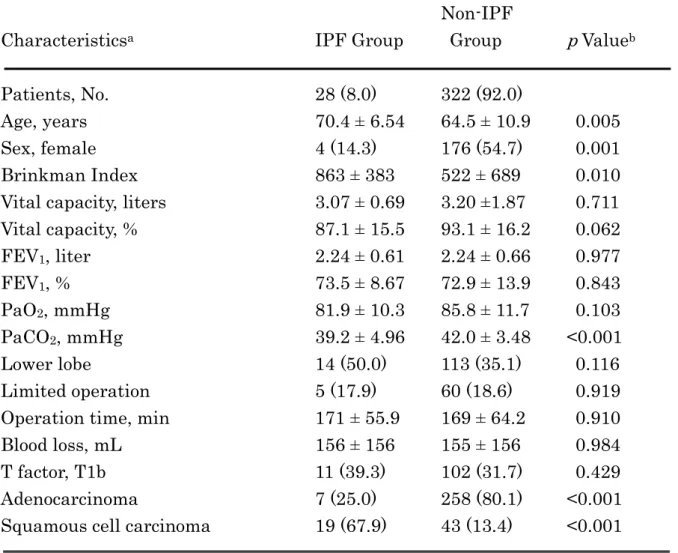
There were 350 patients who met the entry criteria for this study, comprising 28 with IPF and 322 without IPF. Lung cancers with IPF were found more frequently in men (85.7%) and in patients aged older than 70 years (46.4%). Patient characteristics are reported in Table 1. Age, BI, resting partial pressure of arterial carbon dioxide, tumor size, and the percentage of men and squamous cell carcinomas were significantly higher in the lung cancer patients with IPF than in those without IPF.
4-2. Survival rates
Survival data were collected for each patient from the date of the operation, with a median duration of follow-up of 4.8 years (range, 0 to 15.7 years). The 5-year actuarial survival rate for the entire group was 85.4%. The 5-year survival rate of patients with IPF (54.2%) was lower than that of patients without IPF (88.3%), and the difference in survival rates between the two groups was significant (p < 0.0001; Fig. 1).
Univariate and multivariate analyses
Significant differences in overall survival were demonstrated with age (p = 0.001), sex (p = 0.001), BI (p = 0.003), limited operation (p = 0.023), adenocarcinoma (p =0.005), and IPF (p < 0.0001). Use of the variables with a value of p < 0.10 from the univariate analysis in a subsequent multivariate analysis revealed that only IPF was a significant prognostic factor for survival (Table 2).
Morbidity and mortality
Morbidity and mortality rates are listed in Table 3. Postoperative morbidity rates were 40.7% for patients with IPF vs 18.8% for patients without IPF (p = 0.007). The 30-day mortality rate in patients both with and without IPF was 0%; however, patients with IPF showed a 3.6% rate of 90-day mortality vs 0.3% for those without IPF (p =0.028). An acute exacerbation of IPF developed in 3 patients (10.7%). None these patients died within 30 days of resection, but 1 patient died on of acute exacerbation of IPF postoperative day 70.
Causes of death
During the follow-up period, 42 patients (12.0%) died, and the most common cause was cancer-related death; 17 patients (4.9%) died of recurrent lung cancer (Table 4). The rates of cancer-related death were 17.9% for patients with IPF and 3.7% for patients without IPF (p = 0.001). Deaths due to respiratory failure occurred in 14.3% of patients
- 6 -
with IPF and in 1.2% of those without IPF (p <0.0001). Of the 4 patients with IPF who died of respiratory failure, two died of exacerbation of IPF over 90 days postoperatively. One patient died of aspiration pneumonia on postoperative day 128, and the other patient died of gradual progression of IPF at 1595 days.
Propensity score-matching analysis
We performed propensity score-matching analysis to reduce selection bias according to the sample size between patients with IPF and without IPF. Through this method, 28 patients were selected in the non-IPF group and showed similar characteristics (sex, age, BI, pulmonary function, blood gas data, surgical procedure, operation time, blood loss, T factor, and adenocarcinoma) as the IPF group (Table 5). Lower-lobe lung cancer was observed in 50.0% of IPF patients compared with 21.4% in non-IPF patients (p = 0.026). An analysis of the 5-year survival rate and the risk of death showed a rate of 54.2% for patients with IPF, which was significantly lower than that of patients without IPF (p = 0.038; Fig 2).
A multivariate analysis using adenocarcinoma (p = 0.091) and IPF (p = 0.049), the two variables that were deemed significant from a univariate analysis, demonstrated a significant association between overall survival and IPF (Table 6).
5. Discussion
Serious morbidity and a poor prognosis are common after IPF patients undergo pulmonary resection [5, 12–18] which make surgical treatment challenging in such patients. Because surgical indications for early lung cancer are usually considered to be good, it is very important to know the 5-year survival rate, a standard measure of the effectiveness of lung cancer treatment, of IPF patients with early-stage lung cancer after surgical treatment.
Recently, Watanabe and colleagues [5] reported a 5-year survival rate of 61.6%
after pulmonary resection in lung cancer patients with IPF, including pathologic stages IA and IB. However, Fujimoto and colleagues [12] reported extremely poor prognosis and a 5-year survival rate of 0% for patients with IPF. Both studies comprised few patients, so we do not know with certainty whether surgical resection will improve the prognosis of early-stage lung cancer in patients with IPF.
In the present study, the 5-year survival rate was 54.2% after pulmonary resection in pathological stage IA NSCLC patients with IPF. Our results also indicate that IPF had a significant negative effect on survival after surgical resection among these patients, because the multivariate Cox proportional hazards model showed IPF was the only significant independent factor for prognosis after surgical treatment
among these patients. However, we do not believe these data indicate that the presence of IPF with early-stage lung cancer is an absolute contraindication to performing surgical resection, as indicated by the 54.2% survival rate at 5 years; rather, such patients must be evaluated case-by-case. Importantly, these findings imply that it may be necessary for thoracic surgeons to evaluate surgical risk factors more carefully, even in cases of early-stage lung cancer. Our data may help to explain to lung cancer patients the potential difficulties of surgical treatment and the prospective prognosis.
High morbidity and mortality rates after pulmonary resection have been reported in patients with IPF [5, 13–15, 17]. The IPF patients in our study had significantly higher postoperative morbidity and mortality rates than those without IPF (Table 5). The 28 patients with IPF had a 10.7% incidence of postoperative exacerbation of IPF. The exacerbation of IPF was acute in 3 patients: 2 recovered and were discharged from hospital, and the other died during hospitalization. Although the small number of patients with IPF precluded an evaluation of risk factors for the incidence of acute exacerbation, this incidence rate is similar to that reported in the 8.7% to 70.0% of patients with IPF after other anticancer treatments [7, 18–23]. The present study documented a significant difference between the two groups in the frequency of pulmonary complications (p = 0.001); however, there was no significant difference after propensity score matching (p = 0.567). These data may suggest that it was difficult to further reduce the frequency of pulmonary complications, even if we had selected patients with subclinical IPF to tolerate pulmonary resection.
The rate of cancer-related death in our series was higher in patients with IPF than without IPF before propensity score matching, but the difference after propensity score matching was not significant (Table 6). The higher risk of death after pulmonary resection in these patients could be related to factors unrelated to the cancer, because many patients with IPF died of respiratory failure, both before and after propensity score matching.
The present study has some limitations: First, most patients with IPF in this study had subclinical IPF and were selected preoperatively to have pulmonary function adequate to undergo surgical treatment. In fact, the patients with IPF had similar pulmonary function test results as those without IPF. Therefore, postoperative survival of patients with many symptoms of IPF would be expected to be worse than that of our patients. Second, there could be a bias in the location of lung tumors between patients with and without IPF, although we found no significant differences between the two groups. If a patient with interstitial pneumonia under goes an upper lobectomy, the disease, which has a predilection for the basal lung segments, might be difficult to diagnose. However, if we miss patients with IPF who have a tumor in the upper or
- 8 -
middle lobe of the lung, the true postoperative survival rate of patients with IPF would not be expected to be better than 54.2%. Third, this is a retrospective, single-institution study, and the sample size was limited. To confirm these observations, prospective studies that include a larger series of patients are required.
In conclusion, IPF has independent, adverse effects on survival of pathologic stage IA NSCLC patients treated with pulmonary resection, as evidenced by a 5-year survival rate of 54.2% after resection. Although this result indicates that surgical treatment for early-stage lung cancer might not be contraindicated, patients with stage IA NSCLC concomitant with IPF should be carefully selected and should have good respiratory function.
6. Refurences
1. American Thoracic Society; European Respiratory Society. American Thoracic Society/European Respiratory Society International Multidisciplinary Consensus Classification of the Idiopathic Interstitial Pneumonias. This joint statement of the American Thoracic Society (ATS), and the European Respiratory Society (ERS) was adopted by the ATS board of directors, June 2001 and by the ERS Executive Committee, June 2001. Am J Respir Crit Care Med 2002;165:277–304.
2. Olson AL, Swigris JJ, Lezotte DC, Norris JM, Wilson CG, Brown KK. Mortality from pulmonary fibrosis increased in the United States from 1992 to 2003. Am J Respir Crit Care Med 2007;176:277–84.
3. Ozawa Y, Suda T, Naito T, Enomoto N, Hashimoto D, Fujisawa T, et al. Cumulative incidence of and predictive factors for lung cancer in IPF. Respirology 2009; 14:723–8.
4. Turner-Warwick M, Lebowitz M, Burrows B, Johnson A. Cryptogenic fibrosing alveolitis and lung cancer. Thorax 1980;35:496–9.
5. Watanabe A, Higami T, Ohori S, Koyanagi T, Nakashima S, Mawatari T. Is lung cancer resection indicated in patients with idiopathic pulmonary fibrosis? J Thorac Cardiovasc Surg 2008;136:1357–63.
6. Okamoto T, Gotoh M, Masuya D, nakashima T, Liu D, Kameyama K, et al. Clinical analysis of interstitial pneumonia after surgery for lung cancer. Jpn J Thorac Cardiovasc Surg 2004;52:323–9.
7. Isobe K, Hata Y, Sakamoto S, Takai Y, Shibuya K, Homma S. Clinical characteristics of acute respiratory deterioration in pulmonary fibrosis associated with lung cancer following anti-cancer therapy. Respirology 2010;15:88–92.
8. Akira M, Hamada H, Sakatani M, Kobayashi C, Nishioka M, Yamamoto S. CT findings during phase of accelerated deterioration in patients with idiopathic pulmonary fibrosis.
Am J Roentgenol 1997;168:79–83.
9. Sobin LH, Wittekind C, eds. (UICC) TNM classification of malignant tumors. 6th ed.
New York: Wiley; 2002:99–103.
10. Brinkman GL, Coates EO Jr. The effect of bronchitis, smoking, and occupation on ventilation. Am Rev Respir Dis 1963;87:684–93.
11. D’Agostino RB Jr. Propensity score methods for bias reduction in the comparison of a treatment to a non-randomized control group. Stat Med 1998;17:2265–81.
12. Fujimoto T, Okazaki T, Matsukura T, Hanawa T, Yamashita N, Nishimura K, et al.
Operation for lung cancer in patients with idiopathic pulmonary fibrosis: surgical contraindication? Ann Thorac Surg 2003;76: 1674–8.
13. Kawasaki H, Nagai K, Yoshida J, Nishimura M, Nishiwaki Y. Postoperative morbidity,
- 10 -
mortality, and survival in lung cancer associated with idiopathic pulmonary fibrosis. J Surg Oncol 2002; 81:33–7.
14. Kumar P, Goldstraw P, Yamada K, Nicholson AG, Wells AU, Hansell DM, et al.
Pulmonary fibrosis and lung cancer: risk and benefit analysis of pulmonary resection. J Thorac Cardiovasc Surg 2003;125:1321–7.
15. Chiyo M, Sekine Y, Iwata T, Tatsumi K, Yasufuku K, Iyoda A, et al. Impact of interstitial lung disease on surgical morbidity and mortality for lung cancer: analysis of short-term and long-term outcomes. J Thorac Cardiovasc Surg 2003;126:1141–6.
16. Koizumi K, Hirata T, Hirai K, Mikami I, Okada D, Yamagishi S, et al. Surgical treatment of lung cancer combined with interstitial pneumonia: the effect of surgical approach on postoperative acute exacerbation. Ann Thorac Cardiovasc Surg 2004;10:340–6.
17. Kushibe K, Kawaguchi T, Takahama M, Kimura M, Tojo T, Taniguchi S. Operative indications for lung cancer with idiopathic pulmonary fibrosis. Thorac Cardiovasc Surg 2007; 55:505–8.
18. Minegishi Y, Takenaka K, Mizutani H, Sudoh J, Noro R, Okano T, et al. Exacerbation of idiopathic interstitial pneumonias associated with lung cancer therapy. Intern Med 2009;48:665–72.
19. Kudoh S, Kato H, Nishiwaki Y, Fukuoka M, Ichinose Y, Tsuboi M, et al. Interstitial lung disease in Japanese patients with lung cancer: a cohort and nested case-control study.
Am J Respir Crit Care Med 2008;177: 1348–57.
20. 竹内栄治, 山口俊彦, 森 雅秀, 田中茂治, 中川 勝, 横田総一朗, 他:肺癌を合併した特発 性間質性肺炎症例の臨床的検討:日本胸部疾患学会雑誌 1996;34:653–8.
21. 竹中 圭, 吉村明修, 岡野哲也, 清家正博, 神尾孝一郎, 植松和嗣, 他:特発性間質性肺炎
(IIP)合併肺癌における肺癌治療に伴うIIP急性増悪の検討. 肺癌1999;39:955–62.
22. 埴淵昌毅, 山口俊彦, 岡田達也, 中川 勝, 横田総一朗, 伊藤正己, 他:特発性間質性肺炎
(IIP) 合 併 肺 癌 に 対 す る 肺 癌 治 療 後 の IIP 急 性 増 悪 症 例 の 臨 床 的 検 討. 肺 癌 2001;41:281–6.
23. Azuma A, Nukiwa T, Tsuboi E, Suga M, Abe S, Nakata K, et al. Double-blind, placebocontrolled trial of pirfenidone in patients with idiopathic pulmonary fibrosis. Am J Respir Crit Care Med 2005; 171:1040–7.
7. Figure legends
Figure 1. Overall 5-year survival in patients with pathologic stage IA non-small cell lung cancer
Overall 5-year survival in patients with pathologic stage IA non-small cell lung cancer was 54.2% for the 28 patients with idiopathic pulmonary fibrosis (IPF, dashed line) and 83.6%
for the 322 patients without IPF (solid line). The median follow-up was 4.20 years for IPF patients and 4.95 years for non-IPF patients.
Figure 2. Overall 5-year survival in 28 patients, matched by propensity score, with pathologic stage IA non-small cell lung cancer
Propensity score-matching analysis resulted in 54.2% of overall 5-year survival in 28 patients with both pathologic stage IA non-small cell lung cancer and idiopathic pulmonary fibrosis, simultaneously (IPF, dashed line), while it was 82.6% for the 322 patients without IPF (solid line). The median follow-up was 4.20 years for IPF patients and 4.15 years for non-IPF patients group.
- 12 -
8. Figures
Figure 1
Figure 2
9. Tables
Table 1. Patients Characteristics
Non-IPF
Characteristicsa IPF Group Group p Valueb
Patients, No. 28 (8.0) 322 (92.0)
Age, years 70.4 ± 6.54 64.5 ± 10.9 0.005
Sex, female 4 (14.3) 176 (54.7) 0.001
Brinkman Index 863 ± 383 522 ± 689 0.010
Vital capacity, liters 3.07 ± 0.69 3.20 ±1.87 0.711 Vital capacity, % 87.1 ± 15.5 93.1 ± 16.2 0.062
FEV1, liter 2.24 ± 0.61 2.24 ± 0.66 0.977
FEV1, % 73.5 ± 8.67 72.9 ± 13.9 0.843
PaO2, mmHg 81.9 ± 10.3 85.8 ± 11.7 0.103
PaCO2, mmHg 39.2 ± 4.96 42.0 ± 3.48 <0.001
Lower lobe 14 (50.0) 113 (35.1) 0.116
Limited operation 5 (17.9) 60 (18.6) 0.919
Operation time, min 171 ± 55.9 169 ± 64.2 0.910
Blood loss, mL 156 ± 156 155 ± 156 0.984
T factor, T1b 11 (39.3) 102 (31.7) 0.429
Adenocarcinoma 7 (25.0) 258 (80.1) <0.001
Squamous cell carcinoma 19 (67.9) 43 (13.4) <0.001
aCategoric data are expressed as number (%), and continuous data as mean ± standard deviation. bValue of p < 0.05 are significant.
FEV1 = forced expiratory volume in 1 second; IPF = idiopathic pulmonary fibrosis; PaCO2 = partial pressure of carbon dioxide in arterial blood; PaO2 = partial pressure of oxygen in arterial blood.
- 14 -
Table 2. Results of Multivariate Analysis of Preoperative Prognostic Factors Influencing Survival in Pathologic IA Non-Small Cell Lung Cancer Patients
Variable HR (95% CI) p Valueb
Age, years 1.037 (0.996-1.080) 0.080
Sex, male 2.165 (0.778-6.026) 0.139
Brinkman Index 1.000 (1.000-1.001) 0.359
Limited operation 1.261 (0.562-2.826) 0.574
Operation time 0.997 (0.991-1.003) 0.257
Adenocarcinoma 0.922 (0.424-2.005) 0.837
IPF 3.113 (1.373-7.058) 0.007
aValue of p < 0.05 are significant.
CI = confidence interval; HR = hazard ratio; IPF = idiopathic pulmonary fibrosis.
Table 3. Postoperative Morbidity and Mortality
Non-IPF IPF Group Group
Variable No. (%) No. (%) p Valuea
Morbidity 11 (40.7) 60 (18.8) 0.007
Acute exacerbation 3 (10.7) 0 (0) <0.0001 Pulmonary complications 10 (35.7) 41 (12.7) 0.001
Pneumonia 3 (10.7) 5 (1.6) 0.002
Prolonged air leakage 6 (21.4) 27 (8.4) 0.023
Others 1 (3.6) 11 (3.4) 0.965
Cardiac complications 3 (10.7) 20 (6.2) 0.356 Heart failure 1 (3.6) 5 (1.6) 0.430
Arrhythmia 2 (7.1) 16 (5.0) 0.617
Others 2 (7.1) 15 (4.7) 0.557
Mortality
30-day 0 (0) 0 (0)
90-day 1 (3.6) 1 (0.3) 0.028
aValue of p < 0.05 are significant.
IPF = idiopathic pulmonary fibrosis.
- 16 -
Table 4. Causes of Death
Non-IPF IPF Group Group
Cause No. (%) No. (%) p Valuea
Cancer-related death 5 (17.9) 12 (3.7) 0.001 Respiratory failure 4 (14.3) 4 (1.2) <0.0001
Others 3 (10.7) 14 (4.3) 0.133
aValue of p < 0.05 are significant.
IPF = idiopathic pulmonary fibrosis.
Table 5. Patients Characteristics Matched by Propensity Score
Non-IPF
Characteristicsa IPF Group Group p Valueb
Patients, No. 28 28
Age, years 70.4 ± 6.54 70.2 ± 7.66 0.911
Sex, female 4 (14.3) 7 (25.0) 0.313
Brinkman Index 863 ± 383 968 ± 954 0.595
Vital capacity, liters 3.07 ± 0.69 2.87 ±0.85 0.343 Vital capacity, % 87.1 ± 15.5 83.5 ± 17.4 0.419
FEV1, liter 2.24 ± 0.61 2.07 ± 0.64 0.314
FEV1, % 73.5 ± 8.67 74.3 ± 13.1 0.794
PaO2, mmHg 81.9 ± 10.3 87.3 ± 13.1 0.103
PaCO2, mmHg 39.2 ± 4.96 39.8 ± 2.86 0.577
Lower lobe 14 (50.0) 6 (21.4) 0.026
Limited operation 5 (17.9) 5 (17.9) >0.99 Operation time, min 171 ± 55.9 194 ± 88.8 0.272
Blood loss, mL 156 ± 156 168 ± 212 0.806
T factor, T1b 11 (39.3) 8 (28.6) 0.397
Adenocarcinoma 7 (25.0) 7 (25.0) >0.99
Squamous cell carcinoma 19 (67.9) 13 (46.4) 0.105
aCategoric data are expressed as number (%), and continuous data as mean ± standard deviation. bValue of p < 0.05 are significant.
FEV1 = forced expiratory volume in 1 second; IPF = idiopathic pulmonary fibrosis; PaCO2 = partial pressure of carbon dioxide in arterial blood; PaO2 = partial pressure of oxygen in arterial blood.
- 18 -
Table 6. Multivariate Analysis of Preoperative Prognostic Factors Influencing Survival in Pathologic IA Non-Small Cell Lung Cancer Patients Matched by Propensity Score
Variable HR (95% CI) p Valueb
Adenocarcinoma 0.393 (0.141-1.100) 0.075
IPF 0.311 (0.100-0.965) 0.043
aValue of p < 0.05 are significant.
CI = confidence interval; HR = hazard ratio; IPF = idiopathic pulmonary fibrosis.
Acknowledgements
I would like to express my deepest thanks to Eishin Hoshi for all advice and help. And, I would like to express my appreciation to Prof. Yukitoshi Satoh for giving me the opportunity to have an enough time to research some projects about primary lung cancer.