35
Received July 2, 2003
Accepted for Publication August 26, 2003 C2003 Soc. Mater. Eng. Resour. Japan
Synthesis and Guest Binding Properties of
Regioselectively Anthranilate‑Tosyl‑Labeled P‑Cyclodextrins
Tohru KIKUCHI , Naranchimeg DORJPALAM Ken ENDO and Fumio HAMADA
; Aomori Industrial Research Center, Environmental Technology Division, Daini Tonya‑machi, 4‑ I I ‑6, Aomori 030‑01 13, Japan
E‑mai/ .' koug‑kikuchi@net.prefaomorijp
) Department of Materials‑process Engineering and Applied Chemistry for Environments, Faculty of Engineering and Resource Science, Akita University,Tegata, Akita O I O ‑ 8502, Japan
E mcal hamada@lpc aktta‑u.acJp
Flexible hosts, regioselectively hetero‑labeled 6A‑anthranilate‑6x̲Olp‑tosyl‑labeled ‑cyclodextrins (X=B or G, C or F, and D or E for fi‑1, P‑2, and P‑3, respectively), have been synthesized to investigate their chemo‑sensor potential as host compounds for biochemical substances such as bile acids and terpenoids as guest molecules. These hosts showed pure monomer fluorescence, exhibiting increase in fluorescence in‑
tensity on complexation of the all guests examined. The extent of the fluorescence variation with a guest was employed to evaluate the sensing abilities ofthese hosts, and the sensing parameter ( AIllo) was used to de‑
scribe the sensing abilities of the hosts. Hosts fi‑2 and ‑3 recognized bile acids with high sensitivity, whereas fi‑1 detected all guests examined with low sensitivity. On the whole, the sequence of the binding ability of these hosts was P‑3 > p‑2 > p‑1 for bile acids. The behaviors of the appended units ofthese hosts during a formation of the host‑guest complexation were studied by induced circular dichroism (ICD) and fluorescence spectra. The ICD spectra of these hosts showed same patterns. The guest‑induced variations in ICD and fluorescence spectra suggest that anthranilate and tosyl units take movement with a changing their mutual relationship in the place to work as spacers. The parameter values of the titled hosts are much higher than those of homo‑labeled anthranilate ‑cyclodextrin analogs.
Key Words Cyclodextrin, Anthranilate, Molecular sensing, Fluorescent sensor system
1.
Introduction
Macrocyclic hosts molecules such as cyclodextrins, calixarenes and cavitands have been attracted interest as an enzyme model or supramolecular compoundsl), because these hosts can accommo‑
date various compounds such as organic molecules or metals in their cavities.1) 2) We have reported the synthesis of fluorescent ac‑
tive cyclodextrins which were modified with hetero units, which are dansyl and tosyl3) , pyrene and tosyl4)‑5) , or pyrene and cyanobenzene6). These hosts molecules show the molecular recog‑
7) 9)
nition for steroids or endocrine disruptor . It was proved that these analogs show much better sensory system than those of homo units‑labeled cyclodextrins such as bis‑anthranilate modified ‑ and 7 ‑cyclodextrins, bis‑dansyl modified r ‑cyclodextrins 6A , 6B ̲, 6A , 6c̲, and 6A , 6D‑bis‑dansylglysine‑modified ‑ Cyclodextrinslo)‑12) , because of their much more sensitivity and high selectivity. For further extension of our work, we synthesized regioselectively hetero‑labeled ‑cyclodextrins, which are 6A ‑ anthranilate‑6x̲O‑p‑tosyl‑labeled ‑cyclodextrins (X = B or G, C or F, and D or E for fi‑1, P‑2, and fi‑3, respectively) as shown in Scheme I .
2.
Ex perimental
H NMR s pectra were recorded with BURUKER DPX 300
spectrometer using TMS as an internal standard, and mass spectra were obtained with a JMS‑700 mass spectrometer. Elemental analyses were carried out by a Yanagimoto MT‑5 CHN corder.
2. I Preparations of 6A, 6B"e̲, 6A, 6c"*̲, 6^, 6D"=̲anthranilate‑
tosyl‑labeled p‑cyclodextrins ( p‑1, fi‑2 and fi‑3, re‑
spectively)
A mixture of 6^, 6 ‑di‑O‑(p‑tosyl) ‑cyclodextrin*") (500 mg, 0.35mmol) and sodium anthranilate (73 mg, 0.46 mmol) in lO mL of DMF was heated at 80'C for 24 h. After cooling, the reac‑
tion mixture was poured into 500 mL of acetone. The precipitates were filtered off and dissolved in I .5 mL of DMF. The DMF solu‑
ble fraction was applied to reverse‑phase column (Lobar colunm Lichroprep RP‑ 1 8, Merck Ltd., 240 mm x I O mm) . Stepwise elu‑
tion using 250 mL of 10 vol.% and 250 mL of 20 vol.% aqueous CH=CN, and 250 mL of 30 vol.9 ; aqueous CH=CN was applied to obtain P‑1. Yield 1.9 . R* 0.60 (1‑butanol‑ethanol‑water 5:4:3 by volume, TLC; silica gel 60F,5,; Merck Ltd.) and 0.57 (metha‑
nol‑water 2: I by volume, TLC; silica gel RP‑18F,5+s; Merck Ltd.) . H‑NMR (DMSO‑D*): 6 2.34 (3H, s, ‑CH3 Of tosyl), 3.2‑3.8 (42H, m, C'‑C'H ofcyclodextrin) , 3.9‑5.0 (12H, m, 06H and C'H ofcyclodextrin), 5.6‑6.0 (14H, br. 0=H and O'H ofcyclodextrin) , 6.55 (lH, t, J=7.5 Hz, aromatic‑H of anthranilate), 6.63‑6.80 (1H, br, ‑NH ofanthranilate) , 6.78 (1H, d, J= 7.8 Hz, aromatic‑H
Int. J. Soc. Mater. Eng. Resour. Vol. 1 1 , N0.2, (Sept. 2003)
36 Tohru KIKUCHI et al.
of anthranilate), 7.16‑7.48 (3H, m, aromatic‑H of tosyl and anthranilate), 7.61‑7.78 (3H, m, aromatic‑H of tosyl and anthranilate). Calcd. for C=*H**O,*NSNa ' 4H,O: C, 44.77; H, 5.91; N, 0.9391 . Found: C, 44.66; H, 6.07; N, 1.07%. MS (FAB) :
1430 ([M+H]+).
Compound P‑2 was prepared from 6^, 6'‑di‑O‑(p‑tosyl) ‑ cyclodextrin and sodium anthranilate by the same procedure as
p‑1 . Yield 15.1 %. R* 0.60 (1‑butanol‑ethanol‑water 5:4:3 by vol‑
ume, TLC; silica gel 60F,=*;Merck Ltd.) and 0.59 (methanol‑
water 2: I by volume, TLC; silica gel RP‑18F,,+s; Merck Ltd.) . *H‑
NMR (DMSO‑D6) : 6 2.41 (3H, s, ‑CH, oftosyl), 3.2‑3.8 (42H, m, C2‑C6H of cyclodextrin), 3.9‑5.0 (12H, m. O'H and C*H of cyclodextrin) , 5.6‑6.0 ( 1 4H, br, O'H and O'H of cyclodextrin) , 6.48‑6.57 (1H, m, aromatic‑H of anthranilate), 6.60‑6.71 (lH, br, ‑NH of anthranilate), 6.76 (1H, d, J=8.4 Hz, aromatic‑H of anthranilate), 7.08‑7.32 (1H, m. aromatic‑H ofanthranilate) , 7.42
(2H, t, J=8.9Hz, aromatic‑H of tosyl), 7.69‑7.79 (3H, m, aromatic‑H of tosyl and anthranilate) . Calcd. for C,eH**O*8NSNa ' 3H,O: C, 45.31; H, 5.84; N, 0.94 . Found: C, 45.24; H, 5.98; N, l.00 6. MS (FAB): 1430 ([M+H]+).
Compound fi‑3 was prepared from 6^, 6*‑di‑O‑(p‑tosyl) ‑ cyclodextrin and sodium anthranilate by the same procedure as
P‑1 . Yield 6.5 ;. Rf 0.60 (1‑butanol‑ethanol‑water 5:4:3 by vol‑
ume, TLC; silica gel 60F,=+; Merck Ltd.) and 0.71 (methanol‑
water 2: I by volume, TLC; silica gel RP‑18F,=*s; Merck Ltd.) . *H‑
NMR (DMSO‑D*): 6 2.41 (3H, s, ‑CH, oftosyl), 3.2‑3.8 (42H, m. C'‑C'H of cyclodextrin), 3.9‑4.9 (12H, m, O'H and C*H of cyclodextrin), 5.65‑5.9 (14H, br, O'H and O*H ofcyclodextrin), 6.51 (lH, t, J=6.8 Hz, aromatic‑H of anthranilate), 6.57‑6.65 (1H, br, ‑NH of anthranilate), 6.75 (lH, t, J= 7.7 Hz, aromatic‑H of anthranilate), 7.18‑7.28 (1H, m. aromatic‑H of anthranilate), 7.42 (2H, t, J=7.8 Hz, aromatic‑H of tosyl), 7.61‑7.77 (3H, m, aromatic‑H oftosyl and anthranilate). Calcd. for C, H*003sNSNa ' 12H,O: C, 40.86; H, 6.37; N, 0.85 . Found: C, 40.80; H, 5.74; N,
1.05%. MS (FAB): 1430 ([M+H]+).
2.2 Measurements
Fluorescence and circular dichroism spectra were measured at 25"C using a Perkin‑Elmer LS 40B fluorescence spectrophotome‑
ter and a JASCO J‑700 spectropolarimeter, respectively. For the fluorescence measurements, the excitation wavelength of the fluo‑
rescence spectra was 330 nm, and the width of excitation and emission slits were 5 nm. Ethylene glycol aqueous solution ( lO vol.%) was used as a solvent for the host, because the solubility of the host in pure water is poor.10) Five /1L of the guest species
(0.5, 0.05 and O.005 M) in dimethyl sulfoxide (DMSO) or MeOH were injected into a 10 vol.9 ethylene glycol aqueous so‑
lution ofthe host (2.5 mL) to make a sample solution with a host concentration of I .Oxl0‑6 M and guest concentration of I .O, O, I and 0.01 mM, respectively. For the circular dichroism measurements, frve /lL of the guest species (0.05 M) in dimethyl sulfoxide (DMSO) were injected into a I O vol.% ethylene glycol aqueous solution of the host (2.5 mL) to give a sample solution with a host concentration of I .OxIOA M and guest concentration of I .Ox
l0 M.
2.3 Determination of binding constants
The binding constants of three hosts, P‑1, fi‑2 and p‑3, for several guests were obtained from the guest‑induced fluorescence variations at 418 nm by employing a Benesi‑Hildbrand‑type equa‑
tion, as reported previously.') 5)
3. Results and discussion
B.*c t: A , corF ' A I D'*E 3.1 The Preparations of 6A, 6 anthranilate‑tosyl‑labeled P‑cyclodextrins ( ‑1, P‑2 ‑u v ‑u v ‑
and p‑3, respectively)
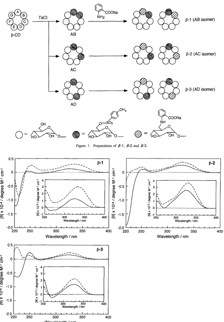
Hosts fi‑1 , fi‑2 and ‑3 were prepared from 6^, 6B‑, 6A, 6c̲ and 6^, 6D‑di‑O‑ (p‑tosyl) ‑cyclodextrin, respectively, using sodium anthranilate at 80'C , as shown in Figure I . These hosts were puri‑
fied by reverse‑phase colunm chromatography. Although, it is sus‑
pected that P‑1, P‑2 and P‑3 are isolated as a mixture of diastereomers, including 6^, 6B‑ and 6A, 6(}‑, 6^, 6c̲ and 6A, 6F‑, and 6A, 6D‑ and 6A, 6E‑anthranilate‑tosyl‑1abeled ‑cyclodextrins, re‑
spectively. This is because each of these diastereomers is insepara‑
ble by reverse‑column chromatography and the existing ratio of these diastereomers was unable to be determined by H‑NMR analysis.3) 6) In this paper, the hosts were assumed to exist as diastereomers and have been named P‑1, P‑2 and fi‑3 for 6A,6B and 6A,6c̲, 6^, 6c̲ and 6A, 6F‑, and 6^, 6D‑ and 6A, 6E‑anthranilate‑
tosyl‑labeled ‑cyclodextrins, respectively.
3.2 Induced circular dichroism (lCD) spectra
The ICD spectra of the three hosts, P‑i, P‑2 and P‑3, alone
GA
F C
ED
‑CD p‑1 (AB isomer) p‑2 (AC isomer) p‑3 (AD isomer)
O = OH Ho OOH o‑ e
pCH'
=
: HO ;;・O‑
( = HO
¥cooNa
NH O
OH o̲
Scheme l Structures of fi‑1, F‑2 and fi‑3.
Int. J. Soc. Mater. Eng. Resour. Vol.1 1 , N0.2, (Sept. 2003)
Synthesis and Guest Binding Properties of Regioselectively Anthranilate‑Tosyl‑Labeled R‑Cyclodextrins
37
GA
C
ED
p‑CD
TsCl
AB
COONa
NH2
AC
p‑1 (AB isomer)
AD 'CH 3
‑S02 O
¥cooNa
NH O
p‑2 (AC isomer)
0= 4 i Ho OOH o 0= I OH
Ho OH ‑ = HO
p‑3 (AD isomer)
OH o̲
Figure l Preparations of ‑1, p‑2 and e‑3.
E
o
2
(D o) o
ID 6
‑x o
0.5
O
‑0.5
*1 .O
.1,5
‑2.0
/ ¥
/ ¥ ¥ ¥
p‑ 1
l , ,
¥l
E o o
o' o
b
x
CD 4 3 2 1 o ‑1 ¥ ¥ ¥ ¥
¥,. ' / / / /
/
/// ¥¥¥
¥ ¥ ¥ ¥ ¥
¥ ¥
250 300 350
Wavelength I nm 400
O
o
{D ‑05 CD
¥ ‑10 ()
‑
.1.5
220 250 300
Wavelength / nm
350 ‑2,0
̲/ ¥
/
/ / ¥ ¥ ¥
¥
p‑2
/ /
‑ 'E
‑ 3 o
= ,D
,D2
c:,
'1' ̲1
,) bo
S ‑1
¥ / ¥ /
¥//
/ /
/ / / / //' ¥¥
¥ ¥ ¥ ¥ ¥ :
250 300 350
Wavelength I nm 400
400 220 250 300
Wavelength / nm
350 400
Eo
= o
o
ID (D
(b
‑x o
05
o
‑o 5
‑1 ,o
‑1 5
‑2. o
l ¥ 1
x ‑¥ t,
p‑3
T4 E T3 o
: 2
o)
1:,1
,1 oo
;(
‑1
1 ¥
/ /
li /
////‑¥¥¥
¥ ¥ ¥
¥
250 300 350
Wavelength / nm 400
220 250 300
Wavelength / nm
350 400
Figure 2 ICD spectra of fi‑1 , B‑2 and p‑3 in a 10 vol. ehtylene glycol aqueous solution ( I .Oxl04 M:‑‑‑, 25'O and containing ursodeoxycholic acid (1 .OxlO M:‑‑‑) .
Int.
J.
Soc. Mater. Eng. Resour. Vol.1 1 , N0.2, (Sept. 2003)
38 Tohru KIKUCHI et. al
and in the presence of ursodeoxycholic acid in a I O vol. ethyl‑
ene glycol aqueous solution are taken to investigate the movement of the appended units when a host‑guest complexation occurs, as shown in Figure 2. The spectrum of P‑1 exhibits a negative Cotton peak around 220‑240 nm and positive Cotton peaks around at 245 and 325 nm, in which these peaks decrease upon addition of a guest. It indicates that the appended units penetrate into the chiral cyclodextrin cavity'o) as it is well known that an increase in the ICD intensity means the appended unit is located in the chiral environment of the cyclodextrin cavity. The ICD spectral patterns of P‑2 and P‑3 are basically similar to that of ‑1, however, changes of [ e I value are not same. The L O I values of P‑1 around at 325 nm increase with much greater than those of fi‑2 and P‑3. On the other hand, ICD intensity of fi‑2 of the negative band at 220‑245 nm with a guest is decreased much more than those of fi‑1 and P‑3, and the negative band at 220‑255 nm of fi‑3 changes to positive one on accorDmodation of a guest. These results suggest that the movements of the anthranilate and tosyl
>
:= co
o c
c o .
, 5
a:
350 400 450
Wavelength / nm
500 550
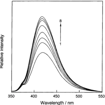
Figure 3 Fliorescence spectra of ‑2 in a 10vol. 6 ethylene glycol aqueous solution (1.0xlO‑' M, 25'O at various concentrations of ursodeoxycholic acid (1: O, 2: 4.0xl0‑6, 3: 8.0xl0‑6, 4: 1.2x 10‑s, 5: 2.4xl0‑5, 6: 4.0xl0‑5, 7: 6.lxl0‑5, 8: 8.3xl0‑5 M).
units associated with a guest are not the same for the three hosts.
It is reported that ICD signs of the spectroscopic active cyclodextrin derivatives indicated a type of inclusion which is equatorial or axial self‑inclusion of the appended unit.*=)‑*') Therefore, opposite of ICD pattern at 220‑255 nm of p‑3 before and after guest addition suggests that the anthranilate and tosyl units of P‑3 may replace each other, probably due to ease of movement of the appended units, because the positions of these appended units of fi‑3, which are A and D or E at C‑6 of seven glucose units, are more distant than those of p‑1 and ‑2. These phenomena should be advantageous for molecular sensing by these hosts, because they might cause differences of sensitivity and se‑
lectivity for the guest molecules.
3.3 Fluorescence spectra
Figure 3 shows fluorescence spectra of fi‑2 in the absence and presence of ursodeoxycholic acid in a I O vol.9 ; ethylene glycol aqueous solution. In the study of anthranilate modified
cyclodextrin system, we found that most effective excitation wave‑
length is 330 nm. o),'o)‑2*) The fluorescence spectra of P‑1 , P‑2 and
‑3 are composed ofpure monomer emission with a peak around 4 1 8 nm, and their intensity increases with increasing of ursodeoxycholic acid concentration. It is reported that a guest‑
induced fluorescence enhancement means that the labeled unit is moving into the cyclodextrin cavity deeply*o) and a decrease means that the labeled unit is moving out of the cavity. ')' *')‑*=) The ICD and fluorescence spectral changed of the three hosts suggest that the anthranilate unit is included into the cyclodextrin cavity upon guest binding and acts as a spacer. On the other hand, we at‑
tempted to examine the guest‑induced behavior of the tosyl unit, which is fluorescent inert, by ROESY *H‑NMR. Unfortunately, our attempt was unsuccessful, because the host in a lOvol.
DMSO‑d* D,O solution was deposited through the NMR measure‑
ment. The host might cause aggregation. The energy‑minimized structures of the three hosts obtained using molecular mechanics in CS Chem 3D (MM2) =)‑*+), as illustrated in Scheme 2. However, the energy‑minimized structures obtained are estimated ones, so it is probably indicates that the tosyl unit of the three hosts are lo‑
cated into the cyclodextrin cavity deeply as self‑inclusion fornra‑
tion. Therefore, as the ICD and fluorescence spectral changed of the hosts, it is speculated that the tosyl units of P‑1 and P‑2 move in the cyclodextrin cavity to play the roll of a spacer when host‑
guest complexation occurs. On the other hand, the tosyl unit of p‑3 is estimated to be excluded from the cyclodextrin cavity and not play the role of a spacer when host‑guest complexation is
p‑ 1 p‑2 p‑3
Scheme 2 Energy‑minimized structures of fi‑1, fi‑2 and fi‑3 obtained using molecular mechanics in CS Chem 3D (MM2)
Int. J. Soc. Mater. Eng. Resour. Vol. 1 1 , N0.2, (Sept. 2003)
Synthesis and Guest Binding Properties of Regioselectively Anthranilate‑Tcsyl‑Labeled p‑Cyclodextrins
39
‑1 and p‑2
p‑3
, anthranilate moiety
: tosyl moiety
Scheme 3 Estimated host‑guest complexation of the host.
O L O E
'U LCC
IL
1 .6
1 .4
1 .2
1 .O
0,8
0.6
0.4
O.2
o i 2 5 6
7 8 9 Io 11 12
Guest
13 14 15 16
Figure 4 Sensing factors of ‑1 ([]), ‑2 ( ]) and p‑3 (1) in a 10 vol. 6 ethylene glycol aqueous solution examined (guest concentrations, 1‑9: 1.0 niM, 10: 0.01 mM, 1 1‑16: 0.1 nlM).
(1.0xlO‑' M, 25 O for all gueats
Int.
J.
Soc. Mater. Eng. Resour. Vol. 1 1 , N0.2, (Sept. 2003)
40 Tohru KIKUCHl et, al
formed, and the anthranilate unit of p‑3 is included into the cyclodextrin cavity, as illustrated in Scheme 3.
3.4 Sensing abilities of p‑1, p‑2 and fi‑3forterpenoids and steroids
As reported previously, the extent of the variation of the fluores‑
cence intensity of these hosts depended on the nature of a guest,
even at low concentration; therefore, those hosts can be used as fluorescent molecular sensors, as seen in the case of anthranilate‑
labeled cyclodextrin analogs reported previously.10) In order to evaluate the sensing ability of modified cyclodextrins, the A Illo value was used as a sensitivity parameter. Here, AI is Illo, where 10 is the fluorescence intensity for the host alone and I is that of a complex. Figure 4 shows the parameter values of p‑1, p‑2 and
'
H20H
l
nerol(1)
ro
(+)‑fenchone(6)
COOH
."
cyclohexanol(2)
r
(‑)‑fenchone(7)
o̲H
cydohexanone(3)
/!¥̲̲'.
(‑)‑bomeol(8)
COOH
( OH eIOH
//¥
(‑)‑menthol(4) cyclooctanol(S)
COOH
)1
adam antane‑1 ‑carboxylic acid(9)
COOH OOH
HO' *
H
lithocholic acid(10)
H O¥'**'
deoxychol ic acid(1 1 )
COOH
O l
HO¥*+* ""'oH
chenodeoxychol ic acid( 1 2)
COOH
HOI *
o
OH
ursodeoxyxholic acid(1 3)
Ho +**
OH
hyodeoxycholic acid(1 4)
HO¥***' ""oH H" *' '
chol ic acid( 1 5) dehydroepiandrosterone( 1 6)
Scheme 4 Guest molecules
10 11 12
Figure 5 Space filling figures of guests 10‑15 afier carring out MM2 energy minimized calculation.
15
Oxygen atoms are shown by dark color.
Int. J. Soc. Mater. Eng. Resour. Vol. 1 1 , N0.2, (Sept. 2003)
Synthesis and Guest Binding Properties of Regioselectively Anthranilate‑Tosyl‑Labeled p‑Cyclodextrins
41
fi‑3 with terpenoids at I .O mM and steroids at 0.1 mM except for lithocholic acid ( I O), which was examined at 0.01 mM because O.1 mM of lithocholic acid is not soluble into a I O vol. 6 ethylene glycol aqueous solution. The structures of 1 6 guest molecules are shown in Scheme 4. Hyodeoxycholic acid (14), which has two hydroxyl groups at C‑3 and C‑6 of the steroidal framework, ursodeoxycholic acid (13) and chenodeoxycholic acid (12), which bear two hydroxyl groups at C‑3 and C‑7 of the steroidal framework and are diastereoisomers each other, were detected by fi‑2 and fi‑3 with the highest sensitivities, exhibiting the sensing values of 1.178 and 1.520 for guest 14, 1.028 and 1.343 for guest 1 3 and O.92 and I .08 for guest 1 2, respectively. Lithocholic acid (10), which has one hydroxyl group at C‑7 of the steroidal frame work at C‑3 of the steroidal framework and its concentration is one tenth of other bile acid concentration, was detected by fi‑2 and fi‑3 with high sensitivities, exhibiting the sensing values of I .22 and 0.94, respectively. It is achieved that the high sensitivity of these hosts for the guests such as 1 2, 1 3 and 1 4. Unfortunately, the selectivity for these guest molecules by P‑2 and fi‑3 is not enough. On the other hand, host fi‑1 detected these guests I O, 1 2, 1 3, and 1 4 with low sensitivities and its sensing parameters were negligible. The sensing factors of the three hosts for deoxycholic acid ( 1 1 ) and cholic acid (・1 5), which have hydroxyl group at C‑
l 2 of the steroidal framework, and dehydroepiandrosterone ( 1 6).
which bears ketone at C‑17 of the steroidal framework, were also negligible. The sensing factors of p‑2 and fi‑3 for bile acids are more sensitive than those of 6A, 6c̲ and 6A, 6D‑di‑anthranilate‑
Table l Binding constants (K/mol‑1 dnf) of P‑1 vol. ethylene glycol aqueous solution
, fi‑2 and P‑3 in a 10 (lOxlO" M, 25"O')
euest
p‑ 1
p‑2 l ‑3
Borneol (8) Lithocholic acid (10) Chenodeoxycholic acid (12) Ursocfeoxycholic acid (13, Hyodeoxycholic acid (14)
1 7,600:!: 520 b) 88,500 8,690 51,400:!:1 110 78,800:1: 5 490 148,000:!:5 OOO
1 2, 800 :t 3 1 O
295 OOOd: 13,000 44, I OO i 870 77,8eO:1: 1 3eO 31 7 OOOi3 360
4 280 1 50 61 5 OOO: 21 800
33=600 i 050 G7 eOO :3 e40
1 O1 ,OOO :!: I ,680
a) The K values were obtained from guest‑induaed fluorescBnce variations
b) The 5talistical errors were values of standerd deviation assessefl by guest‑indueed fluoreseence variations
labeled ‑cyclodextrins reported previously and these of fi‑1 for bile acids are lower than those of 6^, 6*‑di‑anthranilate‑labeled
‑cyclodextrin.'") The sequence of the sensing factors of the three hosts for guests 12, 13 and 14, are fi‑3 > fi‑2> fi‑1 , on the other hand, the sequence of the sensing factors of these hosts for guest 1 O, is fi‑2 > fi‑3 > fi‑1 . These facts mean that the positions of the appended units, which have influence on the flexibility of each ones, and the group at C‑17 of the steroidal framework affect the sensing ability for bile acids. The sensing factors, which are ruled by conformational change, of the three hosts for guests 1 2, 1 3 and 1 4, which have a hydroxyl group on the B ring of themselves, are in inverse proportion to the distance from the C‑17 to the hydroxyl group at the B ring of the steroidal framework as shown in Figure 5. Furthermore, the sensing factors are small by existence of hy‑
droxyl group on the C ring of the steroidal framework. Thus, host
‑1, fi‑2 and P‑3 can recognize the presence of hydroxyl group
2.0
L o 1.5
a) E
(T: L
(o 1.0
Q o)
c a)c 0.5 uD o O
9
‑1
8 7 6 5 ‑Log ([Guest]/M)
4 3
o E
CU *co
, o)
CO o
2.0
1 .5
1 .o
0.5
o
p‑2
9 8
‑Log
6 5
([Guest]/M)
4 3
2,0
L o 1.4 o E
cu
{ 1.0 o) c
'co= co 0.5
(D o
o 9
p‑3
8 7 6 5 ‑Log ([Guest]/M)
4 3
F igure 6 Fluorescence variations of B‑1, p‑2 and fi‑3 (1.0xlO‑' M, 25 O in a 10vol. 6 ehtylene glycol aqueous solution for lithcholic acid chenodeoxycholic acid (A) , ursodeoxycholic acid ([]), and hydeoxycgolic acid (V) as a fimction of guest concentration.
(o),
Int. J. Soc. Mater. Eng. Resour. Vol.1 1 , N0.2, (Sept. 2003)
42 Tohru KIKUCHI et al.
on the B and C ring of the steroidal framework on the cholic acid analogs, and the distance from the C‑17 to the hydroxyl group on the B ring of the steroidal one. These suggest that guests I O and 1 2‑14 enter the cyclodextrin cavity from the site of carboxylic acid and form hydrogen bonding between the carboxylic acid of the guest and the amino group of the anthranilate unit of the host, moreover, A and B rings of the steroidal framework are located on out of the cyclodextrin one. For terpenoid guests, the three hosts exhibited low sensitivities for guests I ‑7, exhibiting lower sensi‑
tivities than homo‑anthranilate‑labeled ‑cyclodextrins. o) On the other hand, for guests 8 and 9, which are bicyclic and tricyclic compounds, respectively, the three hosts detected guest 8 and fi‑3 detected guest 9 with higher sensitivities than other guests, how‑
ever, these sensing factors are not recognized advantage over those of homo‑anthranilate‑1abeled ‑cyclodextrins.'o) These results ob‑
tained as sensing factors suggest that hetero units, which are anthranilate and tosyl, of fi‑2 and fi‑3 contribute to qualitative molecular recognitions.
3.5 Binding conStants
The guest‑induced fluorescence variation at 418 nm was em‑
ployed to calculate the binding constants (K) ofthree hosts. using Eq, l.
l/(Irl f) = l/a [CD] + 1/a [CD]KX l/ [G] Eq. 1 Here, I is the fluorescence intensity at 418 nm (If for complex, 1 f for the host alone), [CD] the total host concentration, [G] the total guest concentration, and a is constant. The binding constants of the three hosts were obtained in order to examine the correlation between the fluorescence variations and the binding abilities ofthe hosts. The results are shown in Table I . In every host, there is no simple correlation between the binding constants and the sensitiv‑
ity factors. This means that the sensitivity values give a relative, but no absolute, measure of the binding abilities of the hosts.
3.6 Response ranges
Figure 6 shows response curves of fi‑1 , p‑2 and P‑3 for guests such as lithocholic cid (10), chenodeoxycholic acid (12), ursodeoxycholic acid (1 3), and hyodeoxycholic acid (14). Host P‑1 detected with response ranges lO‑ j ̲ 104j, 10‑5j‑ 10‑", and 10‑6‑ 104 M to I O, 1 2 and 1 3, and 14, respectively. Host fi‑2 de‑
tected with response ranges I 0‑7 ‑ I 0‑45, I 0‑6 ‑ I 0‑4, and I O‑ ̲ I O M to I O, 1 2 and 1 3, and 1 4, respectively. Host p‑3 detected with response ranges lO‑ 5 ̲ 1045, l0‑5j ‑ 104, 10‑ 5 ‑ l0‑4, and lO‑ 5 ‑ 104 M to I O, 1 2, 1 3, and 14, respectively. These results mean that each host perceives on different guest concentration range, and es‑
pecially host fi‑3 has unique response ranges to each guest. All hosts give almost clear concentration dependency for the guests, reflecting the sensitivities of the system for the guests. Although, it is true that response curves are not simply increased with in‑
crease of guest concentration, sometimes‑different dependency.
This result suggests that two ways of the host‑guest binding pat‑
tem is taking place such as I : I and I :2 complex patterns.
4. ConcIUSion
Three hetero‑functionalized analogs of anthranilate‑ and tosyl‑
labeled ‑cyclodextrins were investigated as new chemo‑sensors for organic guests such as terpenoids and bile acids, which are bio‑
logically significant substances. These hosts show pure monomer fluorescence, the variation of which was used as a parameter to de‑
scribe the sensing ability. The introduction of hetero functional
groups such as antharanilate and tosyl, which are in different posi‑
tions such as 6A and 6X in the cuclodextrin upper rim, gives new sensing factors that impart high sensitivity and selectivity to these hosts. In this system, it is obvious that the collaboration of the anthranilate and tosyl units, which substituted at 6A and 6X (X = C or F, and D or E) in the cyclodextrin upper rim, contrib‑
utes to improve the selective molecular binding ability. The system shown here is a very convenient and useful method, because the chemical modification of a guest, even if it is spectroscopically inert, is not necessary, a guest can be examined directly.
Acknowledgments
This study was supported by a Grant‑in‑Aid for Specially Promoted Research (No. 404: Molecular Synchronization for Design ofNew Materials System) from the Ministry ofEducation Science, Sports and Culture of Japan.
References
l. J. ‑M. Lehn, "Supramolecular Chemisry", 1995, VCH, Weinheim.
2. J. Szejtli, "Cyclodextrin Technolog /", 1 998, Kluwer, Dordrecht.
3 . M. Narita and F. Hamada, "The synthesis of a fluorescent chemo‑sensor system based on regioselectively dansyl‑tosyl‑
modified fi‑ and y‑cyclodextrins", J. Chem. Soc.. Perkin Trans. 2, 2000, 823‑832.
4. A. Makabe, K. Kinoshita, M. Narita, and F. Hamada, "Guest‑
Responsive Fluorescence Variations of 7‑Cyclodextrins la‑
beled with Hetero‑Functionalized Pyrene and Tosyl Moieties", Anal. Sci., 2002, 18, I19‑124.
5. M. Narita, E. Tashiro F. Hamada, "Synthesis of a selective Fluorescent Sensing System Based on y ‑Cyclodextrin Modified with Pyrene and Tosyl on the Hetero Rims", J. Incl.
Phenom. Macrocyclic Chem., 2002, 42, 137‑144.
6. F. Hamada, M, Narita, K. Kinoshita, A. Makabe, and T. Osa,
"Synthesis and fluorescent molecular sensing at exciplex emission of pyrene‑ and cyanobenzene‑modified r ‑ cyclodextrins", J. Chem. Soc., Perkin Trans. 2, 2001, 388‑
3 94 .
7. M. Narita, N. Ogawa and F. Hamada, "Fluorescent Molecular Recognition for Endocrine‑Disrupting Chemicals and Their Analogs by Fluorescent Hetero‑Modified Cyclodextrins", Anal. Sci., 2000, 16, 701‑705.
8. M. Narita, E. Tashiro and F. Hamada, "Selective Recognition of Endocrine Disruptors by Fluorogenic 7 ‑Cyclodextrin Having Pyrene‑Tosyl Substituents on Hetero Rims", Anal.
Sci., 2001, 17, 1453‑1455.
9. M. Narita, A. Makabe, K. Kinoshita, K. Endo, and F. Hamada,
"High Sensitive Molecular Recognition by Monomer and Exciplex Emission for Endocrine‑Disrupting Chemicals and Their Analogs Based on Fluorescent Hetero‑Modified Cyclodextrins", Int. J. Soc. Mater. Eng. Resour., 2001, 9, 6‑
13.
10. M, Narita, F. Hamada, I. Suzuki, and T. Osa, "Variations of fluorescent molecular sensing for organic guests by regioselective anthranilate modified ‑ and r‑cyclodextrins", J. Chem. Soc.. Perkin Trans. 2, 1998, 2751‑2758.
1 l. M. Narita. F. Hamada, M. Sato, I. Suzuki, and T. Osa,
"Fluorescent Molecular Recognition and Sensing System of Brs Dansyl Modified 7 Cyclodextrins". J. Incl. Phenom.
Int. J. Soc. Mater. Eng. Resour. Vol.1 1 , N0.2, (Sept. 2003)
Synthesis and Guest Binding Properties of Regioselectively Anthranilate‑Tosyl‑Labeled p‑Cyclodextrins
Macrocyclic Chem., 1999, 34, 421‑430.
12. M. Sato, M. Narita, N. Ogawa, and F. Hamada, "Fluorescent Chemo‑Sensor for Organic Guests Based on Regioselectively Modified 6A,6B‑, 6^,6c̲, and 6A,6D‑Bis‑dansylglysine‑Modified
‑Cyclodextrins". Anal. Sci., 1999, 15, I 199‑1205.
13, U. Burkert and N. L. Allinger, "Molecular Mechanics", 1982, ACS Monograph, I 17, American Chemical Society, Washington DC. M. J. Dudek and J. W. Ponder, "Accurate Modeling of the Intramolecular Electrostatic Energy of Proteins", J. Comput. Chem., 1995, 16, 791‑816.
14. R. Corradini, A. Dossena, G. Galaverna, R. Marchelli, A.
Panagia, and G. Sartor, "Fluorescent Chemosensor for Organic Guests and Copper (II) Ion Based on
Dansyldiethylenetriamino‑Modified ‑Cyclodextrin". J. Org.
Chem., 1997, 62, 6283‑6289.
15. M. Narita, S. Mima, N. Ogawa, and F. Hamada, "Selective Fluorescent Molecular Sensing by Bis Dansyl‑Modified 7‑
Cyclodextrin Dimer", Anal. Sci., 2000, 16, 865‑869.
16. T. Kikuchi, M. Narita and F. Hamada, "Synthesis of bis dansyl‑modified ‑cyclodextrin linear trimer having multi‑
recognition sites and high hydrophobic environment", Tetrahedron, 2001 , 57, 93 17‑9324.
43
17. M. Narita, J. Itoh, T. Kikuchi, and F. Hamada, "A High Sensitivity Fluorescent Chemo‑sensory System Based on ‑ Cyclodextrin Dimer Modified with Dansyl Moieties", J. Inc/.
Phenom. Macrocyclic Chem., 2002, 42, 107‑1 14.
1 8. A. Makabe, K. Kinoshita, M. Narita, and F. Hamada, "Guest‑
Responsive Fluorescent Variations of 7 ‑Cyclodextrins Labeled with Hetero‑Functionalized Pyrene and Tosyl Moieties", Anal. Sci., 2002, 18, 119‑124.
19. F. Hamada, Y. Kondo, K. Ishikawa, H. Ito, I. Suzuki , T. Osa, and A. Ueno, "Host‑Guest Sensory System of Sodium Anthranilate Modified ‑Cyclodextrin: Molecular
Recognition Properties", J. Inclusion Phenom. Mol. Recognit.
Chem., 1994, 17, 267‑275.
20. F. Hamada, K. Ishikawa, R. Ito, H.Shibuya, S.Hamai, I.
Suznki, T. Osa, and A. Ueno, "Spacer Effect of Appended Moieties for Molecular Recognition in Doubly Sodiurn Anthranilate Modified 7‑Cyclodextrin", J. Inclusion Phenom.
Mol. Recognit. Chem., 1995, 20(1), 43‑51.
2 1 . F. Hamada, K. Ishikawa, I.Tamura, and A. Ueno, "Molecular recognition and sensory system of y‑cyclodextrin capped by a sodium anthranilate", Anal. Sci., 1995, 11, 935‑939.
Int. J. Soc. Mater. Eng. Resour. Vol.1 1 , N0.2, (Sept. 2003)


![Figure 4 Sensing factors of ‑1 ([]), ‑2 ( ]) and p‑3 (1) in a 10 vol. 6 ethylene glycol aqueous solution examined (guest concentrations, 1‑9: 1.0 niM, 10: 0.01 mM, 1 1‑16: 0.1 nlM).](https://thumb-ap.123doks.com/thumbv2/123deta/6674182.2197554/5.892.91.800.53.1208/figure-sensing-factors-ethylene-aqueous-solution-examined-concentrations.webp)