Further Histological Study of the Role of Hepatitis B Infection on the Development of Hepatocellular Carcinoma
Yumiko TODA, Masachika SENBA and Hideyo ITAKURA Department of Pathology, Institute for Tropical Medicine,
Nagasaki University, Nagasaki, Japan
Abstract: A total of 953 cases of various types of liver diseases were selected for examina‑
tion from 6339 autopsies performed in Nagasaki University Hospital from 1964 through 1980. The association between HBs‑Ag in liver tissue and the histology of the liver was studied using Shikata's orcein staining method. Of the 6339 autopsies performed, 488 cases of atrophic liver cirrhosis (7.7%) and 303 cases of primary hepatocellular carci‑
noma (4.8%) were identified. These incidences of liver diseases were statistically sig‑
nificantly higher compared with those of national averages of Japan (5.2% and 3.5%
respectively). A histological analysis of 953 cases indicated that 242 out of 488 cases of atrophic liver cirrhosis were associated with hepatocellular carcinoma (49.6%) and that
242 out of 303 cases of hepatocellular carcinoma were combined with atrophic liver
cirrhosis (78.9%). No HBs‑Ag was detected in the tissues of cases of acute liver atrophy or liver cirrhosis with the exception of atrophic liver cirrhosis. HBs‑Ag was present in 6 out of 22 cases of subacute hepatitis (27.2%) and in 6 out of 37 chronic hepatitis (16.2%). The highest rate of detectable HBs‑Ag was observed in the liver tissues of the subgroup of atrophic liver cirrhosis with hepatocellular carcinoma (60.6%).
Among 196 cases of hepatocellular carcinoma examined, HBs‑Ag was detected in 14 of those cases which did not exhibit atrophic liver cirrhosis. These results suggest that nepatocellular carcinoma can arise without setting of hepatitis B chronic liver cirrhosis and support the hypothesis that HBs‑Ag may be directly oncogenic. Liver cell dysplasia was found in 69 out of 154 cases of atrophic liver cirrhosis with hepatocellular carcinoma (44.8%) and in 22 out of 122 cases of atrophic liver cirrhosis without hapatocellular carcinoma (18.0%). Furthermore, there was a strong correlation between the presence of HBs‑Ag in the liver tissue of Nagayo's type B atrophic liver cirrhosis and the exis‑
tence of cellular dysplasia.
Key words: Hepatitis B surface antigen (HBs‑Ag), Atrophic liver cirrhosis, Hepatocellular
carcinoma
Received for publication. August 31, 1984.
Contridution No・ 1508 from the Institute for Tropical Medicine, Nagasaki University.
INTRODUCTION
A strong correlation between the hepatitis B virus and hepatocellular carcinoma has been reported in various countries (Wu, 1978; Atiyeh etal., 1980; Beasley etal., 1981 ; Keshgegian et al. , 1981). The carrier rate for HBs‑Ag is much higher in a certain part of Africa and in Southeast Asia, East Africa and the Pacific islands (up to lO%) than in North America, Europe or Australia (up to l%) (McCollum et al., 1981). The average carrier rate of HBs‑Ag among blood donors in Japan is reported to be 1.4% to 1.9%, indicating that HBs‑Ag is moderately prevalent in Japan (Halliday et al., 1980). The incidence of hepatocellular carcinoma is roughly concomitant with the incidence of positive serum HBs‑Ag and is observed in 150 cases out of 100,000 in Africa and Asia but in only 1 to 3 cases out of 100,000 in North America and West Europe (Maupus et al., 1981). These reports suggest that the hepatitis B virus may be one of the human onco‑
genie viruses involved in hepatocellular carcinoma. The incidence of the hepatitis B antigenema, however, differs considerably depending upon geographical location. These discrepancies may indicate a true geographical variation in the frequency of HBs‑Ag in
diseased hepatic tissues rather than technical variations in the mesurements of the hepat―
itis B antigen. This study was undertaken as an extention of our previous work (Kanda, 1977) with respect to the role of hepatitis B infection on the development of chronic hepatic diseases, such as liver cirrhosis and hepatocellular carcinoma, in Nagasaki Japan.
MATERIALS AND METHODS
A total of 953 cases of various diseases of the liver were selected for study存om 6339 autopsies performed at Nagasaki University from 1964 through 1980. At the same time a statistical review of the incidences of various hepatic diseases in all autopsies performed in Japan from 1965 through 1980 was conducted. In 652 of the 953 autopsy cases selected for this study from which two to three representative paraffin「blocks were available liver tissues were histologically examined. Sections 5 μm thick were cut and stained with hematoxylin and eosin as well as with orcein using Shikata's method (1973 ; 1974). Other histological staining procedures, such as modified Mallory's collagen stain「
ing (Luna, 1968), Gomori's method for iron (Luna, 1968) and silver staining (Senba,
1983〕 were also conducted when necessary. In each case, the detailed hepati―c histology,
such as the type of liver diseases, the presence of liver cell dysplasia, stromal inflammation and the grade of malignancy, was recorded. The presence of HBs‑Ag in hepatic tissue was expressed according to Kanda's criteria. Anthony's difinition (1973) and Edmond‑
son's criteria (1954〕 were applied in determining whether or not liver cell dysplasia
existed and the grade of malignancy.
t.T
RESULTS
Statistical review
A total of 6339 autopsies were performed at Nagasaki University from 1964 through 1980. Of those 6339 cases, 488 (7.7%) were histologically'diagnosed as atrophic liver cir‑
rhosis and 303 (4.8%) as hepatocellular carcinoma. The incidences of atrophic liver cir‑
rhosis and hepatocellular carcinoma were much higher than those of the national sverage of Japan, as shown in Table 1. The male to female ratio was 3 to 1 for atrophic liver cirrhosis and 5 to 1 for hepatocellular carcinoma. Table 2 shows the incidences of varト ous liver diseases in a total of 953 autopsy cases examined at Nagasaki University. There were 22 cases of acute liver atrophy and 36 cases of subacute liver atrophy. Various types of liver cirrhosis were diagnosed in 561 of those cases which included 488 cases of atrophic liver cirnhosis, 303 were diagnosed as hepatocellular carcinoma, 17 as cholangiocellular
Table 1. Comparative study of incidence of liver cirrhosis and hepatocellular carcinoma between Nagasaki University and all Japan
Total autopsy Atrophic liver Hepatocellular cirrhosis carcinoma
Nagasaki University 1964‑1980 6,339 488 (7・7%) 303 (4.82O All Japan 1965‑1989 386,731 20,072 (5.2%) 13,418 (3.5%)
Table 2. Main liver diseases among autopsy cases (Nagasaki University, 1964〓1980)
・.TVTT^‑>*iPercentageintotal LiverdiseasesMFTotal│ases
Acute liver atrophy Subacute liver atrophy Liver cirrhosis
Atrophic cirrhosis Micronodular cirrhosis Biliary cirrhosis Cardiac cirrhosis Parasitic cirrhosis
―Wilson s disease
Primary liver carcinoma Hepatocellular carcinoma Cholangiocellular carcinoma Combined type
Other malignant tumor
10 12 18 18 (420) (141)
367 121 12 6 24 11 9 3 1 0 7 1 (259) (74)
246 57 6 11 6 6 1 0
2 3 (56 48 1 3 1
(33 30 1 1 2 6 1) 8*1 7 5 2 1 8 3) 3*2 7 2 1
0.4%
0.56%
(8.8 %)
7.7
0.27%
0.55%
0.19%
0.13%
(5.25%) 4.78%
0.27%
0.19%
708 245 953 100%
With or without hepatocellular carcinoma
*2 With or without atrophic liver cirrhosis
carcinoma and 12 as a combination of hepatocellular and cholangiocellular carcinoma・ The correlation between atrophic liver cirrhosis and hepatocellular carcinoma is shown in Table
3. Of 488 cases examined, 242 (49.6%) were atrophic liver cirrhosis combined with he‑
patocellular carcinoma, while 242 out of 303 cases of hepatocellular carcinoma (78.9%〕
were accompanied by atrophic liver cirrhosis.
Table 3. Incidence and correlation between atrophic liver cirrhosis and hepatocellular carcinoma
Atrophic liver cirrhosis M F Total
Atrophic liver cirrhosis alone With hepatocellular carcinoma
With other primary liver carcinoma
159 203 5
77 39 5
236 242 10
367 121 100.0%
Hepatocellular carcinoma M F Total % With atrophic cirrhosis 203 39 242 78.9%
Without atrophic cirrhosis 43 18 61 21.1%
246 57 303 100. 0%
Table 4. Liver diseases examined histologically in this study Liver diseases M
Acute liver atrophy Subacute liver atrophy Chronic hepatitis, active
■ ●
Chronic hepatitis, inactive Fatty liver
Liver cirrhosis
Atrophic liver cirrhosis Micronodular cirrhosis Cardiac cirrhosis Parasitic cirrhosis Wilson s disease Primary liver carcinoma
Hepatocellular carcinoma Cholangiocellular carcinoma Combined type
Kartoffeleber
7 13 17 Il1 3 (257〕
219 13 3 1 4 (172)
157 11 4 0
(8 6
(5 3 1 8 9 3 6 6 0) 7 3 1 0 1
7) 9 1 7 3
(3 2
(2 1
15 22 20 17 9 77〕
86*1 16 14 1 5 29) 96*
22 ll 3
480 172
With or without hepatocellular carcinoma
*2 With or without atrophic liver cirrhosis
48.4%
49.6%
2.0%
Total
652
Histological review
A histological classification of the types of liver diseases was devised for 652 cases of hepatic diseases (Table 4〕 Nagayo's difinition (1914; Miyake, I960) was used to determine the subtypes of atrophic liver cirrhosis. The other types of atrophic liver cir‑
rhosis, with the exception of atrophic liver cirrhosis, were classified according to criteria specified by Gall (I960〕 and Mori (1964). The classification of chronic hepatitis was accomplished according to criteria specified at the lnuyama Symposium (1975〕. Of those 652 cases examined, 15 cases were diagnosed as acute liver atrophy, 22 as subacute liver atrophy, 37 as chronic hepatitis, 337 as liver cirrhosis, of which 286 were identified as atrophic liver cirrhosis, and 229 as primary liver carcinoma which included 196 cases of hepatocellular carcinoma. Nagayo's type B liver cirrhosis was observed in most cases of atrophic liver cirrhosis (93.4%〕 The age distribution of atrophic liver cirrhosis was mostly from 30 to 80 years・ The peak incidence of atrophic liver cirrhosis occured in the 6th decade of life 〔Table 5〕 Biliary or micronodular cirrhosis occured in a younger population than atrophic liver cirrhosis. The 196 cases of hepatocellular carcinoma that were examined were mainly distributed between the ages of 40 to 60 years. The peak age for hepatocellular carcinoma was the 6th decade of life as was the case with atrophic liver cirrhosis. Cholangiocellular carcinoma was observed in an older age group than hepatocellular carcinoma.
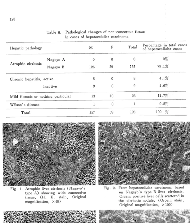
Table 6 presents the histological features of non「cancerous liver tissue observed in cases of hepatocellular carcinoma. Among the 196 cases of hepatocellular carcinoma ex‑
amined, 155 (79.1%) were associated with atrophic liver cirrhosis. No cases exhibiting Nagayo's type A liver cirrhosis were found to have hepatocellular carcinoma (Fig. 1〕.
In addition‑ 23 cases 〔11.7%〕 of hepatocellular carcinoma were either combined with
non‑specific fibrosis or displayed no particular histological changes.
HBs‑Ag was historically detected in 6 of 22 cases of subacute liver atrophy (27.2
2O and in 6 of 37 cases of chronic hepatitis (16.2%). The 15 cases diagnosed as acute
Table 5. Age distribution of atrophic liver cirrhosis and primary liver carcinoma
0‑9 10‑19 20―29 30‑39 40‑49 50‑59 60―69 70―79 80‑
Diseases
Atrophic Na‑‑ A
cirrhosis
Primary liver carcinoma
Nagayo B
2 1 3 4 4 2 3 11 75 86 62 23
Hepatocellular carcinoma Cholangiocellular carcinoma Combined type
19 267
53 67 53 13 196 10 22 液i 皇/ E! 寧 日臼
1
Tota】
Age
Table 6. Pathological changes of non‑cancerous tissue in cases of hepatocelullar carcinoma
Hepatic pathology ・.,T^‑,iPercen MFTotal。」,.違ageintotalcases t。cellularcases
Atrophic cirrhosis
Nagayo A 0 0 0 O%
Nagayo B 126 29 155 79.1%
Chronic hepatitis, active
inactive
4.1%
4.6%
Mild fibr。sis or nothing particular 13 10 23 11.7%
Wilson s disease 0.5%
157 39 196 100
Fig. 1. Atrophic liver cirrhosis (Nagayo s type A) showing wide connective tissue. (H. E. stain, Original
magnification, × 40)
Fig. 3. A: Non‑cancerous tissue showing mild fibrosis observed in a case combined with primary hepatocellular carcino‑
ma. (H. E. stain, Originalmagni‑
fication. ×40〕
Fig. 2. From hepatocellular carcinoma based on Nagayo's type B liver cirrhosis.
)orcein positive liver cells scattered in the cirrhotic nodule. (Orcein stain,
original magnification, × 100)
Fig. 3. B : Orcein staining shows HBs―Ag
in non‑cancerous tissue. (Orcein stain Original magnification,
× 200)
哺清畠
溺鮎磁器批評監護窮議
‑o一〓:,,〓
享喜一4‑i‑x 〓i,三I一I一
^""‑‑‑^*"^‑̲‑
‑‑‑/サ‑‑‑
‑ ^i一一皇一一o一〓と:〓一〓〓O:一子
liver atrophy were all HBs‑Ag negative. The differences in the rates for detectable HBs「
Ag between cases of cirrhosis without hepatocellular carcinoma (30.3%〕 and cases with hepatocellular carcinoma (60.6%, Fig. 2〕 were statistically significant (%2 test, P<
0.005) (Table 7). No HBs‑Ag was detected in any of the cases of liver cirrhosis with the exception of atrophic liver cirrhosis・ Of the 196 cases of hepatocellular carcinoma
examined, 108 were positive for HBs―Ag― As illustrated in Table 8, the incidence of
HBs‑Ag in cases of hepatocellular carcinoma was highly correlated with the presence of
●
nistological features typically found in non‑cancerous liver tissue. The rate of demonstra‑
ble HBs‑Ag was high in cases of hepatocellular carcinoma associatedいvith both atrophic
liver cirrhosis (60.6%) and chronic active hepatitis (75.0%〕 but low in cases 。f chronic inactive hepatitis (22.5%〕, mild fibrosis (Fig. 3〕 and those exhibiting no significant histological changes (26.1%). The prevalence of HBs‑Ag among 286 cases of atrophic
liver cirrhosis as related to age is shown in Figure 4. All groups exhibiting liver cirrhosis were divided into subgroups, namely, those with or without hepatocellular carcinoma.
The highest HBs‑Ag frequency in liver tissue was observed in patients who were in their third decade of life. After 30 years of age, the presence of HBs‑Ag in liver tissue de‑
creased with age in both of the subgroups.
The incidence of HBs「Ag among cases of atrophic liver cirrhosis was invest唱ated in relation to stromal inflammation (Table 9 〕 An entire subgroup of cases of Nagayo's
Table 7. Incidence of HBs‑Ag in liver diseases Diseases
Acute liver atrophy Subacute liver atrophy Chronic hepatitis
Atrophic liver cirrhosis*1
HBs「Ag(+)HBs‑Ag(‑〕Tot exaiエ1cases
lined
0
6 (27.2) 6 (16.2)
15 15 16 22 31 37 without hepatocellular carcinoma 37 (30.3) 85 122
with hepatocellular carcinoma 94 (60.6) with cholangio亡ellular carcinoma
with亡ombined 4 (57.1) Micronodular cirrhosis
Cardiac cirrhosis Biliary cirrhosis Parasitic cirrhosis Wilson's disease
0 0 0 0 0
Primary liver Hepatocellular carcinoma carcinoma Cholangiocellular carcinoma
Combined
61 155 0 2 3 7
16 4 25 1 5
16 16 25
5
108 (55.1) 196 2 ( 9.1) 20 22 4 (36.4) 11 15
〔 ) Parenthesis express percentage of positivity of HBs‑Ag in liver tissue
Average detectable rate for HBs‑Ag in this group is 47.9% (137/286).
Table 8. Incidence of HBs‑Ag in non―cancerous tissue
in cases of hepatocellular carcinoma
♀[epatichistolo
。n‑cancer。us淫of fOIHBs
ssue‑Ag(+〕HBs「Ag(‑〕Positive rati。%
Atrophic cirrhosis Chronic hepatitis, active Chronic hepatitis, inactive
Mild fibrosis or nothing particular Wilson's disease
94
6
2
6
0
61 2 7 17
160.6%
75.0%
22.5%
26.1%
0 % 196(100%)
川巾I HBs‑∧g(+)
Average posiヒive raヒio(30.∃芳)
without liver carcinoma with liver carcinoma Fig. 4. Aging incidence of HBs‑Ag in cases of atrophic liver cirrhosis.
Table 9. Relationship between stromal inflammatory reaction and incidence
of HBs―Ag in cases of atrophic liver cirrhosis
Total cases examined Diseases
Stromal
inflaのmatory HBs‑Ag{+) HBs‑Ag(‑)
reaction
Without
primary liver carcinoma
active 8 (42.4) 11 Nagayo A inactive
undetermined
active 23 (33.8) 45 Nagayo B inactive 6 (20.0) 24
undetermined
With primary liver carcinoma
19 0 0
68 30 5
a亡tive 52 (58.4〕 37 89 inactive 35 (60.3〕 23 58 undetermined 14 (82.4〕 3 17
( ) parenthesis express percentage of positivity of HBs‑Ag in liver tissue
.・
トJ川Iib er
50
Ho
〕0
20
10
,̲;,,一〓̲,̲
「l‑i
"一oo,I,=,
Numbe 6o
5口
∫lo
〕0
20
10
Average posiヒive raヒlo
(61.6芳)
』
一一,二言一3≒‑―一一言壬
108
Table 10. Incidence of liver cell dysplasia in cases of atrophic liver cirrhosis Atrophic liver cirrhosis Dysplasia(+)Dysplasisト〕Positive
ratio%
Nagayo A alone
Nagayo B without liver carcinoma
Nagayo B
/ith hepatocellular carcinoma
2 17 20 83
67 83 with other primary liver carcinoma
Fig. 5. A case of hepatocellular carcinoma alone. Groups of hepatocytes with
nuclear pleomorphism and cytoplastic enlargement are shown (H. E. stain,
Original magnification, × 100)
22/122 (18.0%)
69!154 (44.8%)
Fig. 6. Hepatocellular carcinoma (Grade III) from a case associated with atrophic liver cirrhosis. (H. E. stain, Original magnification × 100)
Table ll. Relationship between dysplasia and HBs‑Ag in atrophic liver cirrhosis Nagayo A
Dysplasia (+) 1/2 (50 %) Dysplasia (「 7/17 (41.2%)
Nagayo B Nagayo B
without with with other hepatocellular hepatocellular primary carcinoma carcinoma carcinoma
Nagayo B
12/20(60 %) 44/66 (66.7%) 3/4(75%) 17/83 (20.5%〕 50/89 (56.2%) 3/5 (60%〕
Table 12. Relationship between HBs‑Ag and grade of malignancy in cases or hepatocellular carcinoma
肝
Grade of malignancy
lll IV
With atrophic liver cirrhosis 39/69 (56.5%) 45/68 (66.2%)‑ 10/18 (55.6%) Without atrophic liver cirrhosis 5/14 (35.7%) 5/20 (25.0%) 3/7 ( 42.9%)
Grade I : No cases
2
type A liver cirrhosis was found to be free of hepatocellular carcinoma but exhibited ac‑
tive stromal inflammation. The frequency of HBs‑Ag was moderately high in the liver tis「
sues of both Nagayo's A and B liver cirrhosis with active stromal inflammation.
IThe frequency of dysplasia among cases of atrophic liver cirrhosis was also studied (Table loo According to Anthony's definition, 2 representative sections were used the evaluation of dysplasia, which included cellular enlargement, nuclear pleomorphism with hyperchromasia and multinucleation (Fig. 5 〕 Dysplasia was most frequently observed in cases of atrophic liver cirrhosis with primary liver carcinoma. Dysplasia was present in
22 cases of atrophic liver cirrhosis without hepatocellular carcinoma (18.0%) and in 69 (44.8%) cases of atrophic liver cirrhosis associated with primary liver carcinoma 〔Table 10). These differences were highly significant statistically (^2 test, P<0.005〕. Table ll lists the correlations among HBs‑Ag in liver tissue, dysplasia and cirrhosis with or without liver cell carcinoma. In Nagayo's type B cirrhosis subgroup alone, there was a difference in the prevalence of HBs「Ag between cases with dysplasia (12 out of 20; 60%) and cases without dysplasia (17 out of 83; 20・5%〕・ In the other subgroups, there was a slightly higher prevalence of HBs一触in those with dysplasia than those without dysplasia. How‑
ever this difference was not statistically significant. Liver tissue with a high preva一ence
of HBs‑Ag was noted in cases of hepatocellular carcinoma with or without dysplasia.
Malignancies in all cases of hepatocellular carcinoma were graded according to Ed‑
mondson's criteria. The results indicated that the incidence of grade III hepatocellular carcinoma was the highest in this group (Fig. 6〕 No cases of grade I hepatocellular carcinoma were observed. No significant association was observed between the presence of HBs‑Ag and the grade of malignancy (Table 12〕・
DISCUSSION
The incidence of atrophic liver cirrhosis and hepatocellular carcinoma among the total number of autopsies performed at Nagasaki University were 7.7% and 4.8% resp‑
ectively. The incidence of these 2 diseases were much higher in Nagasaki than in other districts of Japan. Accordi喝to statistical data collected from 8 universities (Miyaji, 1965〕
in Japan, a correlation between atrophic 一iver cirrhosis and hepatocellular carcinoma was
quite evident : 42.2% of the liver cirrhosis cases had developed hepatocellular carcinoma and 84.3% of the cases of hepatocellular carcinoma were associated with atrophic liver cirrhosis. In the present study, 242 out of 488 cases (49.6%) of atrophic liver cirrhosis
had also developed hepatocellular carcinoma, whereas 242 out of 303 hepatocellular carcinoma cases were associated with atrophic liver cirrhosis (78.9%). All the liver cirrhosis cases that were combined with hepatocellular carcinoma were identified as Nagayo's type B liver cirrhosis.
studies on the association of hepatocellular carcinoma and HBs‑Ag in the tissue have advantages over serum studies in that they can be used for large scale retrospective
rl̲
investigation. Shikata et al. , (1973, 1974) showed that an orcein staining technique specifically stains HBs‑Ag in fixed paraffin embedded tissue. Another researcher (Huang, 1975) has reported that the sensitivity and specificity of the orcein stain are almost equal to those or immunological methods of mesurement. The present study indicated that
HBs‑Ag was present at a rate of 47.9% in cases of atrophic liver cirrhosis and that the incidence of HBs‑Ag in liver tissue was quite high, especially in cases of atrophic liver cirrhosis with hepatocelluler carcinoma. HBs‑Ag was detected in 108 of 196 hepatocellular carcinoma cases (55.1%). The HBs‑Ag rate in the tissue among cases of hepatocellular carcinoma tended to be highly dependent on the histology of non‑cancerous tissue. That is. the incidence of HBs‑Ag in liver tissues was higher in hepatocellular carcinoma sub‑
groups combined with atrophic liver cirrhosis or chronic active hepatitis than in hep‑
atocellular carcinoma subgroups associated with mild fibrosis or in those exhibiting no histological significant changes.
●
These results were almost identical to those obtained in Hong‑Kong (Wu, 1978〕
where the carrier rate for the hepatitis B virus is as high as sA%. A similar investigation was carried out on the east coast of the United States (Keshegegian et al,. 1981) using the orcein staining technique. Although the incidence of HBs‑Ag was as low as 15.1%
in cases of hepatocellular carcinoma, it was also highly correlated with the presence of atrophic liver cirrhosis. The highest incidence of HBs「Ag in liver tissue occured in the second decade of life in cases of hepatocellular carcinoma and atrophic liver cirrhosis.
The rate then decreased gradually with age. These morphological phenomenon might best be explained by the concept of seroconversion proposed by Kubo et al., (1977) and
Szmness 〔1975〕. These researchers also indicated that the carrier rate for HBs―Ag among
blood donors in New York city was highest at 50 years of age then decreased with increasing age.
It has been reported by Almeida et a/., (1969), Shulman (1969), Edinton, et al., (1975〕 that a hepatitis B virus‑induced immune reaction may be responsible for causing the hepatocyte injury which leads to chronic liver diseases such as chronic hepatitis liver cirrhosis and hepatocellular carcinoma. Our present data suggests that a long term in‑
cubation of HBs‑Ag may cause a stromal inflammatory reaction. Patients with detectable levels of both serum HBs‑Ag and core‑antigen reportedly ran the highest risk of developing liver cirrhosis (Gudat et al., 1975). Controversies exists as to whether or not the hepatitis B virus has a direct oncogenic role in the etiology of hepatocellular carcinoma (Atiyeh et at. , 1980) and whether or not a hepatitis B infection causes liver cirrhosis predisposing patients to hepatocellular carcinoma (Zukerman, 1977). In 1969, Harris, et al., first reported the following hypothesis concerning the role of the hepatitis B virus on oncogen‑
esis : The hepatitis B virus induces malignant changes in infected liver cells which then
fuse with the neighboring normal liver cells to form large, abnormal dysplastic cells. Some
of these dysplastic cells then develop into cancer cells. In 1977, Lutwick et al. found
the HBV「DNA complex in a DNA fraction extracted from a chronically infected liver by
means of a DNA‑DNA hybridization technique. Their results indicated that the hepatitis B virus in possessed of an essential candidation of oncogen. At almost the same time, Macnab et al., (1976) derived a human cell line (Alexander cell PLC/PRF/5) which
produced hepatitis B virus surface antigen in the culture supernatant. They then sue「
ceeded in producing neopiasia in nude mouse by transplantation of that cell line. Later,
another cell line derived by Aden (Hep 3B〕 was found to produce α―fetoprotein as well
as HBs‑Ag (Alexander et al., 1978〕 These results lend support to the theory that the
hepatitis B virus plays a direct role in oncogenesis青
In the present study, the incidence of HBs‑Ag in cases of atrophic liver cirrhosis associated with hepatocellular carcinoma was found to be remarkably greater than in cases without hepatocellular carcinoma. Furthermore, HBs‑Ag was detected in 14 cases of hepatocellular carcinoma which did not exhibit atrophic liver cirrhosis : In 6 of those cases, a histological examination of the non‑cancerous tissue revealed only mild fibrosis or no significant changes. These results indicate that hepatocellular carcinoma can arise as a direct result of a hepatitis B virus infection without the development of atrophic liver cirrhosis.
In 1973, Anthony et a/. , defined liver cell dysplasia using cytological features such as cellular enlargement, nuclear pleomorphysm with hyperchromasia and multinucleation.
The present study has shown a strong correlation between the presence of the hepatitis B virus and liver cell dysplasia, indicating a possible carcinogenic role of the long term in‑
cubation of the hepatitis B virus in the development of hepatocellular carcinoma.
AcKNOWLEDGEMENTS
Thanks are tendered to the directors and staff of every Department of Pathology in Nagasaki University and Radiation Effects Research Foundation for generous supply of materials.
Thanks are also due to Dr. Takayoshi Toda and Mrs. Virginia Yamamoto for their advice and suggestion.
REFERENCES
1) Alexander J., Macnab, G. and Saunders, R. (1978): Studies on in vitro production of hepatitis B surface antigen by a human hepatome cell line・ Perspect. Virol., 10, 103=120
2) Almeida, J. D. and Waterson, A. P. (1969): Immune complexes in hepatitis. Lancet, II, 983
‑986
3) Anthony, P. P., Vogal, C. L. and Baker, L. F. (1973): Liver cell dysplasia: a malignant condition. J. Clin. Pathol., 36, 217‑223
4) Atiyeh, M. and AH, M. A. (1980): Primary hepatocellular carcinoma in Saudi Arabia: A clini‑
copathological study of 54 cases. Am. J. Gastroenterol., 74 (l), 25―29
5) Beasley, R. P., Hwang, L. Y. Lin, C. C. and Chein, C. S. (1981): Hapatocellular carcinoma
and hepatitis B virus‥ A prospective study of 22707 men in Taiwan. Lancet, 21, 1129‑1133
6) Edginton, T. ,S. and Chisari F. V. (1975): Immunological aspects of hepatitis B virus infection.
Am. J. Med. Sci., 270, 213「227
7) Edmondson, H. A. and Steiner, P. E. (1954)‥ Primary carcinoma of the liver: A study of 100
cases among 48,900 necropsies. Cancer, 7, 462‑503
8) Gall, E. A. (1960): Posthepatitic, postnecrotic and nutritional cirrhosis: a pathological analysis.
Am. J. Pathol., 36, 241‑259
9) Gudat, F., Bianchi, L., Sonnabend, W., Thiel. G., Aenishaenalin, W. and Stalder, G. A.
(1975〕 Pattern of core and surface expression in liver tissue reflects state of specific immune
responce in hepatitis B. Lab. Invest., 32, 1―9
10) Halliday, J. W. and Oda, T. (1980): Report of survey of hepatitis antigens in‑ the Asian‑Pacific
region. Ann. Acad. Med., 9(2), 133―135
ll) Harris, H., Miller, D. J., Klein, G., Worst, P. and Tachibana, T. (1969): Suppressionof
malignancy by cell fusion. Nature, 223, 363―368
12) Huang, S. N. (1975): Immunohistochemical demonstration of hepatitis B core and surface antigens in paraffin sections. Lab. Invest., 33, 88‑95
13) Kanda, T. (1977〕: Hepatitis B surface antigen in cirrhosis of the liver and primary liver carcinoma in Nagasaki area: A histopathological study. Jpn. J. Trop. Med. Hyg., 5(2) 117‑139(Japanese with English abstract)
14) Keshgegian, A. A. and Ochs, R. H. (1981): Orcein positive hepatitis B surface antigen and liver carcinoma: Their association in an eastern US population. Arch. Pathol. Lab. Med. 105
190―193
15) Kubo, Y. and Okuda, K. (1977): Antibody to hepatitis B core antigen in patients with hepato‑
cellular carcinoma. Gastroenterol. , 72, 1217‑1220
16) Luna, L. G. (1968): Manual of histologic staining methods of the Armed Forces Institute of Pathol咽y. 3rd ed., McGraw‑Hill, New York.
17) Lutwick, L. I. and Robinson, W. S. (1977): DNA synthesized in the hepatitis B Dane particle:
DNA polymerase reaction. J. Viro1 21, 96‑104
18) Macnab, G. N., Alexander, J. J., Lecatsas, G., Bey, E. M. and Urobanowicz, J. M. (1976):
Hepatitis B surface antigen produced by a human hepatoma cell line. Br. J. Cancer, 34, 509‑515 19) Maupus, P. and Melnick, J. L. (1981): Hepatitis B infection and liver cancer. Prog. Med.
Virol., 27, 1‑5
20) McCollum, R. W. and Zukerman, A. J. (1981): Viral hepatitis‥ Report on a WHO informal
consultation. J. Med. Virol., 8, 1‑29
21〕 Miyaji, T. (1965): Hepatocellular carcinoma in Japan: especially its relation to liver cirrhosis, Tr. Soc. Pathol. Jpn,, 54, 23‑36 (in Japanese〕
22) Miyake, H. (1960): Pathology of the liver. Tr. Soc. Pathol. Jpn., 49, 589‑613'(in Japanese) 23) Mori, I. (1964): Classification of liver cirrhosis. Igaku no Ayumi, 48, 37ト381 (in Japanese)
24〕 Nagayo, M. (1914)‥ Liver cirrhosis. Tr. Soc. Pathol. Jpn., 4, 31‑72 (in Japanese)