TNF- ␣ Suppressed FSH-Induced LH Receptor Expression Through Transcriptional Regulation in Rat Granulosa Cells
Kohshiro Nakao, Hiroshi Kishi, Fumiharu Imai, Hiroto Suwa, Takashi Hirakawa, and Takashi Minegishi
Department of Obstetrics and Gynecology, Gunma University Graduate School of Medicine, Maebashi, Gunma 371-8511, Japan
Several inflammatory cytokines regulate ovarian function. TNF-␣is produced in granulosa cells under physiological conditions and has a reciprocal action on follicle development. In contrast, in pelvic inflammatory diseases, TNF-␣is excessively produced in the pelvic cavity and has an adverse effect on reproductive functions. The objective of this study was to elucidate the mechanism of action of TNF-␣on the expression of LH receptor (LHR) in immature rat granulosa cells. TNF-␣ suppressed FSH-induced LHR mRNA and protein expression and was not associated with cAMP accumulation. By using a luciferase assay, the construct containing base pairs⫺1389 to⫺1 of the ratLhcgrpromoter revealed that TNF-␣decreased FSH-induced promoter activity. In response to TNF-␣, nuclear factor (NF)-B p65 was translocated to the nucleus, and the suppressive effect of TNF-␣on LHR mRNA expression was abrogated by an NF-B inhibitor. In a chromatin immuno- precipitation assay, TNF-␣induced the association of NF-B p65 with the ratLhcgrtranscriptional promoter region. NF-B p65 and histone deacetylase (HDAC) interact to mediate expression of several genes at a transcriptional level. HDAC activity is thought to induce tight connections within local chromatin structures and repress gene transcription. Furthermore, the TNF-␣–induced sup- pression of LHR mRNA expression was blocked by an HDAC inhibitor. Taken together, these results suggest that the interaction of NF-B p65 with HDAC in the promoter region of ratLhcgrmight be responsible for TNF-␣action on the regulation of LHR.(Endocrinology156: 3192–3202, 2015)
T
NF-␣, a 17-kDa homotrimeric soluble protein, is an inflammatory cytokine that is identified as a factor induced by the hemorrhagic necrosis of tumors and is pri- marily produced by activated macrophages, monocytes, and lymphocytes (1, 2). The effects of TNF-␣, including cell proliferation, apoptosis, and antiapoptosis, are ex- erted through binding to its cell surface receptors, and TNF-␣activates the inhibitor of nuclear factor-B (IB) kinases, resulting in phosphorylation and degradation of IB␣and translocation of the nuclear factor-B (NF-B) dimer to the nucleus. NF-B belongs to the Rel family of transcription factors, which includes RelA (p65), RelB, c-Rel, NF-B1 (p50/p105), and NF-B2 (p52/p100) (3, 4).Intranuclear NF-B regulates the expression of various genes by binding directly to the DNA binding domain or by modifications of a variety of other transcription fac- tors. In particular, the p50 and p65 subunits, which con- tain a potent transcription regulator function, have been most widely studied (5–9).
The pituitary gonadotropins are essential hormones for follicular growth, acting through stimulatory G protein–
coupled receptors. The LH receptor (LHR) is a G protein–
coupled receptor considered necessary for reproductive function, including steroidogenesis and ovarian folliculo- genesis. LHR expression is a major marker of FSH-in- duced granulosa cell (GC) differentiation. FSH acts
ISSN Print 0013-7227 ISSN Online 1945-7170 Printed in USA
Copyright © 2015 by the Endocrine Society Received March 15, 2015. Accepted June 22, 2015.
First Published Online June 30, 2015
Abbreviations: 8-Br-cAMP, 8-bromo-cAMP; ChIP, Chromatin immunoprecipitation; CREB, cAMP response element– binding protein;CT, cycle threshold; DES, diethylstilbestrol; GC, granulosa cell; hCG, human chorionic gonadotropin; HDAC, histone deacetylase; IB, inhibitor of nuclear factor-B; LHR, LH receptor; NF-B, nuclear factor-B; NIH, National Institutes of Health; PKA, protein kinase A; SF1, steroidogenic factor 1; Sp1, specificity protein 1; TSA, trichostatin A.
3192 press.endocrine.org/journal/endo Endocrinology, September 2015, 156(9):3192–3202 doi: 10.1210/EN.2015-1238
through the FSH receptor and induces LHR expression primarily through adenylyl cyclase activation and cAMP production (10). Chen et al (11) reported that the 5⬘-flank- ing region of the ratLhcgrpromoter contained specificity protein 1 (Sp1), steroidogenic factor 1 (SF1), and activat- ing protein 2 binding sites, and they also reported that Sp1 is essential for theLhcgrgene transcriptional activity in- duced by 8-bromo-cAMP (8-Br-cAMP). Law et al (12) indicated that the expression of FSH-induced LHR is pro- moted by phosphorylation of forkhead box O1 via phos- phatidylinositol 3-kinase/Akt and by phosphorylation of
-catenin through the protein kinase A pathway. In GCs, it has been reported that FSH activates several signaling pathways including cAMP response element– binding protein (CREB), phosphatidylinositol 3-kinase/Akt, and ERK1/2 (13, 14). However, the pathways downstream of FSH leading to the activation of LHR expression have not been fully elucidated. In addition, many local factors in- cluding cytokines and hormones are probably involved in the modulation of LHR expression (14 –16).
TNF-␣has been confirmed to be present in the ovaries of a variety of species, including human GCs (17, 18) and corpora lutea (18) and rat GCs (19). It has been reported that TNF-␣levels were lower in the sera of patients with premature ovarian failure than in women with normal menstrual cycles (20). Compared with wild-type mice, TNF type 1 receptor knockout mice have been shown to exhibit earlier vaginal opening and human chorionic go- nadotropin (hCG)–induced ovulation even in the prepu- bertal period (21), indicating that TNF type 1 receptors might prevent the ovarian response to gonadotropins to avoid early senescence; therefore, TNF-␣ might have a protective effect on ovarian aging. In an in vitro study, high concentrations of TNF-␣ inhibited gonadotropin- stimulated estrogen production (22–24), whereas TNF-␣ differentially affects steroidogenesis, depending on the fol- licular phase (25). Furthermore, TNF-␣ drastically re- duces FSH-induced LHR expression at a post-cAMP site (26), but the molecular mechanisms of this effect have not been clarified.
From a pathophysiological perspective, it has been re- ported that TNF-␣concentrations in the peritoneal fluid and follicular fluid were elevated in patients with endo- metriosis (27, 28), and TNF-␣ was also elevated in GC cultures derived from patients with endometriosis (29).
Patients with endometriosis are often infertile, but details of the causal relationship between endometriosis-related infertility and high TNF-␣ concentrations are not yet known (28 –31).
In the present study, we examined the effect of TNF-␣ on FSH-induced LHR mRNA and protein expression lev- els and mechanisms of regulation in rat primary GCs. We
found that the inhibitory effect of TNF-␣on FSH-induced LHR expression was attributable to the transport of p65 to the nucleus. Furthermore, intracellular p65 may inter- act with histone deacetylase (HDAC) to repress theLhcgr promoter.
Materials and Methods Hormones and reagents
Recombinant human FSH was supplied by Dr A. Parlow and the National Hormone and Pituitary Program (National Insti- tute of Diabetes and Digestive and Kidney Diseases, National Institutes of Health [NIH]). DMEM/Ham’s nutrient mixture F-12, diethylstilbestrol (DES), and 8-Br-cAMP were purchased from Sigma-Aldrich, Inc. TNF-␣was purchased from R&D Sys- tems, Inc. SN50, a cell-permeable NF-B-inhibitory peptide, was purchased from Enzo Life Sciences, Inc. Trichostatin A (TSA) was purchased from Sigma-Aldrich. Gentamicin sulfate and Fungizone were purchased from Invitrogen Corp. The RNA la- beling kit and nucleic acid detection kit were purchased from Roche Diagnostics.
Animals
Immature female Wistar rats (Japan SLC, Inc.) were main- tained at all times according to the NIHGuide for the Care and Use of Laboratory Animalsand the policies of the Gunma Uni- versity Animal Care and Use Committee. Animals were housed in a temperature- and light-controlled room (12-hour light/12- hour dark cycle; lights on at 6:00AM) with food and water pro- vided ad libitum.
GC culture
GCs were obtained from immature female Wistar rats in- jected daily for 4 days with 2 mg of DES in 0.2 mL of sesame oil.
The ovaries were then excised, and the GCs were released by puncturing the follicles with 26-gauge needles. GCs were washed and collected by brief centrifugation, and cell viability was de- termined by trypan blue exclusion. The GCs were then cultured in DMEM/Ham’s nutrient mixture F-12 supplemented with 20 mg/L gentamicin sulfate, 500g/L Fungizone, and 1 g/L bovine serum albumin on collagen-coated plates in a humidified atmo- sphere under 5% CO2at 37°C.
RNA isolation and Northern blot analysis
GCs were cultured in 60-mm dishes containing 5⫻106viable cells in 5 mL of medium. Twenty-four hours later, GCs were incubated with or without inhibitors for 1 hour at 37°C and then stimulated with each reagent. GCs were further incubated, and the cultures were stopped at selected times using Isogen (Nippon Gene). The final RNA pellet was dissolved in diethylpyrocar- bonate-treated H2O. Total RNA was quantified by measuring the absorbance of samples at 260 nm. For the Northern blot analysis, 15g of total RNA from each dish was separated by electrophoresis on denaturing agarose gels and subsequently transferred to a nylon membrane (Biodyne; Pall Corp). Rat LHR cDNA was prepared as described previously and linearized with BglII (32). Digoxigenin-labeled LHR cRNA probes correspond- ing to bases 440 to 2560 were produced by in vitro transcription
with T3 RNA polymerase and an RNA labeling kit (Roche Di- agnostics). A digoxigenin-labeledGAPDHprobe was obtained by the same method. In accordance with the standard protocol for the nucleic acid detection kit (Roche Diagnostics), the membranes were then exposed on Kodak XAR film (Eastman Kodak Co).
Reverse transcription and quantitative RT-PCR Isolated RNAs (2g of each sample) from the GC cultures were treated with DNase I (Invitrogen) to eliminate residual genomic DNA. These RNAs were reverse transcribed with ran- dom primers, 10 mM deoxynucleoside triphosphate mix, and SuperScript III reverse transcriptase (Invitrogen) according to the manufacturer’s protocol. The reactions were incubated for 5 minutes at 25°C, 60 minutes at 50°C, and 15 minutes at 70°C in a thermal cycler. To remove cRNA, ribonuclease H was added to the cDNAs and incubated for 20 minutes at 37°C. The quanti- tative RT-PCR was performed according to the manufacturer’s instructions using EagleTaq Universal Master Mix with ROX (Roche Diagnostics) and an ABI Prism 7000 sequence detection system (Applied Biosystems). The quantitative PCR condi- tions were initial denaturation at 95°C for 10 minutes, fol- lowed by 40 cycles of 95°C for 15 seconds and 60°C for 1 minute. The primers and probes for each gene were purchased from Applied Biosystems (LH/choriogonadotropin receptor Rn00564309_m1 and eukaryotic18SrRNA Hs99999901_s1 as an internal control). Relative quantifications of mRNA species were performed using the comparative threshold cycle (CT) method. For each sample, we normalized the geneCTvalue using the formula⌬CT⫽CTgene⫺CT18S. For relative expression levels, the following formula was used:⌬⌬CT⫽ ⌬CTsample⫺
⌬CTcalibrator. This value was then used to plot gene expression with the formula 2⫺⌬⌬CT.
Receptor binding assay
GCs were cultured in 24-well plates containing 5⫻105viable cells in 0.5 mL of medium. After 24 hours of incubation, reagents were added to the medium for 72 hours. Thereafter, the cells were placed on ice and quickly washed 3 times with 0.4 mL of cold medium. The GCs were then incubated in a 1:1 (vol/vol) mixture of DMEM/Ham’s F-12 medium containing 0.1% bo- vine serum albumin (pH 7.4) at 37°C with 1.5⫻105cpm125I- hCG (5 ng, 30 000 cpm/ng). The hCG was iodinated according to the chloramine-T method (33). The incubation medium was removed after 1 hour of incubation, and the cells were washed twice with 0.4 mL of medium. The amount of radioactivity re- maining in the well (cell-bound hormone) was then quantified by
-spectrometry. Nonspecific binding was determined by adding excess unlabeled hCG (50 IU/well).
Intracellular cAMP assay
The intracellular accumulation of cAMP was measured using the cyclic AMP EIA kit (Cayman Chemical Co). GCs were cul- tured in 24-well culture plates containing 5⫻105viable cells per well in 0.5 mL of medium. After 24 hours of cell culture, the cells were treated with reagents for 30 minutes, and then intracellular accumulation of cAMP was measured according to the manu- facturer’s instructions.
Luciferase reporter assay
Plasmid pGL3-basic is a luciferase vector lacking the eukary- otic promoter and enhancer sequences (Promega Corp). The pGL3-control contains a simian virus 40 promoter and a simian virus 40 enhancer inserted into the structure of pGL3-basic (Pro- mega). The fragment of the 5⬘-flanking region from base pairs
⫺1389 to⫺1 (relative to the translational initiation site), which included the transcriptional regulating region of rat LHR (34), was generated from genomic DNA as described previously (16).
To evaluate promoter activity, these fragments were ligated to a luciferase reporter vector (pGL3-basic) and named LH-R(1389)- Luc. Using FuGENE 6 reagent (Promega), we transfected 200 ng of plasmid DNA into the primary rat GC culture plates (2⫻105 cells per 0.5 mL of medium in a 24-well culture plate). To assay the regulatory elements, the GCs were cultured for 24 hours in a hormone-free medium before transfection. Twenty-four hours after transfection, the cells were treated with hormones for 3 hours. The cells were then harvested, and the luciferase activity was measured. The luciferase assay was performed using the dual-luciferase reporter system (Promega), in which the trans- fection efficiency was monitored by cotransfected pRL-CMV- Rluc, an expression vector ofRenillaluciferase.
Preparation of whole-cell extracts
GCs were cultured in 3.5-mm dishes containing 2.5⫻106 viable cells in 2.5 mL of medium, and the reagents were added to the medium after 24 hours of cell culture. After incubation, the cells were washed twice with 0.5 mL of cold buffer (20 mM HEPES and 0.15 M NaCl [pH 7.4]). Then they were lysed in radioimmunoprecipitation assay buffer (150 mM NaCl, 50 mM Tris, 1 mM EDTA, 1% Nonidet P-40, 0.5% sodium deoxy- cholate, 0.1% SDS, and protease complete inhibitor [pH 7.4]) supplemented with 200 mM NaF and 200 mM sodium or- thovanadate for 40 minutes with rotation at 4°C. Lysates were clarified by centrifugation at 20 000⫻gfor 10 minutes at 4°C and stored at⫺80°C.
Preparation of nuclear and cytosolic fractions GCs were cultured in 60-mm dishes containing 7⫻106viable cells in 5 mL of medium. After 24 hours of cell culture, the cells were incubated with or without inhibitors for 1 hour at 37°C, and then reagents were added to the medium. The cells were washed twice with 1 mL of cold PBS, scraped, and collected by centrifugation at 1500⫻gfor 3 minutes at 4°C. The pellet was resuspended in freshly prepared cold extraction buffer A (10 mM HEPES [pH 7.9], 10 mM KCl, 0.1 mM EDTA, 1 mM dithio- threitol, 0.5 mM phenylmethylsulfonyl fluoride, and protease complete inhibitor) and incubated for 15 minutes on ice. After addition of 1% Nonidet P-40, cells were vigorously vortexed for 15 seconds and centrifuged at 500⫻gfor 3 minutes at 4°C. The supernatants were cytosolic fractions. The pellet containing nu- clear extract was washed twice in buffer A. The pellet was then resuspended in extraction buffer B (20 mM HEPES [pH 7.9], 400 mM NaCl, 0.1 mM EDTA, 1 mM dithiothreitol, 0.5 mM phe- nylmethylsulfonyl fluoride, and protease complete inhibitor) and vortexed for 15 minutes at 4°C. The nuclear extracts were obtained from supernatants by centrifugation at 18 000⫻gfor 15 minutes and stored at⫺80°C.
SDS-PAGE and Western blot analysis
Protein lysate (20 – 40g) was resolved on 10% SDS gels and electrophoretically transferred to a polyvinylidene difluoride membrane. After blocking, expression of the different proteins was determined overnight at 4°C with the following antibodies purchased from Cell Signaling Technology: rabbit anti-phospho- CREB (1:2000), rabbit anti-CREB (1:2000), rabbit anti-phos- pho-Akt (1:2000), rabbit anti-Akt (1:2000), rabbit anti-phos- pho-p44/42 MAPK (ERK1/2) (1:3000), rabbit anti-p44/42 MAPK (ERK1/2) (1:2000), rabbit anti-NF-B p65 (1:2000), rabbit anti-lamin A/C (1:2000), and rabbit anti--tubulin (1:
2000) (Table 1). After membranes were washed in Tris-buffered saline with Tween 20, they were incubated at room temperature for 60 minutes with horseradish peroxidase-conjugated goat an- ti-rabbit IgG antibody (no. 170-6515, 1:40 000; Bio-Rad Lab- oratories). The proteins were finally visualized using enhanced chemiluminescence (Immobilon Western; Millipore). Lumines- cence was quantified by scanning the films with a CCD camera and digitizing the data using NIH ImageJ (version 1.60).
Chromatin immunoprecipitation (ChIP) assay ChIP assays were performed using the ChIP-IT High Sensi- tivity Kit (Active Motif) according to the manufacturer’s proto- col with some modifications. GCs were cultured in 90-mm dishes containing 1⫻107viable cells in 10 mL of medium. After 48 hours of cell culture, the cells were treated with reagents for 1 hour at 37°C. Cells were then fixed with formaldehyde at a final concentration of 1% for 10 minutes at room temperature. After this reaction was stopped, the cells were pelleted, resuspended, and passed through a 26-gauge syringe 30 times to aid nucleus release. Samples were sonicated 20 times, with pulses of 30 sec- onds on and 30 seconds off, using a Bioruptor (Cosmo Bio Co) set at high power. Chromatin fragments (average size 500 –1500 bp) were used for the ChIP assay with 10g of a rabbit anti-p65 polyclonal antibody. As a negative control, normal rabbit IgG was used (Santa Cruz Biotechnology). The DNA fragments were analyzed by quantitative PCR according to the manufacturer’s
instructions using SYBR Green Real-Time PCR Master Mix (Toyobo Co) and an ABI Prism 7000 sequence detection system.
The quantitative PCR conditions were initial denaturation at 95°C for 1 minute, followed by 40 cycles of 95°C for 15 seconds and 65°C for 1 minute. The primer sequences were as follows: for rat Lhcgr DNA, the proximal promoter detects 5⬘,⫺314 to
⫺156 nucleotides from the transcription start site with forward primer 5⬘-CCTTGGCAGTGCATAGTTCA-3⬘ and reverse primer 5⬘-AGTCCCTGCTTTCCTTGACC-3⬘; the distal primer de- tects 5⬘,⫺2612 to⫺2393 nucleotides from the transcription start site with forward primer 5⬘-ACCTTTGGGGGAAAATCTTG-3⬘and re- verse primer 5⬘-GGCCTCTGTGGTTTGGTATG-3⬘; and the trans- lated region primer detects 5⬘,⫹811 to⫹950 nucleotides from the transcription start site with forward primer 5⬘-AATTCCAGCGGTC- CTTCCAT-3⬘ and reverse primer 5⬘-CCCACTGCTGCTTAC- CCTTG-3⬘. The values were corrected using the input values and are presented as percent input.
Data analysis
The data represent the means⫾SEM from at least 3 inde- pendent experiments. Comparisons between groups were per- formed by one-way ANOVA. The significance of the differences between the mean values of the control group, and each treated group was determined using a Scheffé multiple-comparison test.
APvalue of⬍.05 was considered significant.
Results
TNF-␣–suppressed FSH-induced LHR mRNA and protein expression
We investigated the effect of FSH and TNF-␣on LHR mRNA in primary rat GC cultures. GCs were cultured for 24 hours after plating on a cell culture dish, and then the cells were treated with FSH (30 ng/mL) and TNF-␣(0.1 Table 1. Antibody Table
Peptide/Protein Target
Antigen Sequence (if
Known) Name of Antibody
Manufacturer, Catalog No., and/or Name
of Individual Providing the Antibody
Species Raised in; Monoclonal
or Polyclonal Dilution Used
Phospho-CREB Phospho-CREB (Ser133)
antibody
Cell Signaling, catalog no. 9197 Rabbit, polyclonal 1:2000
CREB CREB antibody Cell Signaling, catalog no. 9198 Rabbit, polyclonal 1:2000
Phospho-Akt Phospho-Akt (Ser473) antibody Cell Signaling, catalog no. 9271 Rabbit, polyclonal 1:2000
Akt Akt antibody Cell Signaling, catalog no. 9272 Rabbit, polyclonal 1:2000
Phospho-ERK1/2 Phospho-p44/42 MAPK (Erk1/
2) (Thr202/Tyr204) antibody
Cell Signaling, catalog no. 9101 Rabbit, polyclonal 1:3000
ERK1/2 p44/42 MAPK (Erk1/2)
antibody
Cell Signaling, catalog no. 9102 Rabbit, polyclonal 1:2000 NF-B p65 NF-B p65 (D14E12) XP
antibody
Cell Signaling, catalog no. 8242 Rabbit, polyclonal 1:2000 Lamin Lamin A/C antibody Cell Signaling, catalog no. 2032 Rabbit, polyclonal 1:2000
-Tubulin -Tubulin (9F3) Cell Signaling, catalog no. 2128 Rabbit, polyclonal 1:2000 NF-B p65 NF-B p65 antibody (C-20) x Santa Cruz Biotechnology,
catalog no. sc-372 x
Rabbit, polyclonal Normal rabbit IgG normal rabbit IgG antibody x Santa Cruz Biotechnology,
catalog no. sc-2027 x
Rabbit, polyclonal
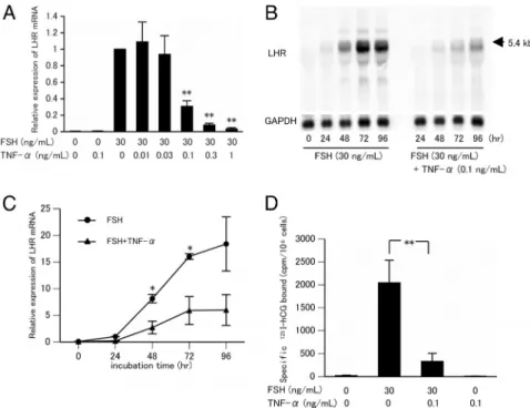
ng/mL) alone or with FSH and increasing concentrations of TNF-␣(0.01–1 ng/mL) for 72 hours (Figure 1A). FSH- induced LHR mRNA was suppressed in a dose-dependent manner by TNF-␣. LHR mRNA expression induced by treatment with FSH and 0.1 ng/mL TNF-␣was approxi- mately one-third less than that induced by FSH alone.
However, LHR mRNAs were undetected in the GCs in- cubated with TNF-␣alone or receiving no treatment. GCs were cultured for 24 to 96 hours in the presence of FSH (30 ng/mL) with or without TNF-␣(0.1 ng/mL) (Figure 1, B and C). LHR mRNA expression has been shown to reach a plateau at 72 hours with FSH treatment (32). Concur- rent treatment with FSH and TNF-␣ significantly sup- pressed LHR mRNA expression by approximately one- third at 48 and 72 hours, respectively.
To measure the effect of TNF-␣on membrane LHR protein levels, a125I-hCG binding assay was undertaken.
GCs were cultured in the absence or presence of FSH (30 ng/mL) with or without TNF-␣(0.1 ng/mL) for 72 hours.
Basal-specific LH binding was negligible and remained
unchanged in response to treatment with TNF-␣ alone, whereas treat- ment with FSH produced, as ex- pected, a substantial increase in spe- cific LH binding, and concurrent treatment with TNF-␣ produced a suppressive effect compared with that produced by FSH alone (Figure 1D).
TNF-␣had no effect on intracellular cAMP concentration
It is known that the binding of FSH to its receptor causes cAMP syn- thesis in GCs (35). Therefore, we ex- amined whether there was a change in the amount of cAMP with the ad- dition of FSH alone or with FSH and TNF-␣. The data presented in Table 2 show that TNF-␣, either alone or in combination with FSH, had no effect on intracellular cAMP levels.
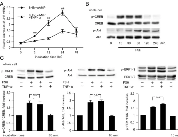
TNF-␣suppressed 8-Br-cAMP- induced LHR mRNA and had no effect on the CREB and Akt phosphorylation
It is also known that the actions of FSH to regulate LHR expression are mimicked by cAMP (35). We next examined the effect of TNF-␣on the expression of 8-Br-cAMP-induced LHR mRNA. GCs were cultured for 6 to 48 hours in the presence of 8-Br-cAMP (1 mM) with or without TNF-␣ (0.1 ng/mL). 8-Br-cAMP increased the LHR mRNA level, an effect that was suppressed by concurrent treatment with TNF-␣(Figure 2A). Furthermore, we confirmed the effect of FSH alone or of FSH and TNF-␣on the phosphorylation of
Figure 1. Effect of TNF-␣on FSH-induced LHR mRNA and LHR expression in rat GCs. A, GCs from DES-primed immature rats were cultured for 24 hours. These cells were then further incubated with FSH (30 ng/mL) and TNF-␣(0.1 ng/mL) alone or with FSH plus increasing concentrations of TNF-␣for 72 hours. Results show rat LHR mRNA accumulation, relative to that of18SmRNA, as measured by quantitative RT-PCR. The amount of LHR mRNA with FSH alone was set as 1. Data in the bar graphs represent the means⫾SEM from 4 independent experiments. **,P⬍.01 compared with the control (FSH alone). B and C, GCs were incubated with FSH alone or with FSH plus TNF-␣(0.1 ng/mL) for the indicated times. B, Northern blot representative of 3 experiments. C, rat LHR mRNA accumulation, relative to that of18SmRNA, as measured by quantitative RT-PCR. The amount of LHR mRNA with FSH alone at 24 hours was set as 1. Data in the bar graphs represent the means⫾SEM from 3 independent experiments. *, P⬍.05 compared with the control (FSH alone) at the same time. D, GCs were incubated with FSH alone or with FSH plus TNF-␣for 72 hours. LHR levels were determined by125I-hCG binding assays. Data in the bar graphs represent the means⫾SEM from 3 independent experiments. **, P⬍.01 compared with the control (FSH alone).
Table 2. Effect of FSH and TNF-␣on Intracellular cAMP Accumulation in Rat GCs
Treatment cAMP Accumulation,
pmol/5ⴛ105cells
Control 0.08⫾0.01
FSH 39.10⫾1.22a
FSH⫹TNF-␣(0.01 ng/mL) 40.23⫾3.46a FSH⫹TNF-␣(0.1 ng/mL) 46.27⫾2.19a FSH⫹TNF-␣(0.3 ng/mL) 38.03⫾0.69a FSH⫹TNF-␣(1 ng/mL) 39.90⫾1.23a
TNF-␣(1 ng/mL) 0.09⫾0.01
Each data point represents the mean⫾SEM from 3 replicate cultures.
FSH was used at a concentration of 30 ng/mL.
aP⬍.01 compared with the control value.
CREB, Akt, and ERK1/2, which is downstream of cAMP actions. Western blot analysis showed that FSH induced maximal phosphorylation of CREB and Akt after 60 minutes (Figure 2B) and that maximal phosphorylation of ERK1/2 occurs after 15 minutes (14). Phosphorylation of CREB, Akt, or ERK1/2 was not detected after exposure to TNF-␣alone (data not shown). After concurrent treatment of FSH with TNF-␣(60 minutes for CREB and Akt and 15 minutes for ERK1/2), there was no change in CREB, Akt, or ERK1/2 phosphorylation (Figure 2C). These observations suggest that TNF-␣inhibited the action of FSH at sites distal to cAMP action and did not affect CREB, Akt, and ERK1/2 phosphorylation in GCs.
TNF-␣suppressed FSH-induced activity of the Lhcgrpromoter
To understand the mechanism through which TNF-␣ suppressed FSH-induced LHR expression, we next inves- tigated the effect of TNF-␣onLhcgrpromoter activity.
Based on a previous study in which the same region of the rat Lhcgr gene was studied and well analyzed (34), we selected the proximal 1389 bp of the 5⬘-flanking region for our experiment. GCs transiently transfected with lu- ciferase-reporting plasmids [LH-R(1389)-Luc] responded to FSH (30 ng/mL) with significantly enhanced activity.
Moreover, treatment with TNF-␣(0.1 ng/mL) degraded the FSH-induced activity of theLhcgrpromoter (Figure 3).
Effect of TNF-␣to suppress LHR mRNA expression is mediated by p65 in GCs
It was previously reported that, in several cell types, TNF-␣ activates the IB kinases, and these kinases de- grade IB␣and translocate the NF-B p50/65 heterodimer to the nucleus. We investigated whether TNF-␣ caused p65 translocation to the nucleus in our culture system.
Western blot analysis showed that TNF-␣ caused p65 translocation to the nucleus after 30 minutes of exposure
Figure 2. Effect of TNF-␣on 8-Br-cAMP-induced LHR mRNA expression and on FSH-induced CREB, Akt, and ERK1/2 phosphorylation in rat GCs.
A, GCs were cultured for 24 hours and then incubated with 8-Br-cAMP (1 mM) alone or with 8-Br-cAMP plus TNF-␣(0.1 ng/mL) for the indicated times. Results show rat LHR mRNA accumulation, relative to that of18SmRNA, as measured by quantitative RT-PCR. The amount of LHR mRNA in 8-Br-cAMP alone at 6 hours was set as 1. Data in the bar graphs represent the means⫾SEM from 3 independent experiments. **,P⬍.01 compared with the control (8-Br-cAMP alone) at the same time. B, For Western blot analysis, GCs were cultured for 24 hours and then incubated with FSH (30 ng/mL) alone for the indicated times. C, GCs were incubated with FSH (30 ng/mL) alone or with TNF-␣(0.1 ng/mL) for 60 (for CREB and Akt) or 15 minutes (for ERK1/2). CREB, Akt, and ERK1/2 protein served as loading controls. Relative ratios of phosphorylated proteins to total proteins were quantified by scanning the films obtained from Western blotting, and digitized data were analyzed with NIH ImageJ (version 1.60).
The digitized data of untreated groups were set as 1. The absorbance values in the bar graphs represent the means⫾SEM from 3 independent experiments. n.s., not significant.
(Figure 4A), but it was not detected with FSH alone (data not shown). After concurrent treatment of FSH with TNF-␣for 30 minutes, there was also translocation of p65 to the nucleus (Figure 4B). To identify the signal pathways involved in down-regulation of LHR mRNA by TNF-␣, we assessed the effect of SN50 (inhibitor of translocation of the NF-B active complex into the nucleus) by measur- ing LHR mRNA. GCs were pretreated for 1 hour with SN50 (0.5g/mL), and the cells were then cultured for 72 hours in the presence of FSH (30 ng/mL) with TNF-␣(0.1 ng/mL). SN50 suppressed nuclear translocation of p65 (Figure 4C) and attenuated the inhibitory effect of TNF-␣ (Figure 4D). These results suggest that the effect of TNF-␣ is mediated by p65.
NF-B p65 is recruited to the ratLhcgr gene proximal promoter by TNF-␣
It was demonstrated that TNF-␣induced repression of the target gene expression as a result of p65 binding to the gene promoter region (36, 37). We considered the possi- bility of p65 recruitment to the 5⬘ region ofLhcgr and performed a ChIP assay using chromatin isolated from GCs. As shown in Figure 5, p65 protein was recruited to the Lhcgr proximal promoter region (⫺314/⫺156) but not to theLhcgrdistal promoter region (⫺2612/⫺2393)
or the translated region (⫹811/⫹950) in response to TNF-␣. Although p65 recruitment to the proximal pro- motor region tended to increase in the presence of both FSH and TNF-␣, this was not statistically significant com-
Figure 3. Effects of FSH with TNF-␣on LH-R-Luc expression in rat GCs. GCs were cultured for 24 hours under hormone-free conditions and then were cotransfected with LH-R(1389)-Luc and pRL-CMV-Rluc.
At 24 hours after transfection, cells were treated with FSH (30 ng/mL) alone or with TNF-␣(0.1 ng/mL) for 3 hours and then were lysed and assayed for luciferase activity. Luciferase activities were corrected for the amount ofRenillaluciferase activity detected in each lysate. The luciferase activity of the untreated sample was set as 100%. Data in the bar graph represent the means⫾SEM from 5 independent experiments. a– c, Significant differences (P⬍.01).
Figure 4. Effects of TNF-␣on translocation of p65 and effect of NF-
B inhibitor on FSH-induced LHR mRNA expression in rat GCs. A, For Western blot analysis, GCs were cultured for 24 hours and then incubated with TNF-␣(0.1 ng/mL) alone. B, GCs were incubated with TNF-␣(0.1 ng/mL) alone or with FSH (30 ng/mL) for 30 minutes. C, GCs were pretreated with NF-B translocation inhibitor SN50 (0.5g/
mL) for 1 hour and then treated for 30 minutes with TNF-␣(0.1 ng/
mL). The nuclear fraction and cytoplasmic fraction lysates were used to detect p65 levels. Lamin A/C and-tubulin protein served as loading controls. The blots are representative of 3 (A) or 5 (B and C) independent experiments. Relative ratios of p65 to lamin A/C translocation were quantified in the same way as described for Figure 2C. For each treatment, statistically significant differences in protein expression are indicated with asterisks: **,P⬍.01; *,P⬍.05. D, GCs were pretreated with the NF-B translocation inhibitor SN50 for 1 hour and then treated for 72 hours with FSH alone or with FSH plus TNF-␣. Results show rat LHR mRNA accumulation, relative to that of18S mRNA, as measured by quantitative RT-PCR. The amount of LHR mRNA with FSH alone was set as 1. Data in the bar graphs represent the means⫾SEM from 5 independent experiments. For each treatment, statistically significant differences in mRNA expression are indicated with asterisks: **,P⬍.01; *,P⬍.05.
pared with the control. These results suggest that p65 as- sociated with the Lhcgr promoter region, and this p65 association tended to be attenuated by the presence of FSH.
HDAC inhibitor rescued the effect of TNF-␣on down-regulation of LHR mRNA
As previously noted, p65 has been recruited by HDAC to repress target gene expression (38, 39). We assessed the effects of TSA (HDAC inhibitor) by measuring LHR mRNA in rat GCs. GCs were pretreated for 1 hour with TSA (50 nM), and the cells were then cultured for 72 hours in the presence of FSH (30 ng/mL) alone and with TNF-␣ (0.1 ng/mL). TSA enhanced the effect of FSH on LHR mRNA expression approximately 5-fold and counter- acted the TNF-␣–induced suppression of LHR mRNA ex- pression (Figure 6). This result supports the idea that HDAC might interact with p65 and suppress promoter activity of theLhcgrgene.
Discussion
In the present study, we have shown that TNF-␣inhibited FSH-induced LHR expression without affecting either cAMP production or LHR mRNA stability. TNF-␣has been shown to induce apoptosis in several cell types, but in our culture system, caspase activity was not activated by TNF-␣, probably because this concentration of TNF-␣ may be sufficient to reduce LHR mRNA expression but not caspase activity (Supplemental Figure 1). We also demonstrated for the first time that TNF-␣ down-regu- lated FSH-induced LHR expression through transcrip- tional activity in immature rat GCs. TNF-␣, through the NF-B signal transduction pathway, repressed Lhcgr
Figure 5. TNF-␣–induced transcriptional factor NF-B p65 binding to theLhcgrproximal promoter region. A–C, GCs were cultured for 48 hours and then incubated with FSH (30 ng/mL) alone or with TNF-␣ (0.1 ng/mL) for 1 hour. Chromatin was immunoprecipitated with p65 antibody, and normal rabbit IgG was used as a negative control.
Immunoprecipitated DNA was then analyzed by quantitative PCR using LHR distal (A) and proximal (B) promoter-specific primers and the translated region primer (C) as described inMaterials and Methods.
Data in the bar graph represent percent input means⫾SEM from 4 independent experiments. *,P⬍.05 compared with untreated groups.
Figure 6. Effect of HDAC inhibitor on FSH-induced LHR mRNA expression in rat GCs. GCs were cultured for 24 hours, pretreated with TSA (50 nM) for 1 hour, and treated with FSH (30 ng/mL) alone or with FSH plus TNF-␣(0.1 ng/mL) for 72 hours. Results show rat LHR mRNA accumulation, relative to that of18SmRNA, as measured by quantitative RT-PCR. The amount of LHR mRNA with FSH alone was set as 1. Data in the bar graphs represent the means⫾SEM from 4 independent experiments. *,P⬍.01 compared with the control (FSH alone). n.s., not significant.
transcription, and this might be mediated by the binding of p65 at theLhcgrpromoter region.
In the present study, TNF-␣ decreased FSH-induced LHR mRNA expression and LHR protein in a dose- and time-dependent manner. We examined whether there was a change in the intracellular cAMP accumulation in the presence of TNF-␣. Table 1 shows that FSH-induced cAMP accumulation was not changed by the addition of TNF-␣. The actions of FSH to regulate GC differentiation and LHR expression are mimicked by cAMP and are de- pendent on the protein kinase A pathway (40, 41). There- fore, we examined the inhibitory effect of TNF-␣on 8-Br- cAMP-induced LHR mRNA expression and found it to be similar to its effect on FSH-stimulated LHR mRNA ex- pression (Figure 2A). In addition, because CREB, Akt, and ERK1/2 are potential downstream targets of cAMP action and affect the expression of many cAMP-responsive genes, including some steroidogenic genes (13, 42, 43) and are also potentially activated by TNF-␣ (44), we evaluated CREB, Akt, and ERK1/2 phosphorylation after treatment with FSH and TNF-␣. FSH, but not TNF-␣, induced CREB, Akt, and ERK1/2 phosphorylation, and concur- rent treatment with both FSH and TNF-␣did not affect the FSH-induced phosphorylation (Figure 2C). These results support the fact that the FSH-induced cAMP activation that caused CREB, Akt, and ERK1/2 phosphorylation was not affected by TNF-␣.
Previously, it was reported that 3 Sp1 (11), SF1, and T-cell factor binding sites (12) upstream of the proximal 5⬘-flanking region of ratLhcgrprobably play an essential role in LHR transcription. Therefore, we measured lu- ciferase activity using LHR promoter constructs contain- ing these binding sites (34). The luciferase assay revealed that FSH enhanced the activity of the 1389-bp LHR 5⬘- flanking region, an effect significantly suppressed by TNF-␣treatment (Figure 3). We also studied LHR mRNA stability using actinomycin D (15), and TNF-␣treatment did not affect the rate of LHR mRNA degeneration (Sup- plemental Figure 2). These data suggest that TNF-␣in- hibited the action of FSH at the transcriptional level.
NF-B regulates transcription of various genes by di- rectly binding to the DNA binding domain, and the major form of NF-B is a heterodimer of the p65 and p50 sub- units. To determine whether NF-B translocation is es- sential for the inhibitory action of TNF-␣, we evaluated the effect of the NF-B translocation inhibitor SN50 in our GC culture system. SN50 incompletely, but significantly, blocked the effects of TNF-␣ on FSH-induced LHR mRNA expression (Figure 4D). These results support the idea that the inhibitory effect of TNF-␣is due to NF-B translocation from the cytosol to the nucleus.
TheLhcgrproximal promoter sequence has several es- sential binding sites but no consensus NF-B binding site.
However, some reports indicated that transcriptional reg- ulation by NF-B did not require binding sites but instead required a protein-protein interaction. For example, NF-B repressed renin gene transcription by interacting with the cAMP-responsive element (5), type I collagen gene transcription by interacting with Sp1 and Sp3 (7), and P450c17 gene transcription by interacting with SF1 (9). We also investigated the association of p65 with the Lhcgr promoter in GCs in the presence or absence of TNF-␣treatment, using ChIP assays that showed TNF-
␣–induced recruitment of p65 to the LHR proximal pro- moter but not to the distal promoter (Figure 5). These results suggest that the p65 protein associates with the proximal promoter region of Lhcgr, essential for LHR expression, in the presence of TNF-␣. Because the exact promoter region is not yet identified, we cannot clarify the mechanism of action of p65, but we speculate that this protein might facilitate assembly of transcriptional core- pressors and interfere with the promoter activation in- duced by FSH.
Histone modifications have been recognized as impor- tant mechanisms in the regulation of target gene expres- sion. HDAC is thought to induce tight connections at local chromatin structures and repress gene transcription. It has been reported that activated NF-B translocates to the nucleus and forms the NF-B–HDAC complex, which in- duces the repression of gene expression (38, 39). There- fore, we assessed the effect of the HDAC inhibitor TSA on the TNF-␣–mediated inhibition of LHR mRNA expres- sion. Although TSA did not affect LHR mRNA expression in the absence of FSH, TSA enhanced the effect of FSH on LHR mRNA expression approximately 5-fold. In addi- tion, TNF-␣inhibited FSH-induced LHR mRNA expres- sion, and this effect was diminished in the presence of TSA (Figure 6). Furthermore, because the presence of TSA en- hanced the action of FSH on LHR mRNA expression, the Lhcgr gene might be sensitive to the degree of histone acetylation in chromatin. With respect to humanLhcgr, an approximately 40-fold induction was reported in the presence of TSA without a stimulant (45). These data sug- gest that forLhcgrgene expression, recruitment of HDAC may contribute to FSH action and the TNF-␣–mediated inhibition.
Healthy follicular maturation and ovulation require appropriate interactions between gonadotropins and sev- eral local ovarian factors. During follicle selection in the ovary, with the exception of 1 dominant follicle, follicular growth is arrested. Although the exact mechanisms of this process are not yet known, it has been shown that the atretic process begins with apoptotic cell death in the gran-
ulosa, and local TNF-␣is suspected to be involved in the apoptosis of GCs in atretic follicles (46). In earlier follicle stages, it has been suggested that TNF-␣protects imma- ture follicles from the effect of gonadotropins by means of inhibitory effects on the expression of gonadotropin re- ceptors. These inhibitory effects might be important for the maintenance of follicles for future ovulations (20, 21).
Our investigation suggests that NF-B p65 plays an im- portant role in the epigenetic regulation of pathophysio- logical processes responsible for the effects of TNF-␣, in- cluding those on LHR expression. Thus, TNF-␣–induced epigenetic alteration may potentially be involved in follicle selection and follicle development and protection.
Based on our observations, we speculate that cross talk of p65 exists between the specific intracellular signaling pathways triggered by TNF-␣and FSH. However, verifi- cation of the interaction of p65 with other transcriptional factors has not yet been established. In this study, we dem- onstrated that TNF-␣reduced FSH-induced LHR mRNA and protein expression by suppressing gene transcription, but it did not influence cAMP accumulation. We also found that p65 was involved in the regulation of LHR transcriptional activity in rat GCs.
Acknowledgments
We thank Dr Kaoru Miyamoto and Dr Tetsuya Mizutani for much technical advice, Hiroko Matsuda and Junko Sakurai for their excellent technical assistance, and Rika Kuroda for her excellent secretarial assistance.
Address all correspondence and requests for reprints to:
Takashi Minegishi, MD, PhD, Department of Obstetrics and Gynecology, Gunma University Graduate School of Medicine, Maebashi, Gunma 371-8511, Japan. E-mail: tminegis@gunma- u.ac.jp.
This work was supported by the Ministry of Education, Cul- ture, Sports, Science, and Technology of Japan [Grant-in-Aid for Scientific Research (B) 21390448].
Disclosure Summary: The authors have nothing to disclose.
References
1. Carswell EA, Old LJ, Kassel RL, Green S, Fiore N, Williamson B.An endotoxin-induced serum factor that causes necrosis of tumors.Proc Natl Acad Sci USA. 1975;72:3666 –3670.
2. Wang AM, Creasey AA, Ladner MB, et al.Molecular cloning of the complementary DNA for human tumor necrosis factor.Science.
1985;228:149 –154.
3. Bazzoni F, Beutler B.The tumor necrosis factor ligand and receptor families.N Engl J Med. 1996;334:1717–1725.
4. Zandi E, Rothwarf DM, Delhase M, Hayakawa M, Karin M.The IB kinase complex (IKK) contains two kinase subunits, IKK␣and
IKK, necessary for IB phosphorylation and NF-B activation.
Cell. 1997;91:243–252.
5. Todorov VT, Völkl S, Müller M, et al.Tumor necrosis factor-␣
activates NFB to inhibit renin transcription by targeting cAMP- responsive element.J Biol Chem. 2004;279:1458 –1467.
6. Choo KB, Hsu MC, Tsai YH, Lin WY, Huang CJ.Nuclear factorB and tumor necrosis factor-␣ modulation of transcription of the mouse testis- and pre-implantation development-specific Rnf33/
Trim60 gene.FEBS J. 2011;278:837– 850.
7. Beauchef G, Bigot N, Kypriotou M, et al.The p65 subunit of NF-B inhibits COL1A1 gene transcription in human dermal and sclero- derma fibroblasts through its recruitment on promoter by protein interaction with transcriptional activators (c-Krox, Sp1, and Sp3).
J Biol Chem. 2012;287:3462–3478.
8. Du Y, Teng X, Wang N, et al.NF-B and enhancer-binding CREB protein scaffolded by CREB-binding protein (CBP)/p300 proteins regulate CD59 protein expression to protect cells from complement attack.J Biol Chem. 2014;289:2711–2724.
9. Hong CY, Park JH, Ahn RS, et al.Molecular mechanism of sup- pression of testicular steroidogenesis by proinflammatory cytokine tumor necrosis factor␣.Mol Cell Biol. 2004;24:2593–2604.
10. Knecht M, Catt KJ.Induction of luteinizing hormone receptors by adenosine 3⬘,5⬘-monophosphate in cultured granulosa cells.Endo- crinology. 1982;111:1192–1200.
11. Chen S, Liu X, Segaloff DL.A novel cyclic adenosine 3⬘,5⬘-mono- phosphate-responsive element involved in the transcriptional regu- lation of the lutropin receptor gene in granulosa cells.Mol Endo- crinol. 2000;14:1498 –1508.
12. Law NC, Weck J, Kyriss B, et al.Lhcgrexpression in granulosa cells:
roles for PKA-phosphorylated-catenin, TCF3, and FOXO1.Mol Endocrinol. 2013;27:1295–1310.
13. Zhou P, Baumgarten SC, Wu Y, et al.IGF-I signaling is essential for FSH stimulation of AKT and steroidogenic genes in granulosa cells.
Mol Endocrinol. 2013;27:511–523.
14. Imai F, Kishi H, Nakao K, Nishimura T, Minegishi T.IL-6 up- regulates the expression of rat LH receptors during granulosa cell differentiation.Endocrinology. 2014;155:1436 –1444.
15. Ikeda S, Nakamura K, Kogure K, et al.Effect of estrogen on the expression of luteinizing hormone-human chorionic gonadotropin receptor messenger ribonucleic acid in cultured rat granulosa cells.
Endocrinology. 2008;149:1524 –1533.
16. Inoue K, Nakamura K, Abe K, et al.Effect of transforming growth factoron the expression of luteinizing hormone receptor in cul- tured rat granulosa cells.Biol Reprod. 2002;67:610 – 615.
17. Zolti M, Meirom R, Shemesh M, et al.Granulosa cells as a source and target organ for tumor necrosis factor-␣.FEBS Lett. 1990;261:
253–255.
18. Roby KF, Weed J, Lyles R, Terranova PF.Immunological evidence for a human ovarian tumor necrosis factor-␣.J Clin Endocrinol Metab. 1990;71:1096 –1102.
19. Roby KF, Terranova PF. Localization of tumor necrosis factor (TNF) in the rat and bovine ovary using immunocytochemistry and cell blot: evidence for granulosal production. In Hirshfield AN, ed.
Growth Factors and the Ovary.New York: Plenum Press; 1989:
273–278.
20. Naz RK, Thurston D, Santoro N.Circulating tumor necrosis factor (TNF)-␣in normally cycling women and patients with premature ovarian failure and polycystic ovaries.Am J Reprod Immunol. 1995;
34:170 –175.
21. Roby KF, Son DS, Terranova PF.Alterations of events related to ovarian function in tumor necrosis factor receptor type I knockout mice.Biol Reprod. 1999;61:1616 –1621.
22. Adashi EY, Resnick CE, Croft CS, Payne DW.Tumor necrosis factor
␣inhibits gonadotropin hormonal action in nontransformed ovar- ian granulosa cells. A modulatory noncytotoxic property.J Biol Chem. 1989;264:11591–11597.
23. Montgomery Rice V, Limback SD, Roby KF, Terranova PF.Tumor
necrosis factor␣inhibition of follicle-stimulating hormone-induced granulosa cell estradiol secretion in the human does not involve reduction of cAMP secretion but inhibition at post-cAMP site(s).
Endocrine.1999;10:19 –23.
24. Ghersevich S, Isomaa V, Vihko P.Cytokine regulation of the ex- pression of estrogenic biosynthetic enzymes in cultured rat granu- losa cells.Mol Cell Endocrinol. 2001;172:21–30.
25. Montgomery Rice V, Limback SD, Roby KF, Terranova PF.Dif- ferential responses of granulosa cells from small and large follicles to follicle stimulating hormone (FSH) during the menstrual cycle and acyclicity: effects of tumour necrosis factor-␣.Hum Reprod. 1998;
13:1285–1291.
26. Darbon JM, Oury F, Laredo J, Bayard F.Tumor necrosis factor-␣ inhibits follicle-stimulating hormone-induced differentiation in cul- tured rat granulosa cells.Biochem Biophys Res Commun. 1989;
163:1038 –1046.
27. Mori H, Sawairi M, Nakagawa M, Itoh N, Wada K, Tamaya T.
Peritoneal fluid interleukin-1and tumor necrosis factor in patients with benign gynecologic disease.Am J Reprod Immunol. 1991;26:
62– 67.
28. Sarapik A, Velthut A, Haller-Kikkatalo K, et al.Follicular proin- flammatory cytokines and chemokines as markers of IVF success.
Clin Dev Immunol.2012;2012:606459.
29. Carlberg M, Nejaty J, Fröysa B, Guan Y, Söder O, Bergqvist A.
Elevated expression of tumour necrosis factor␣in cultured granu- losa cells from women with endometriosis.Hum Reprod. 2000;15:
1250 –1255.
30. Rönnberg L, Kauppila A, Rajaniemi H.Luteinizing hormone recep- tor disorder in endometriosis.Fertil Steril. 1984;42:64 – 68.
31. Cahill DJ, Wardle PG, Maile LA, Harlow CR, Hull MG.Pituitary- ovarian dysfunction as a cause for endometriosis-associated and unexplained infertility.Hum Reprod. 1995;10:3142–3146.
32. Nakamura K, Nakamura M, Igarashi S, et al.Effect of activin on luteinizing hormone-human chorionic gonadotropin receptor mes- senger ribonucleic acid in granulosa cells.Endocrinology. 1994;
134:2329 –2335.
33. Greenwood FC, Hunter WM, Glover JS.The preparation of I-131- labelled human growth hormone of high specific radioactivity.
Biochem J. 1963;89:114 –123.
34. Wang H, Nelson S, Ascoli M, Segaloff DL.The 5⬘-flanking region of the rat luteinizing hormone/chorionic gonadotropin receptor gene confers Leydig cell expression and negative regulation of gene transcription by 3⬘,5⬘-cyclic adenosine monophosphate.Mol Endo- crinol. 1992;6:320 –326.
35. Amsterdam A, Knecht M, Catt KJ.Hormonal regulation of cytod-
ifferentiation and intercellular communication in cultured granu- losa cells.Proc Natl Acad Sci USA. 1981;78:3000 –3004.
36. Kumar A, Lin Z, SenBanerjee S, Jain MK.Tumor necrosis factor
␣-mediated reduction of KLF2 is due to inhibition of MEF2 by NF-B and histone deacetylases. Mol Cell Biol. 2005;25:5893–
5903.
37. Doig CL, Bashir J, Zielinska AE, Cooper MS, Stewart PM, Lavery GG.TNF␣-mediated Hsd11b1 binding of NF-B p65 is associated with suppression of 11-HSD1 in muscle.J Endocrinol. 2014;220:
389 –396.
38. Hong CY, Park JH, Seo KH, Kim JM, Im SY, Lee JW, Choi HS, Lee K.Expression of MIS in the testis is downregulated by tumor ne- crosis factor␣through the negative regulation of SF-1 transactiva- tion by NF-B.Mol Cell Biol. 2003;23:6000 – 6012.
39. Wang Y, Ding M, Chaudhari S, et al.Nuclear factorB mediates suppression of canonical transient receptor potential 6 expression by reactive oxygen species and protein kinase C in kidney cells.J Biol Chem. 2013;288:12852–12865.
40. Escamilla-Hernandez R, Little-Ihrig L, Orwig KE, Yue J, Chandran U, Zeleznik AJ.Constitutively active protein kinase A qualitatively mimics the effects of follicle-stimulating hormone on granulosa cell differentiation.Mol Endocrinol. 2008;22:1842–1852.
41. Hunzicker-Dunn M, Maizels ET.FSH signaling pathways in im- mature granulosa cells that regulate target gene expression: branch- ing out from protein kinase A.Cell Signal. 2006;18:1351–1359.
42. Carlone DL, Richards JS.Functional interactions, phosphorylation, and levels of 3⬘,5⬘-cyclic adenosine monophosphate-regulatory el- ement binding protein and steroidogenic factor-1 mediate hormone- regulated and constitutive expression of aromatase in gonadal cells.
Mol Endocrinol. 1997;11:292–304.
43. Fang WL, Lee MT, Wu LS, et al.CREB coactivator CRTC2/TORC2 and its regulator calcineurin crucially mediate follicle-stimulating hormone and transforming growth factor1 upregulation of ste- roidogenesis.J Cell Physiol. 2012;227:2430 –2440.
44. Lee SE, Chung WJ, Kwak HB, et al.Tumor necrosis factor-␣sup- ports the survival of osteoclasts through the activation of Akt and ERK.J Biol Chem. 2001;276:49343– 49349.
45. Zhang Y, Dufau ML.Dual mechanisms of regulation of transcrip- tion of luteinizing hormone receptor gene by nuclear orphan recep- tors and histone deacetylase complexes.J Steroid Biochem Mol Biol.
2003;85:401– 414.
46. Soboloff J, Sasaki H, Tsang BK.Follicular stage-dependent tumor necrosis factor␣-induced hen granulosa cell integrin production and survival in the presence of transforming growth factor␣in vitro.Biol Reprod. 2001;65:477– 487.