Title
Superficial Temporal Artery to Middle Cerebral Artery Double
Bypass Via a Small Craniotomy : Technical Note( 本文(Fulltext)
)
Author(s)
YOSHIMURA, Shinichi; EGASHIRA, Yusuke; ENOMOTO,
Yukiko; YAMADA, Kiyofumi; YANO, Hirohito; IWAMA, Toru
Citation
[Neurologia medico-chirurgica = 神経外科] vol.[50] no.[10]
p.[956]-[959]
Issue Date
2010-10-15
Rights
The Japan Neurosurgical Society (社団法人日本脳神経外科学
会)
Version
出版社版 (publisher version) postprint
URL
http://hdl.handle.net/20.500.12099/37843
Received December 10, 2009; Accepted May 13, 2010
Superficial Temporal Artery to Middle Cerebral
Artery Double Bypass Via a Small Craniotomy
—Technical Note—
Shinichi Y
OSHIMURA, Yusuke E
GASHIRA, Yukiko E
NOMOTO,
Kiyofumi Y
AMADA, Hirohito Y
ANO, and Toru I
WAMADepartment of Neurosurgery, Graduate School of Medicine, Gifu University, Gifu, Gifu
Abstract
Frontotemporal craniotomy is usually necessary to perform superficial temporal artery (STA)-middle cerebral artery (MCA) double bypass for cerebrovascular occlusive disease. This report describes a less invasive technique of double bypass through a small craniotomy with minimum skin incision. Thirty-four consecutive patients underwent an elective STA-MCA double bypass via a small craniotomy from January 2006 to October 2009. The parietal and frontal branches of the STA were divided through a minimum linear or y-shaped skin incision, and these branches were anastomosed to the supra- and in-frasylvian portions of the MCA. No periprocedural complication such as subdural hematoma or cutane-ous necrosis occurred. Postoperative cerebral angiography within 6 months showed that the bypasses were patent in all 34 patients. Double STA-MCA bypass via a small craniotomy might be less invasive, especially for patients at high risk for postoperative hemorrhagic complication or cutaneous necrosis. Key words: bypass, cerebral ischemia, less invasive technique
Introduction
The International Extracranial-Intracranial (EC-IC) By-pass Trial failed to show a benefit following surgery in patients with various degrees of angiographic internal carotid artery stenosis,2)but other studies have suggested
benefits in selected patients based on cerebral blood flow (CBF).10,12,14) A recent systematic review indicated that
patients with severe hemodynamic failure have higher risk of cerebral infarction and respond better to surgery than those with mild disease.3)However, the indication for
by-pass is still controversial,1,4,8,10–12,14) because no
ran-domized trial has shown the beneficial effect of EC-IC by-pass for cerebral ischemia. A randomized controlled trial, such as the Japanese EC-IC Bypass Trial (JET), will define the efficacy of this surgery in cerebrovascular occlusive disease with impaired CBF.5,6) However, EC-IC bypass
continues to be performed to prevent cerebral ischemic brain damage in various conditions that obstruct CBF, such as moyamoya disease, or parent artery occlusion to treat either a giant or large aneurysm.11)When the
superfi-cial (M4) segment of the middle cerebral artery (MCA) is
chosen as a recipient artery, two types of craniotomy are available, regular frontotemporal craniotomy via a hairline skin incision and a small craniotomy via a mini-mum linear skin incision. The double bypass is theoreti-cally useful if the MCA bifurcation is occluded or CBF in both the frontal and temporal lobes are impaired.
However, this procedure requires a regular craniotomy to expose both superficial temporal artery (STA) branches, and postoperative scalp necrosis occurs more frequently.7)
To avoid this complication, a linear incision approach may be a better choice.
This report describes a less invasive STA-MCA double bypass technique through a small craniotomy.
Materials and Methods
Thirty-four consecutive patients, 24 males and 10 females aged 14 to 79 years (mean 65.5 ± 11.4 years), underwent elective STA-MCA double bypass via a small craniotomy with a minimum skin incision between January 2006 and October 2009. The indications for STA-MCA bypass were determined by the clinical symptoms and preoperative single photon emission computed tomography according to the inclusion criteria of the JET.5,6)
Before the induction of general anesthesia, the courses of the parietal and frontal branches of the STA are marked on the skin using portable continuous-wave Doppler ultrasonography (Bidop Es-100V2; Hayashi Denki Co., Ltd., Kawasaki, Kanagawa). The patient is placed in the su-pine position with the head turned to the contralateral side and the ipsilateral shoulder raised with a mattress. The hair shaved to a width of 2 cm along the courses of the STA branches. A linear skin incision is made directly over the parietal branch, approximately 6 cm from the tragus to its distal portion (Fig. 1A). After dividing the parietal branch in the linear incision, the frontal branch is divided
957
Fig. 1 Schematic drawings of the superficial temporal artery-middle cerebral artery (STA-MCA) double bypass via a small craniotomy. A: A linear incision made on the parietal branch of the STA. B: An additional incision is made on the frontal branch at least 1 cm from the original incision. C: Both the frontal and parietal branches are sufficiently long for a double bypass through this linear incision. D: The frontal branch is exposed in the original linear incision and an additional short incision is made on the frontal branch. E: The frontal and parietal branches of the STA are anastomosed to the superficial portions of the MCA, usually both the supra-sylvian and infra-sylvian portions, with 10–0 nylon suture.
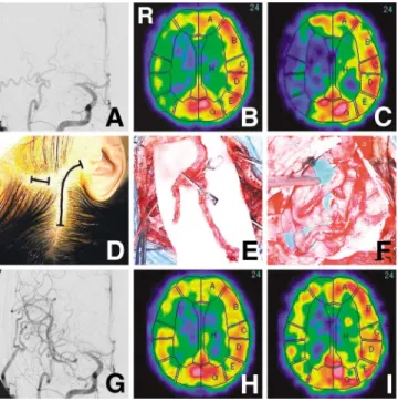
Fig. 2 Representative case. A 42-year-old male with left mild hemiparesis due to recent cerebral infarction was introduced to our clinic for cerebral revascularization. Right carotid angio-gram disclosing occlusion of the right middle cerebral artery (MCA) origin (A). Single photon emission computed tomography (SPECT) scans showing severely decreased cerebral blood flow (CBF) in the resting state (B) and after Dia-mox challenge (C) in the right MCA territory. Photographs showing the y-shaped incision (D), dissection of both the frontal and parietal branches of the superficial temporal artery (STA) (E), and the double STA-MCA bypass via a small craniotomy (F). No postoperative events occurred, such as intracranial hemorrhage or cutaneous necrosis. Postoperative right carotid angiogram showing patency of both bypasses (G) and postprocedural SPECT scans revealing improvement of the CBF in the resting state (H) and after Diamox challenge (I) in the right MCA territory.
957
Neurol Med Chir (Tokyo) 50, October, 2010
Double STA-MCA Bypass Via a Small Craniotomy
under the scalp, with careful pulling of the vessel. There-after, a small craniotomy is performed and the dura opened to inspect the recipient arteries. If the two candi-date recipient arteries (supra- and infra-sylvian portions) are not exposed, the bone window is widened. If the fron-tal and pariefron-tal branches are long enough for a double by-pass through this linear incision (Fig. 1C), the distal parts of both branches are cut as long as possible. If not, another short incision (2–3 cm) is made over the frontal branch, at least 1 cm from the original incision (Fig. 1B). Thereafter,
the frontal branch is divided carefully (Fig. 1D), ligated and cut in this short incision, and withdrawn into the original incision through the subcutaneous tunnel be-tween these two incisions. Usually, the shorter branch is anastomosed to the infrasylvian portion of the MCA and the longer branch to the suprasylvian portion. This small craniotomy, measuring approximately 3 × 4 cm in size, is usually sufficient for the double bypass. After division of the recipient artery, both branches of the STA are inter-mittently anastomosed to the infra- and suprasylvian M4
branches in end-to-side manner using 10–0 nylon suture (Fig. 1E). After anastomosis, the patency of both branches is confirmed by Doppler ultrasonography (DVM4200P; Hayashi Denki Co., Ltd.), the anastomotic sites are co-vered with fibrin glue, the bone flap is fixed with titanium plates, and the wound is closed with absorbable sutures.
Results
11 of the 34 patients and a y-shaped incision in 23 patients. The two branches of the STA were difficult to dissect in one of the patients because the frontal branch was hypoplastic. Therefore, the distal part of the STA was cut and anastomosed to the STA in end-to-side manner, and STA-MCA double anastomosis was performed.
All bypass procedures were performed uneventfully and no subdural hematoma or cutaneous necrosis occurred in this series. The craniotomy was widened via the same skin incision to expose the recipient arteries in 6 patients. Blood loss during the operation was 5 to 260 ml (mean 59 ± 54 ml). No periprocedural neurological complication occurred, but one patient developed wound infection, which was quickly cured by intravenous administration of antibiotics. No ischemic or hemorrhagic events were de-tected in the follow-up period from 6 to 50 months (mean 24.6 ± 13.8 months). Postoperative cerebral angiography was performed in all patients within 6 months and showed all bypasses were patent. A representative case of STA-MCA double bypass through a small craniotomy is shown (Fig. 2).
Discussion
The less invasive STA-MCA double bypass technique through a small craniotomy was effective in the present series, but some problems are discussed below.
Two types of skin incision can be made for preparation of the STA. Both the parietal and frontal branches of the STA can be exposed via a linear incision made on the parietal branch in almost half of all patients. However, an additional incision allows dissection of a longer section of the frontal branch. Therefore, the use of y-shaped incision is becoming routine, but we did not experience any cutaneous necrosis using this method. Additional incision of the frontal branch separate from the original linear inci-sion on the parietal branch may be the key to avoid cutane-ous ischemia. On the other hand, the two branches of STA were difficult to dissect in one patient of this series be-cause of a hypoplastic frontal branch. Therefore, the distal part of the STA was cut and anastomosed to the STA trunk, and STA-MCA double bypass was performed. This method might be one of the options if two branches of the STA are difficult to obtain.
The two MCA branches may not be found in the small craniotomy. In the present series, the craniotomy was wi-dened in 6 patients to expose the two recipient arteries and obtain sufficient space for bypass, but additional skin incision was not necessary in any patient. Double bypass could be performed in all the patients in the current study. Computed tomography (CT) angiography might be used to determine the appropriate position of craniotomy to ob-tain two recipient arteries before the operation. We are now trying to establish this method for more accurate de-termination of the craniotomy position using CT an-giography and navigation systems such as the `target by-pass' for moyamoya disease.9)
The necessity for double bypass should be discussed, as the procedure may not be essential in all patients. However, a single bypass was not sufficient to increase
the CBF and failed to prevent ischemic events in some patients of our previous series, possibly due to limited selection of a single recipient in the small craniotomy. Therefore, we introduced the double bypass procedure, which seemed to improve impaired cerebrovascular reac-tivity in our preliminary study (data not shown). However, this result should be confirmed statistically by comparing the single and double bypass groups, because no sig-nificant difference was reported between these groups.13)
In this series, intraoperative bleeding was minimum and no patient required a regular blood transfusion. The mean operating time was around 3.5 hours (212 ± 48 minutes). These results suggest that this surgical technique is relatively less invasive than the regular double bypass, performed by the so-called flap method.
This operation requires a bypass procedure in a rela-tively deep and narrow space. It is important to make the craniotomy in the appropriate position and to use small hooks to obtain wider and lower retraction of the scalp and the temporal muscle. Preparation of the MCA bran-ches requiring an arachnoid incision should be performed after preparation of both STA branches to avoid brain sinking. If these critical procedures are appropriately per-formed, this operation is not difficult, as confirmed by the observation that all bypassed branches were patent on postoperative angiography.
The present less invasive method of STA-MCA double bypass through a small craniotomy may be effective to avoid some complications related to wider craniotomy such as cutaneous necrosis or hemorrhagic events. Fur-ther experience is necessary to determine if the double by-pass is superior to a single byby-pass in patients with im-paired CBF due to cerebrovascular occlusive diseases.
Acknowledgment
We thank Mr. Nobuharu Kimura for his excellent illustra-tions.
References
1) Amin-Hanjani S, Charbel FT: Is extracranial-intracranial by-pass surgery effective in certain patients? Neurol Clin 24: 729–743, 2006
2) The EC/IC Bypass Study Group: Failure of extracranial-in-tracranial arterial bypass to reduce the risk of ischemic stroke. Results of an international randomized trial. N Engl J Med313: 1191–1200, 1985
3) Garrett MC, Komotar RJ, Starke RM, Merkow MB, Otten ML, Sciacca RR, Connolly ES: The efficacy of direct ex-tracranial-intracranial bypass in the treatment of sympto-matic hemodynamic failure secondary to athero-occlusive disease: a systematic review. Clin Neurol Neurosurg 111: 319–326, 2009
4) Ishikawa T, Yasui N, Suzuki A, Hadeishi H, Shishido F, Ue-mura K: STA-MCA bypass surgery for internal carotid ar-tery occlusion—comparative follow-up study. Neurol Med Chir (Tokyo)32: 5–9, 1992
5) JET Study Group: [Japanese EC-IC Bypass Trial (JET study): Study design and interim analysis]. Surgery for Cerebral Stroke30: 97–100, 2002 (Japanese)
959 959
Neurol Med Chir (Tokyo) 50, October, 2010
Double STA-MCA Bypass Via a Small Craniotomy
6) JET Study Group: [Japanese EC-IC Bypass Trial (JET study): The second interim analysis]. Surgery for Cerebral Stroke 30: 434–437, 2002 (Japanese)
7) Katsuta T, Inoue T, Arakawa S, Uda K: Cutaneous necrosis after superficial temporal artery-to-middle cerebral artery anastomosis: is it predictable or avoidable? Neurosurgery 49: 879–882, 2001
8) Kawaguchi S, Sakaki T, Uranishi R: Effects of bypass on CO2 cerebrovascular reactivity in ischaemic cerebrovascu-lar diseases—based on the intra-operative LCBF and CO2 cerebrovascular reactivity studies. Acta Neurochir (Wien) 141: 369–374, 1999
9) Kikuta K, Takagi Y, Fushimi Y, Ishizu K, Okada T, Hanaka-wa T, Miki Y, Fukuyama H, Nozaki K, Hashimoto N: ``Tar-get bypass'': a method for preoperative tar``Tar-geting of a recipient artery in superficial temporal artery-to-middle cerebral artery anastomoses. Neurosurgery 62(6 Suppl 3): 1434–1441, 2008
10) Neff KW, Horn P, Dinter D, Vajkoczy P, Schmiedek P, D äuber C: Extracranial-intracranial arterial bypass surgery im-proves total brain blood supply in selected symptomatic patients with unilateral internal carotid artery occlusion and insufficient collateralization. Neuroradiology 46: 730–737, 2004
11) Newell DW, Vilela MD: Superficial temporal artery to mid-dle cerebral artery bypass. Neurosurgery 54: 1441–1448, 2004
12) Przybylski GJ, Yonas H, Smith HA: Reduced stroke risk in patients with compromised cerebral blood flow reactivity treated with superficial temporal artery to distal middle cerebral artery bypass surgery. J Stroke Cerebrovasc Dis 7: 302–329, 1998
13) Sayama T, Inoue T, Uda K, Fujimoto S, Okada Y: [Cerebral blood flow after superficial temporal artery-middle cerebral artery anastomosis]. No Shinkei Geka Journal 17: 533–537, 2008 (Japanese)
14) Yasui N, Suzuki A, Sayama I, Kawamura S, Shishido F, Ue-mura K: Comparison of the clinical results of STA-MCA anastomosis and the medical treatment in the cerebral low perfusion patients with viable brain tissue. Neurol Res 13: 84–88, 1991
Address reprint request to: Shinichi Yoshimura, M.D., Ph.D., Clin-ical Professor, Department of Neurosurgery, Graduate School of Medicine, Gifu University, 1–1 Yanagido, Gifu, Gifu 501–1194, Japan.