PRODUCTION OF AN UNUSUALLY STABLE ANTHOCYANIN
BY
DIOSCOREA CIRRHOSA CULTURED CELLS
Hiroshi
FUKUI,
Satoru KADORIKU* AND Masakatsu IWAMOTO* A main anthocyanin accumulated in tubers of Dioscorea czrrhosa was identified as alatanin C isolated from purple yam Dzoscorea alata. The accumulation of anthocyanins inside the tubers of the former suggests that the cultured cells are capable of producing anthocyanins without il- lumination. The cultured cells were found to produce the same anthocyanins as the tubers in the dark at a content of ca. 3% of the dried cells. The anthocyanins were unusually stable in a phosphate buffer (0 05M, pH 6 ) due probably to the cooperation of self-association and intramolecular stacking between anthocyanindins and aromatic nuclei of phenolic acids combined with the anthocyanin glycoside moiety. It was found that the anthocyanins combined with more sinapic acids were more stable in the same bufferKey Words : Dzoscorea czrrhosa, Dioscoreaceae, anthocyanin, stability, biotechnological pro- duction
Introduction
( 1 )
Anthocyanins are very important natural pigments used for food and drink coloration
.
However, most of the pigments are known to be unstable in neutral or weakly acidic aqueous conditions. It has been re-(2 3)
ported that some of acylated anthocyanin pigments are very stable to decoloration
.
In addition, the biotechnological production of the pigments by cultured cells usually needs the costly illumination to in-(4 5 )
duce and enhance the productivity
,
because many of the pigments are formed in plant tissues exposed to sun light that is an important inducing factor of the biosynthesis.In this study, in order to biosynthesize the red pigments in plant cultured cells in the dark, we selected
Dzoscorea cirrhosa that is known to accumulate the pigments inside the tubers used as stable colorant in the Southeast Asia. This paper reports that the cultured cells have the capability of producing the red pig- ments in the dark, and that the pigments are very stable even in a weakly acidic condition.
Materials and Methods
Plant MaterialThe plant used was a perennial, twining plant, Dzoscorea czrrhosa (~ioscoreaceae, Japanese name: SOMEMONOIMO). The plant was cultivated in a green house of our Department and harvested in late autumn. The young purple petioles were used for callus induction on Linsmaier-Skoog agar medium con- taining 3% sucrose, I ,u M 2,4-D and 1 ,u m kinetin in the dark. The dedifferentiated cultured cells were subcultured on various agar media containing a combination of auxin (1 ,U M 2,4-D, I ,u M and 1 0 , ~ M NAA, and 10,u M IAA) and cytokinin (I/* M kinetin) in the dark at 25°C.
The red-colored cell aggregates in the callus on the agar medium containing lO,u M NAA and 1 ,u M kinetin were selected and transferred onto the agar medium containing l o o p M NAA and 1 ,u M kinetin for more effective pigment production. The dark-red cells were harvested and kept in a freezer ( -
20°C) for further chemical investigation. Isolation of anthocyanins
The red cells (120g fresh wt) were freeze-dried and extracted, 3 times, with MeOH containing 1% HCI (500 m1)for 24 hrs. The combined extracts were concentrated under reduced pressure at ca. 4 0 C . The concentrate was subjected to polyvinylpolypyrolidone (PVPP) column ( 3 x 5 cm) eluted with water con- taining 1% AcOH. The fractions were collected in the order of elution of 4 red pigments. Each eluate was re-chromatographed on ODs column (10 ,u m, 8 X 300 mm, elution: 0 , s mllmin of AcOH: CH,CN: H, 0 : TFA=14: 17.5: 120: 0.5) detected at 520 nm to p u r i ~ the red pigment.
The tubers (200g fresh wt) of the original plant grown in a green house of our Department were sliced and treated with almost the same procedures with the cultured cells to give the red pigments.
Chemical analysis
The isolated pigment was subjected to
k-NMR
( 4 0 0 ~ ~ 2 , JEOL JNM-~$00) and ',C-NMR (100 MHz, JEOL JMN-A400) in 10%TFA-MeOH-d, containing TMS as an internal standard and to MS (JOEL, JMS-SXlOZAQQ Hybrid Mass Spectrometer, positive FAB in a mixture of acetic acid and glyc- erine). UVIvisible spectra were measured with Hitachi A2000 Recording Spectrometer.Results and Discussion
Growth and pigment formationThe cultured cells of D czrrhosa grew very slowly on the Linsmaier-Skoog agar medium tested. The
red pigments were produced on the medium containing NAA as an auxin in the dark; a combination of 100 ,u M NAA and 1 ,u M kinetin w a e best for pigment production among the combinations tested,
Chemical analysis of the pigments
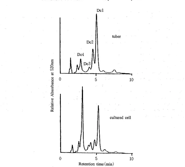
The MeOH-1%HCI extract of the cultured cells was compared with that of the tubers of the original plants by HPLC ( ~ i ~ . 1 ). The HPLC profile of the former was qualitatively identical with that of the lat- ter, indicating the presence of several red pigments in both tissues. The total contents of the red pigments in the cultured cells were estimated to be ca. 3% of the dried weight as keracyanin. The MS and NMR spectral data of the main pigment ( ~ c l ) isolated was identical with those of alatanin C ( ~ i g . 2 ) isolated
(6)
from Dzoscorea alata by Yoshida et a1
.
The other three pigments were estimated to be the derivatives of alatanin C based on the NMR data, although the chemical structures of these pigments were not elu- cidated. Their NMR spectra in the aromatic region ( ~ i ~ . 3 ) indicate that, while Dcl contains one residue of sinapate per anthocyanidin nucleus, Dc2 and Dc3 are combined with two sinapate residues per anthocyanidin nucleus and Dc4 has no sinapate residue in the molecule; the signals characteristic for a group of 2,6-2H of sinapate were assigned as a broad signal at 6 6.28 for Dcl, two broad singlets at 6 6.04 and 6 6.12 for Dc2, and two signals at 6 6.05 and 6 6.10 for Dc3, and no signal for Dc4, though their positions in the molecule were not determined. The tubers were treated with almost the same methods with the cultured cells to give a main pigment. The NMR data were identical with that of Dcl. These data suggest that most of the anthocyanins in the tubers of D czrrhosa are esterified with sinapicDcl
I
cultured cell I' Dc2 0 510
Retention time (min)
Fig. 1
.
HPLC profiles of the MeOH -1%HCl extracts from the cultured cells and the tubers of Dcirrhosa.
HO
0-
OH
OCH3
Fig. 2.
The chemical structure of Dcl (alatanin C) isolated from the tubers of D cirrhosa.f
0 cu LC Y mI
tuberStability of the pigments in the tubers
The MeOH-1%HCl extract of the tubers was concentrated and dissolved in a phosphate buffer (0.05M, pH 6.0) and the absorbancy of the red solution at 520 nm was measured at some intervals ( ~ i ~ . 4 )
.
The crude anthocyanin solution of the tubers was found very stable compared with keracyanin (cyanidin-3-0 -rutinoside) as a standard. Although keracyanin decomposed in one hour, the crude anthocyanin solution from the tubers kept the absorbancy at 520 nrn very high for over 20 hours, suggesting that the anthocyanins in the tubers are very stable even in a weakly acidic solution.Each anthocyanin ( ~ c l - 4 ) isolated from the tubers was also dissolved in the same phosphate buffer (0.05M, pH 6.0) and the absorbancy was compared for over 40 hours ( ~ i ~ . 5 ). Dc4 and keracyanin which are not esterified with phenolic acids decomposed quickly. However, the anthocyanins ( D C ~ , Dc2 and D C ~ ) , the glycoside moiety of which is esterified with sinapic acid, were found to more stable than keracyanin and Dc4; Dcl containing one molecule of sinapic acid as phenolic acids were medium stable, and Dc2 and Dc3 including two molecules of sinapic acid based on the NMR data were highly stable
(4)
among the anthocyanins tested here. It is reported, by Yoshida et al. that phenolic residues combined
with the glycoside moiety are able to make anthocyanins highly stable through the cooperation of unique self-association and intramolecular stacking between anthocyanidin and phenolic nucleus. It is suggested that the esterification of anthocyanin glycoside moiety with more phenolic acids like sinapic acid makes anthocyanins more stable.
8 ' 9 ~ " ' 1 ' ~ ~ " ' ~ " " 8 7 6 Fig. 3 . NMR spectra of Dcl, Dc2, Dc3 and
Dc4 purified from the tubers of D
cirrhosa.
tuber crude
80 extract
time (hr)
Fig. 4
.
The stability of keracyanin and the crude extract from the tubers of Dczrrhosa in a phosphate buffer (0.05
M, pH 6 ) .
+ keracyanin
* V
0 20 40
2
time (hr) 60Fig. 5 . The stability of the red pigments ( ~ c l
-
Dc4 and keracyanin) in a phosphate ebuffer (0,05M, pH 6).References
O TANIMURA A. , KAIAYAMA 0. , ENDO H. ,
KUROKAWA K . , and YOSIZUMI 'I. : Handbook of Nat-
ural Pigments (in ~apanese) , pp. 247-369, Korin, Tokyo, 1979.
(2) Go10 T, and KONDO 1. : Angew, Chem., 103,
17-33 (1991); Angew. Chem. Int. Ed. Eng ,
30, 17-33(1991);
(3) YOSHIDA K . , KONDO T., and Goro 1. : Tetrahedron
Len , 32 (401, 5579-5580 (1991)
(4) YAMAMOIO Y . , KINOSHIIA Y . , WAIANABE S., and
YAMADA Y . : Agric. Biol. Chem., 53 (2). 417-
423 (1989).
(5) IBRAHIM R . K : Regulation of Synthesis of
Phenolics. In Constabel F. C. and Vasil I. K. (eds), Cell Culture and Somatic Cell Genetics of Plants, Vol 4, Cell Culture in Phytochemistry. Ac- ademic Press, San Diego, 1987.
(6) YOSHIDA K . , KONDO I . , KAMEDA K , KAWAKISHI
S., LUBAG A. J. M . , MENDOZA E M. T , and
Go10 1. : Tetrahedron Lett., 32 (401, 5575-5578
(1991)
(Received November 29, 1996)
'I 2 .El I'
*
(Dioscorea cirrhosa, ?''TI I' -'f#,) tC;f%%C:b%@-/ 2 b 9'7.2 >%.$ X % % @ L ,.irtr,+.R-?=RRfi'7Y,7PC&%#
k L , T @ ; h h ' r b l b b b ?a),&,&% Ci, z&.$YI'? 3 (Dioscorea alata, $%
:
purple yam) $ 6 .&E 3 h?: alatanin C km g
S h?:. 7 2~511'~5tak~xa,3~~5a~\%za,rnac=.72
P
> . 7 . = 2 e % a . i t a z k s b , +a,%%aRti@lT,T.XJiiU&Q2~zk11Tl:7
2 1. 9'7.2 2 %&,%T3
b k%$OS hk.. BE%v e % L t : a a c i m m i 3 7 : 1 1 ~ 1 3
P
9'7.=:/%&.ljfi.~ B L ,
.ea,72P
L'7.z. 2 Ci, I) 2@@@% (pH6. 0 . . 0 5 ~ ) + T 4 0 R M M l . % g T & 3 f:,, C h i & , C 0 . 7
2
P
' / ' 7 . 2 2 a , i g p ~ C : % $ . ~ i f s ' J t . r " 2 @ k 7 2 1 . ' / ' 7 = . ? 2 @ 7 5 I $ ~ ~ ~ & b b ~ t i ~ .b \ C : x P r 3 2 3 ' L , 2 ? 2 j G ? b l : & k % ; i b h , S b l : , %$$@a,tGE.h~b, %$?if& 9.f.