Tottori University research result repository
タイトルTitle Wood-mimetic skins prepared using horseradishperoxidase catalysis to induce surface wrinkling of chitosan film upon drying
著者
Auther(s) Izawa, Hironori; Dote, Yuki; Okuda, Noriko ; Sumita,Masato ; Ifuku, Shinsuke; Morimoto, Minoru; Saimoto, Hiroyuki
掲載誌・巻号・ページ
Citation Carbohydrate polymers , 173 (1) : 519 - 525
刊行日
Issue Date 2017
資源タイプ
Resource Type 学術雑誌論文 / Journal Article
版区分
Resource Version 著者版 / Author
権利
Rights Copyright © 2017 Elsevier B.V. All rights reserved.This manuscript version is made available under the CC-BY-NC-ND 4.0 license https://creativecommons.org/ licenses/by-nc-nd/4.0/
DOI 10.1016/j.carbpol.2017.06.034
Carbohydrate Polymers, Original full-length research papers
1 2
Wood-mimetic skins prepared using horseradish peroxidase catalysis to
3
induce surface wrinkling of chitosan film upon drying
4
Hironori Izawaa,*, Yuki Doteb, Noriko Okudaa, Masato Sumitac, Shinsuke Ifukua, Minoru 5
Morimotod, Hiroyuki Saimoto a,* 6
a Graduate School of Engineering, Tottori University, 4-101 Koyama-Minami, Tottori
680-7
8550, Japan 8
b Faculty of Engineering, Tottori University, 4-101 Koyama-Minami, Tottori 680-8550, Japan
9
c Department of Chemistry, Graduate School of Pure and Applied Sciences, University of
10
Tsukuba, 1-1-1 Tennoudai, Tsukuba, Ibaraki 305-8571, Japan 11
d Division of Instrumental Analysis, Research Center for Bioscience and Technology, Tottori
12
University, Tottori 680-8550, Japan 13
14
*Correspondence to: Hironori Izawa and Hiroyuki Saimoto. 15
Postal Address: Department of Science and Biotechnology, Graduate School of Engineering, 16
Tottori University, 4-101 Koyama-Minami, Tottori 680-8550, Japan. 17
HI: Phone: +81-857-31-5813. Fax: +81-857-31-5813. E-mail: [email protected] 18
HS: Phone: +81-857-31-5693. Fax: +81-857-31-5813. E-mail: [email protected] 19
20 21
Abstract
22
We previously developed bio-based wrinkled surfaces induced by wood-mimetic skins upon 23
drying in which microscopic wrinkles were fabricated on a chitosan (CS) film by immersing it 24
in a phenolic acid solution, followed by horseradish peroxidase (HRP)-catalyzed surface 25
reaction and drying. However, the detailed structure of the resulting wood-mimetic skins, 26
including crosslinking mode and thickness, has not been clarified due to the difficulty of the 27
analysis. Here, we prepare wrinkled films using ferulic acid (FE), vanillic acid (VA), and 28
homovanillic acid (HO) and characterize their structures to clarify the unknown 29
characteristics of wood-mimetic skin. Chemical and structural analyses of wood-mimetic 30
skins prepared using VA and HO indicate that the crosslinking structure in the skin is 31
composed of ionic bonds between CS and an oligophenolic residue generated by the HRP-32
catalyzed reaction on the CS surface. Moreover, the quantity of these ionic bonds is related to 33
the skin hardness and wrinkle size. Finally, SEM and TOF-SIMS analyses indicate that the 34
skin thickness is on the submicron order (<200 nm). 35
36
Key Words: Chitosan, Surface wrinkling, Skin layer, Horseradish peroxidase, Phenolic acid,
37 Biomimetic materials 38 39
1. Introduction
40Biomimetic systems that imitate the design principles of nature are key technologies in the 41
progress toward environmentally benign and high-performance materials (Bhushan, 2009; 42
Bhushan & Jung, 2011). Many functional materials inspired by nature have been developed 43
(Aizenberg & Fratzl, 2013; Kawamura, Kohri, Morimoto, Nannichi, Taniguchi & Kishikawa, 44
2016; Kawamura, Kohri, Yoshioka, Taniguchi & Kishikawa, 2017; Otsuka, Fujikawa, 45
Yamane & Kobayashi, 2017; Pandian & Sugiyama, 2016; Sedo, Saiz-Poseu, Busque & Ruiz-46
Molina, 2013). 47
Surface-wrinkling is a ubiquitous physical process that creates macro/microscopic 48
wrinkles in nature (Genzer & Groenewold, 2006; Ionov, 2012). This spontaneous process is 49
the result of inhomogeneous changes triggered by internal stresses and swelling/shrinking of 50
tissue layers possessing different elastic moduli (Ionov, 2012). The formation of fine wrinkles 51
in human skin is closely associated with decreases in the water content of the stratum 52
corneum by aging; i.e., fine wrinkles are formed by drying, and as a result of inhomogeneous 53
shrinkage (Barel, Paye & Maibach, 2009; Imokawa & Takema, 1993; Tsukahara, Hotta, 54
Fujimura, Haketa & Kitahara, 2007). 55
Nano/microscopic wrinkled surfaces inspired by nature-mimetic surface designs have 56
been developed for optical (Ohzono, Suzuki, Yamaguchi & Fukuda, 2013) and electronic 57
devices (Lee et al., 2013), the realization of tunable wettability (Li, Dai, John & Carter, 2013) 58
and adhesion (Davis, Martina, Creton, Lindner & Crosby, 2012), and the synthesis of cell 59
culture scaffolds (Zhao, Gu, Zhao, Guan, Zhu & Zhang, 2014). Basically, a skin layer is 60
fabricated on a soft substrate via dry processing methods, including chemical vapor deposition 61
(Bowden, Brittain, Evans, Hutchinson & Whitesides, 1998), photo-crosslinking (Chen, Reed 62
& Yang, 2013), and UV/O3 oxidation (Efimenko, Rackaitis, Manias, Vaziri, Mahadevan &
63
Genzer, 2005). The wrinkling event can be caused/controlled by mechanical stress (Efimenko, 64
Rackaitis, Manias, Vaziri, Mahadevan & Genzer, 2005), thermal expansion (Bowden, Brittain, 65
Evans, Hutchinson & Whitesides, 1998), and/or swelling-shrinking (Huraux, Narita, Bresson, 66
Fretigny & Lequeux, 2012; Rizzieri, Mahadevan, Vaziri & Donald, 2006; Zhao, Gu, Zhao, 67
Guan, Zhu & Zhang, 2014). 68
We previously developed a surface-wrinkling system inspired by the fine wrinkles and 69
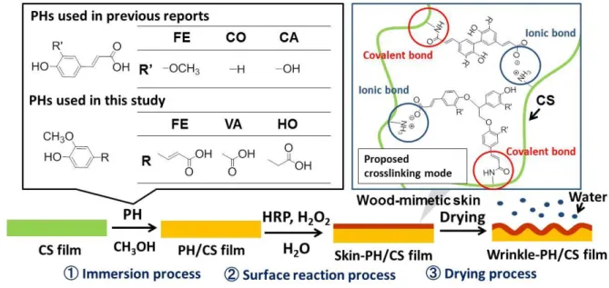
design principles of wood-cell walls (Fig. 1) (Izawa, Okuda, Ifuku, Morimoto, Saimoto & 70
Rojas, 2015; Izawa et al., 2016). In this method, wood-mimetic skins are fabricated by 71
immersing chitosan (CS) film in a phenolic acid (PH)-methanol solution, then treated with 72
horseradish peroxidase (HRP) to catalyze a surface reaction. Finally, surface wrinkling is 73
induced by water evaporation during drying. The wrinkle wavelength and amplitude can be 74
controlled by the choice of a phenolic acid (ferulic acid, FE; p-coumaric acid, CO; or caffeic 75
acid, CA) and by varying the temperature of the immersion process. Using this system, we 76
found that the wrinkle size was predominately determined by the hardness of the wood-77
mimetic skins (Izawa et al., 2016). However, the structure of the wood-mimetic skin layer has 78
not been fully elucidated. 79
80
Fig. 1. Illustration of the wood-inspired surface wrinkling systems used in this study and in
81
previous reports. 82
83
Using this surface-wrinkling system, a dehydration-condensation reaction was observed 84
between CS and the PHs during the immersion process (Izawa, Okuda, Ifuku, Morimoto, 85
Saimoto & Rojas, 2015). Therefore, we hypothesized that the covalently bound PHs on the 86
CS film acted as reaction sites for chemical and/or ionic crosslinking via the HRP-catalyzed 87
oligomerization of the precursor molecules to yield a skin layer. However, the role of 88
covalently bound PH in skin layer formation has not yet been proved due to the difficulty of 89
analyzing insoluble skin layers. In addition, the detailed crosslinking mode between CS and 90
phenolic oligomers has not been fully clarified. 91
In common wrinkled surfaces, the wavelength (λ) of the wrinkle is dependent on skin 92
thickness (d), and the mechanical properties of the film are described as follows (Chung, 93
Nolte & Stafford, 2011; Genzer & Groenewold, 2006): 94 λ = 2πd (Ē𝑠 3Ē𝑓) 1 3 , (1) 95
where the subscripts s and f refer to the skin layer and the foundation (substrate), respectively; 96
Ē is the plane-strain modulus given by E/(1-ν2), where E is the elastic modulus, and ν is the
97
Poisson’s ratio. Thus, the skin thickness is important information for understanding surface 98
wrinkling. However, the skin thickness has not been fully investigated because it is not 99
distinguishable due to its very small thickness, and also because of the similar electron density. 100
Here, we investigate the unknown characteristics of wood-mimetic skin. To clarify the 101
detailed structure of the skin layers, we use surface wrinkling induced by ferulic acid (FE), 102
vanillic acid (VA), and homovanillic acid (HO), which are PHs having different substituents 103
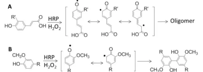
(R), although always including a carboxyl group (Fig. 1). The previously used HRP-catalyzed 104
reaction of FE generates a phenoxide radical that can resonate with the 5’-position or the β-105
position (Fig. 2A) (Oudgenoeg et al., 2002). The coupling reaction of these radicals provides 106
oligomers (Izawa, Miyazaki, Ifuku, Morimoto & Saimoto, 2016). However, the HRP-107
catalyzed reactions of VA and HO provide only dimers (Fig. 2B) (Ci & Wang, 1991; Foppoli, 108
Coccia, Blarzino & Rosei, 2000; Tai, Sawano & Ito, 2012). In addition, the carboxyl groups 109
in FE and VA are conjugated to styryl and phenyl groups, respectively, while that in HO is 110
not conjugated. These differences in the HRP-catalyzed reactions and reactivity of the 111
carboxyl groups provide important information regarding wood-mimetic skin. In addition, we 112
conducted scanning electron microscopic (SEM) and Time-of-Flight secondary ion mass 113
spectrometry (TOF-SIMS) analysis of the wrinkled surface to estimate the skin thickness and 114
structure. 115
116
Fig. 2. Previously reported HRP-catalyzed reactions of FE (A) and VA or HO (B).
117 118
2. Experiments
119 2.1. Materials 120CS (Mn: 5.6 x 10⁴; Mw/Mn: 2.36; GPC analysis with Pullulan standards) was supplied
121
by the Koyo Chemical Co., Ltd. (Tottori, Japan), with an undeacetylated 23.5% fraction of CS 122
(elemental analysis). FE, HO, and VA were purchased from the Tokyo Chemical Industry Co., 123
Ltd. (Tokyo, Japan). HRP (274 U/mg) was purchased from Toyobo Co., Ltd. (Osaka, Japan). 124
Other reagents were commercial grade and used without further purification. 125
126
2.2. Instrumentation
127
SEM images of film surfaces were recorded by a TM303Plus (Hitachi, Japan) without 128
coating. SEM cross-sectional images were recorded with a JSM-6700F (JEOL, Japan). The 129
sample was coated with an approximately 5 nm layer of Pt with an ion sputter coater. The 130
wrinkle amplitudes of the wrinkled films were obtained with a NanoCute-NanoNavi IIs 131
(Seiko Instruments, Japan). Elemental analysis data were recorded on a Perkin Elmer 2400 II 132
CHNS/O (Perkin Elmer, US). Infrared (IR) spectra of the samples were recorded by a 133
Spectrum 65 (Perkin-Elmer Japan Co., Ltd., Japan) equipped with an ATR attachment. TOF-134
SIMS measurement was performed with a PHI TRIFT V nanoTOF (ULVAC-PHI, Japan). 135
The pulsed primary ion source was Bi32+, and the ion beam was operated at 30 kV (50 fA AC)
136
with a 50 µm x 50 µm rastering area at an incident angle of 45°. The sputtering was done with 137
an Ar+ ion beam operated at 300 V and 150 nA with a 0.1 mm x 0.1 mm rastering area at an 138
incident angle of 45°. 139
140
2.3. Preparation of the CS film
141
CS (2.0 g) was dissolved in 100 mL of an acidic aqueous solution containing 0.5 mL 142
acetic acid. Then, 10 mL of the CS solution was added to a Teflon Petri dish (φ=50 mm) and 143
degassed under reduced pressure. The CS solution was heated at 50˚C for 24 h to yield a CS 144
film after evaporation. The film was then heated at 50˚C under reduced pressure for 12 h. The 145
inhomogeneous edge of the film was cut down with scissors. The weight and thickness of the 146
CS film were ca 0.15 g and 111±12 µm, respectively. 147
148
2.4. Surface wrinkling of films
149
In a typical experiment, a CS film was immersed in 20 mL methanol containing 0.05 150
g/mL FE at 30˚C for 24 h. The resulting film (hereafter, FE/CS film) was removed and soaked 151
in 10 mL water, followed by the prompt addition of the HRP (1 mL, 137 U) and H2O2 (200
152
µL, 30% concentration). The system was kept at 30˚C for 12 h, after which the film was 153
removed and dried at 40˚C under for 12 h. 154
155
3. Results and Discussion
156
3.1 Morphology of the obtained films
157
Figures 3A-F show plane-view SEM images of the surface of the wrinkled films. A 158
detailed characterization of the wrinkles formed is provided in Fig. 3G. In the case of FE, the 159
results were almost the same as that in our previous report (Izawa, Okuda, Ifuku, Morimoto, 160
Saimoto & Rojas, 2015), in which the mean wrinkle wavelengths and amplitudes under 161
immersion treatments at 30˚C and 40˚C were 1.42±0.34 and 0.97±0.22 m, respectively, and 162
0.52±0.15 and 0.39±0.10 m, respectively. The mean wrinkle wavelengths and amplitudes at 163
40˚C immersion were smaller than those at 30˚C. In this wrinkling system, higher 164
temperatures on the immersion process led to the formation of softer skins, due to the harder 165
decomposition of the CS around the film surface, inducing smaller wrinkles (Izawa et al., 166
2016). Note that wrinkled surfaces were not observed on the FE/CS films, the control CS film 167
prepared by the adsorption of oligomeric FE, and the original CS film (Figures S1A-B, S1C, 168
and S1D, respectively). For VA, surprisingly, wrinkling occurred during both the 30˚C and 169
40˚C immersion treatments, even though the HRP-catalyzed reaction of VA only provided the 170
dimer as described above. The mean wrinkle wavelengths and amplitudes at 30˚C and 40˚C 171
were 3.07±0.17 and 1.61±0.25 m, respectively, and 0.62±0.24 and 0.30±0.10 m, 172
respectively, which were larger than those of the FE/CS system. This result suggests that VA 173
produces harder skin, even though the HRP-catalyzed reaction of VA provides just the dimer. 174
When HO was used, wrinkling was observed on the wrinkle-HO/CS film at an immersion 175
temperature of 40˚C. The mean wrinkle wavelength and amplitude under treatments at 40˚C 176
were 2.96±0.16 m and 0.68±0.18 m, respectively, which were larger than those for the 177
VA/CS system at 40˚C. No wrinkling occurred at 30˚C. The same phenomenon was observed 178
in the CA/CS system. There was the suggestion that no wrinkling is due to a lack of 179
crosslinking reaction sites on the CA/CS film (Izawa, Okuda, Ifuku, Morimoto, Saimoto & 180
Rojas, 2015). The results obtained with VA and HO clearly indicated that the vinyl moiety in 181
FE is not needed to induce surface wrinkling. Note that we additionally confirmed the absence 182
of wrinkling by using 2-methoxyphenol, without a carboxyl group, for the treatment at 30˚C 183
and 40˚C. 184
186
Fig. 3. Plane-view SEM images of the films obtained via immersion treatment at 30˚C (A) or
187
40˚C (B) using FE, via immersion treatment at 30˚C (C) or 40˚C (D) using VA, and via 188
immersion treatment at 30˚C (E) or 40˚C (F) using HO and their mean wavelength and 189
amplitude of wrinkles (G). 190
191
3.2 Characterization of the film surfaces
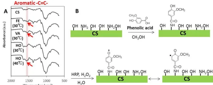
To confirm the presence of covalently bound PHs on the CS film, the PH/CS films were 193
Soxhlet-extracted with methanol for 1 week in order to remove any unreacted PH, and the IR 194
spectra of the film surfaces were measured (Fig. 4A). In the 30˚C treatment, the absorption 195
peaks attributed to aromatic -C=C- (Swislocka, Kowczyk-Sadowy, Kalinowska & 196
Lewandowski, 2012) appeared at around 1540 cm-1 for the FE/CS and VA/CS systems. The 197
absorption peaks observed even after Soxhlet-extraction indicated the presence of the 198
covalently bound PH via an amide bond on the CS film. In contrast, this was not seen in the 199
HO/CS system at this temperature. We considered that the lack of a peak observed on the 200
HO/CS system was due to the lower reactivity of unconjugated carboxyl groups than in FE 201
and VA. On the other hand, the absorption peak due to aromatic -C=C- was observed in the 202
HO/CS system in the 40˚C treatment. These observations are consistent with the wrinkling 203
results. Thus, this result confirmed our previous speculation that the carboxyl group in PH 204
plays a critical role in the formation of covalently bound PH, acting as a reaction site for the 205
HRP-catalyzed reaction to form the skin layer (Fig. 4B). 206
207
Fig. 4. IR spectra of the extracted FE/CS, VA/CS, HO/CS films and CS (A), and illustration
208
of the confirmed role of covalently bound PH (B). 209
In order to analyze the chemical structure of the skin, we performed IR analysis of the 211
FE/CS, VA/CS, and HO/CS films from the 40˚C treatment (Fig. 5). In the wrinkle-212
FE/CS film spectrum, the absorption peaks attributed to -COO- and -NH3+ (Hu, Jiang, Ding,
213
Ge, Yuan & Yang, 2002) were observed at 1565 cm-1 and 1630 cm-1, respectively.Meanwhile,
214
absorption peaks due to aromatic alkene and glycosidic ether were observed as significant 215
ones at 1511 cm-1 and 1024 cm-1, respectively, indicating the skin layer was composed of both 216
CS and oligomeric FE. By using VA, the absorption peak due to -COO- was slightly enhanced 217
compared to that of the wrinkle-FE/CS film. Interestingly, using HO further enhanced the 218
absorption peak attributed to -COO-, to the point where the absorption peak attributed to the
219
aromatic alkene was completely overlapped by it. These results suggested a higher quantity of 220
the ionic bonds in the case of VA and HO than in FE. Indeed, the HRP-catalyzed reaction of 221
FE involves decarboxylation that reduces the quantity of the carboxyl group in the system 222
(Oudgenoeg et al., 2002). In addition, it was suggested that there was a higher quantity of the 223
ionic bonds in the HO/CS system than in the VA/CS system. As described above, the 224
products from the HRP-catalyzed reaction of VA and HO are dimers with a biphenyl 225
framework (Fig. 2B). The only structural difference between those dimers is whether the 226
methylene spacer is present or not. The carboxyl group in HO has higher mobility than that of 227
the VA by virtue of the methylene spacer. We consider that the higher mobility facilitates 228
ionic bonding in the HO/CS system. 229
Fig. 5. IR spectra of surfaces of the wrinkle-FE/CS, VA/CS, and HO/CS films.
231 232
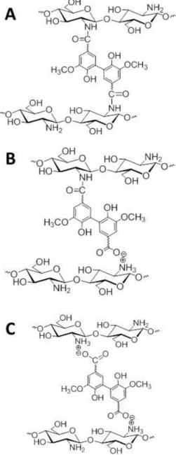
There are three possible crosslinking modes in the VA/CS and HO/CS systems. The 233
first is the covalently crosslinked structure generated by the radical coupling of each of the 234
covalently bound PHs (Fig. 6A). The second is the ionically crosslinked structure between a 235
dimeric side chain, generated by radical coupling of PH and the covalently bound PH, and 236
CS (Fig. 6B). The third and final possible modes are the ionically crosslinked structure 237
composed of CS and dimeric PH (Fig. 6C). When covalently bound HO was not formed, 238
surface wrinkling did not occur, as mentioned above. Therefore, this third crosslinking mode 239
is not important for skin layer formation. Indeed, the top layers fabricated by the adsorption 240
of oligomers on the CS film could not induce surface wrinkling (Fig. S1C). Elemental 241
analysis of the VA/CS and HO/CS films and the extracted VA/CS and HO/CS films 242
provided evidence that the VA/CS and HO/CS films included ca 50-fold greater amounts of 243
VA and HO than covalently bound VA and HO, respectively. Under this condition, the 244
radical coupling of PH and the covalently bound PH proceeds more readily than that 245
between the covalently bound PHs themselves. In addition, the HRP-catalyzed reaction of 246
the extracted VA/CS and HO/CS films did not provide wrinkled surfaces upon drying. Thus, 247
the second crosslinking mode is the most likely crosslinking structure for the VA/CS and 248
HO/CS systems. This is important information which suggests that ionic crosslinking by the 249
dimeric/oligomeric side chain is capable of surface wrinkling upon drying. In addition, we 250
observed a crucial phenomenon that underscored the importance of the ionic bonding for the 251
skin formation. Namely, the wrinkles were maintained in water even after 1 week, while 252
they disappeared in 100 mM NaOH aqueous solution due to the dissociation of the ionic 253
bond (Fig. S2), indicating that the wood-mimetic skins were formed by ionic crosslinking. 254
Our theoretical calculations (see Table S1) show that the ionic bonds between 255
production of larger wrinkles in the VA/CS and HO/CS systems could be explained by the 257
quantity of the ionic bonds. The order of the speculated quantity of the ionic bonds by IR 258
analysis was as follows: the HO/CS system > VA/CS system > FE/CS system. The wrinkle 259
wavelengths and amplitudes decreased in the following order: the HO/CS system > the 260
VA/CS > the FE/CS. This relation indicates that a higher quantity of the ionic bonds results 261
in a harder skin layer, leading to larger wrinkling (Izawa et al., 2016). 262
263
Fig. 6. Possible crosslinking structure in the VA/CS and HO/CS systems.
264 265
3.3 SEM and TOF-SIMS analysis for the skin layer
Figure 7 shows an SEM image of the cross-section of the wrinkle-FE/CS film. The 267
topmost layer, which is considered to be the skin layer, is shown. This topmost layer was 268
approximately 120 nm thickness. Fig. 8A shows TOF-SIMS spectra of positively charged 269
secondary ions produced from the wrinkled surface. Characteristic fragment ions for the α,β-270
unsaturated carboxylic acid groups of oligomeric FE moieties in the skin layer were observed 271
at m/z 41 (C3H5+), 55 (C3H3O+), and 69 (C4H5O+) (Lawrence, Tripathi & Jeyakumar, 2009;
272
Pati, Crupi, Benucci, Antonacci, Di Luccia & Esti, 2014). The depth profiles of the 273
characteristic fragment ions are illustrated in Fig. 8B. The intensities of these peaks were 274
linearly decreased with the increase in the sputter time. The negative slope increased after 420 275
s. The depth profile for impurities (Na+ and K+) were completely different from those for the 276
characteristic peaks, which were exponential decay curves. These results imply that the 277
component of the topmost layer changes after 420 s. The depth at 420 s under the sputter 278
condition for SiO2 was ca 40 nm. The sputter rate for the organic materials was 2-4 times
279
higher than for the SiO2 (Fearn, 2015). This observation shows good agreement with the
280
observed thickness of the topmost layer. Thus, this result indicates the topmost layer observed 281
in the SEM analysis is the wood-mimetic skin, and the thickness of the skin layer is on the 282
submicron order (<200 nm). Note that the thickness of wood-mimetic skins produced by the 283
HRP-catalyzed reaction does not depend on the choice of PH or the conditions of the 284
immersion process, because the previously reported correlation between the wrinkle sizes and 285
mechanical properties indicated that there was no large difference in the skin thicknesses of 286
the wrinkled films prepared under different immersion conditions (Izawa et al., 2016). 287
288
Fig. 7. SEM cross-section image of the wrinkle-FE/CS film. Scale bar is 1 µm.
289 290
291
Fig. 8. TOF-SIMS spectrum (A) of the wrinkle-FE/CS film and depth profile of the
292
characteristic fragment ions (B). 293
294
4. Conclusion
295
We have analyzed the chemical and structural characteristics of wood-mimetic skins that 296
were produced by a horseradish peroxidase (HRP)-catalyzed reaction of ferulic acid (FE), 297
vanillic acid (VA), and homovanillic acid (HO) on a chitosan (CS) film and that exhibited 298
surface wrinkling upon drying. When HO was used, covalently bound HO was not observed 299
on immersion treatment at 30˚C. This means that no wrinkling occurred at this temperature. In 300
contrast, wrinkled surfaces were observed when covalently bound FE, VA, and HO were 301
formed. Therefore, we determined that the carboxyl group in PH plays a critical role in that 302
the formation of covalently bound PH acts as a reaction site for the HRP-catalyzed reaction to 303
form the skin layer. In addition, the observation of surface wrinkling using VA and HO 304
revealed that an ionic crosslinking structure composed of CS and dimeric phenolic acid 305
residues on CS enables skin layer formation, and induces surface wrinkling upon drying. 306
Furthermore, SEM and TOF-SIMS analyses indicated that the thickness of the skin layer was 307
on the order of submicrons (<200 nm). This study underscores the importance of ionic 308
crosslinking for skin layer formation. This perspective should result in novel polysaccharide-309
based wrinkled materials being created for application to various fields. 310
311
Acknowledgements
312
This work was supported in part by JSPS KAKENHI Grant Number 16K05916. Partial 313
support was also provided by the NIMS Microstructural Characterization Platform, which is a 314
program of the Nanotechnology Platform of the Ministry of Education, Culture, Sports, 315
Science and Technology (MEXT), Japan. 316
317
References
318
Aizenberg, J., & Fratzl, P. (2013). New Materials through Bioinspiration and Nanoscience. 319
Advanced Functional Materials, 23(36), 4398-4399.
320
Barel, A. O., Paye, M., & Maibach, H. (2009). Handbook of Cosmetic Science and 321
Technology. London: CRC Press.
322
Bhushan, B. (2009). Biomimetics: lessons from nature - an overview. Philosophical 323
Transactions of the Royal Society a-Mathematical Physical and Engineering Sciences,
324
367(1893), 1445-1486.
325
Bhushan, B., & Jung, Y. C. (2011). Natural and biomimetic artificial surfaces for 326
superhydrophobicity, self-cleaning, low adhesion, and drag reduction. Progress in 327
Materials Science, 56(1), 1-108.
328
Bowden, N., Brittain, S., Evans, A. G., Hutchinson, J. W., & Whitesides, G. M. (1998). 329
Spontaneous formation of ordered structures in thin films of metals supported on an 330
elastomeric polymer. Nature, 393(6681), 146-149. 331
Chen, C. M., Reed, J. C., & Yang, S. (2013). Guided wrinkling in swollen, pre-patterned 332
photoresist thin films with a crosslinking gradient. Soft Matter, 9(46), 11007-11013. 333
Chung, J. Y., Nolte, A. J., & Stafford, C. M. (2011). Surface Wrinkling: A Versatile Platform 334
for Measuring Thin-Film Properties. Advanced Materials, 23(3), 349-368. 335
Ci, Y. X., & Wang, F. (1991). Catalytic Effects of Peroxidase-Like Metalloporphyrins on the 336
Fluorescence Reaction of Homovanillic-Acid with Hydrogen-Peroxide. Fresenius Journal 337
of Analytical Chemistry, 339(1), 46-49.
Davis, C. S., Martina, D., Creton, C., Lindner, A., & Crosby, A. J. (2012). Enhanced 339
Adhesion of Elastic Materials to Small-Scale Wrinkles. Langmuir, 28(42), 14899-14908. 340
Efimenko, K., Rackaitis, M., Manias, E., Vaziri, A., Mahadevan, L., & Genzer, J. (2005). 341
Nested self-similar wrinkling patterns in skins. Nature Materials, 4(4), 293-297. 342
Fearn, S. (2015). An Introduction to Time-of-Flight Secondary Ion Mass Spectrometry Bristol: 343
IOP Publishing. 344
Foppoli, C., Coccia, R., Blarzino, C., & Rosei, M. A. (2000). Formation of homovanillic acid 345
dimer by enzymatic or Fenton system - catalyzed oxidation. The International Journal of 346
Biochemistry & Cell Biology, 32(6), 657-663.
347
Genzer, J., & Groenewold, J. (2006). Soft matter with hard skin: From skin wrinkles to 348
templating and material characterization. Soft Matter, 2(4), 310-323. 349
Hu, Y., Jiang, X. Q., Ding, Y., Ge, H. X., Yuan, Y. Y., & Yang, C. Z. (2002). Synthesis and 350
characterization of chitosan-poly(acrylic acid) nanoparticles. Biomaterials, 23(15), 3193-351
3201. 352
Huraux, K., Narita, T., Bresson, B., Fretigny, C., & Lequeux, F. (2012). Wrinkling of a 353
nanometric glassy skin/crust induced by drying in poly(vinyl alcohol) gels. Soft Matter, 354
8(31), 8075-8081.
355
Imokawa, G., & Takema, Y. (1993). Fine wrinkle formation. Etiology and prevention. Cosmet 356
Toilet, 108, 65-77.
357
Ionov, L. (2012). Biomimetic 3D self-assembling biomicroconstructs by spontaneous 358
deformation of thin polymer films. Journal of Materials Chemistry, 22(37), 19366-19375. 359
Izawa, H., Miyazaki, Y., Ifuku, S., Morimoto, M., & Saimoto, H. (2016). Fully Biobased 360
Oligophenolic Nanoparticle Prepared by Horseradish Peroxidase-catalyzed 361
Polymerization. Chemistry Letters, 45(6), 631-633. 362
Izawa, H., Okuda, N., Ifuku, S., Morimoto, M., Saimoto, H., & Rojas, O. J. (2015). Bio-based 363
Wrinkled Surfaces Harnessed from Biological Design Principles of Wood and Peroxidase 364
Activity. ChemSusChem, 8(22), 3892-3896. 365
Izawa, H., Okuda, N., Moriyama, A., Miyazaki, Y., Ifuku, S., Morimoto, M., & Saimoto, H. 366
(2016). Biobased Wrinkled Surfaces Induced by Wood Mimetic Skins upon Drying: 367
Effect of Mechanical Properties on Wrinkle Morphology. Langmuir, 32(48), 12799-12804. 368
Kawamura, A., Kohri, M., Morimoto, G., Nannichi, Y., Taniguchi, T., & Kishikawa, K. 369
(2016). Full-Color Biomimetic Photonic Materials with Iridescent and Non-Iridescent 370
Structural Colors. Scientific Reports, 6. 371
Kawamura, A., Kohri, M., Yoshioka, S., Taniguchi, T., & Kishikawa, K. (2017). Structural 372
Color Tuning: Mixing Melanin-Like Particles with Different Diameters to Create Neutral 373
Colors. Langmuir, 33(15), 3824–3830. 374
Lawrence, R., Tripathi, P., & Jeyakumar, E. (2009). Isolation, Purification and Evaluation of 375
Antibacterial Agents from Aloe Vera. Brazilian Journal of Microbiology, 40(4), 906-915. 376
Lee, S. G., Kim, H., Choi, H. H., Bong, H., Park, Y. D., Lee, W. H., & Cho, K. (2013). 377
Evaporation-Induced Self-Alignment and Transfer of Semiconductor Nanowires by 378
Wrinkled Elastomeric Templates. Advanced Materials, 25(15), 2162-2166. 379
Li, Y. Y., Dai, S. X., John, J., & Carter, K. R. (2013). Superhydrophobic Surfaces from 380
Hierarchically Structured Wrinkled Polymers. Acs Applied Materials & Interfaces, 5(21), 381
11066-11073. 382
Ohzono, T., Suzuki, K., Yamaguchi, T., & Fukuda, N. (2013). Tunable Optical Diffuser 383
Based on Deformable Wrinkles. Advanced Optical Materials, 1(5), 374-380. 384
Otsuka, T., Fujikawa, S., Yamane, H., & Kobayashi, S. (2017). Green polymer chemistry: the 385
biomimetic oxidative polymerization of cardanol for a synthetic approach to 'artificial 386
urushi'. Polymer Journal, 49(3), 335-343. 387
Oudgenoeg, G., Dirksen, E., Ingemann, S., Hilhorst, R., Gruppen, H., Boeriu, C. G., Sr, P., 388
van Berkel, W. J. H., Laane, C., & Voragen, A. G. J. (2002). Horseradish peroxidase-389
catalyzed oligomerization of ferulic acid on a template of a tyrosine-containing tripeptide. 390
Journal of Biological Chemistry, 277(24), 21332-21340.
391
Pandian, G. N., & Sugiyama, H. (2016). Nature-Inspired Design of Smart Biomaterials Using 392
the Chemical Biology of Nucleic Acids. Bulletin of the Chemical Society of Japan, 89(8), 393
843-868. 394
Pati, S., Crupi, P., Benucci, I., Antonacci, D., Di Luccia, A., & Esti, M. (2014). HPLC-DAD-395
MS/MS characterization of phenolic compounds in white wine stored without added 396
sulfite. Food Research International, 66, 207-215. 397
Rizzieri, R., Mahadevan, L., Vaziri, A., & Donald, A. (2006). Superficial wrinkles in 398
stretched, drying gelatin films. Langmuir, 22(8), 3622-3626. 399
Sedo, J., Saiz-Poseu, J., Busque, F., & Ruiz-Molina, D. (2013). Catechol-Based Biomimetic 400
Functional Materials. Advanced Materials, 25(5), 653-701. 401
Swislocka, R., Kowczyk-Sadowy, M., Kalinowska, M., & Lewandowski, W. (2012). 402
Spectroscopic (FT-IR, FT-Raman, H-1 and C-13 NMR) and theoretical studies of p-403
coumaric acid and alkali metal p-coumarates. Spectroscopy-an International Journal, 404
27(1), 35-48.
405
Tai, A., Sawano, T., & Ito, H. (2012). Antioxidative Properties of Vanillic Acid Esters in 406
Multiple Antioxidant Assays. Bioscience Biotechnology and Biochemistry, 76(2), 314-318. 407
Tsukahara, K., Hotta, M., Fujimura, T., Haketa, K., & Kitahara, T. (2007). Effect of room 408
humidity on the formation of fine wrinkles in the facial skin of Japanese. Skin Research 409
and Technology, 13(2), 184-188.
410
Zhao, Z. Q., Gu, J. J., Zhao, Y. N., Guan, Y., Zhu, X. X., & Zhang, Y. J. (2014). Hydrogel 411
Thin Film with Swelling-Induced Wrinkling Patterns for High-Throughput Generation of 412
Multicellular Spheroids. Biomacromolecules, 15(9), 3306-3312. 413
414 415
Figure Captions
416
Fig. 1. Illustration of the wood-inspired surface wrinkling systems used in this study and in
417
previous reports. 418
Fig. 2. Previously reported HRP-catalyzed reactions of FE (A) and VA or HO (B).
419
Fig. 3. Plane-view SEM images of the films obtained via immersion treatment at 30˚C (A) or
420
40˚C (B) using FE, via immersion treatment at 30˚C (C) or 40˚C (D) using VA, and via 421
immersion treatment at 30˚C (E) or 40˚C (F) using HO and their mean wavelength and 422
amplitude of wrinkles (G). 423
Fig. 4. IR spectra of the extracted FE/CS, VA/CS, HO/CS films and CS, and illustration of
424
the confirmed role of covalently bound PH (B). 425
Fig. 5. IR spectra of surfaces of the wrinkle-FE/CS, VA/CS, and HO/CS films.
426
Fig. 6. Possible crosslinking structure in the VA/CS and HO/CS systems.
427
Fig. 7. SEM cross-section image of the wrinkle-FE/CS film. Scale bar is 1 µm.
428
Fig. 8. TOF-SIMS spectrum (A) of the wrinkle-FE/CS film and depth profile of the
429
characteristic fragment ions (B). 430