Immunohistochemical Detection of Occult Serosal
Micro-invasion in Primary Lesions of Gastric Cancer with
Sub-serosal Invasion
Zheng Qiang Wei, Michio Maeta*, Haruhiko Yoshida**, Masahide Ikeguchi, Shunichi
Tsujitani and Nobuaki Kaibara
Division of Surgical Oncology, Department of Surgery, School of Medicine, *Department of Fun-damental Nursing and **Department of Pathobiological Science and Technology, School of Health Sciences, Tottori University Faculty of Medicine, Yonago 683-8503, Japan
In gastric cancer, the presence or absence of serosal invasion by cancer in the primary lesion is an important prognostic factor. Pathological findings are routinely determined by hematoxylin-eosin (H&E) staining, but it is well known that micrometastasis or micro-invasion are easily overlooked by H&E staining. Cytokeratin (CK) proteins serve as reliable markers for cells from epithelial origins. The purpose of this study was to clarify the usefulness of CK immunohistochemical staining in the detection of serosal microinvasion in gastric cancer with subserosal invasion. We examined 50 primary lesions from 50 gastric cancer patients with subserosal invasion. Two consecutive sections were prepared for simultaneous staining with ordinary H&E and CK immunostaining with anticytokeratin antibody (CAM 5.2), respectively. Although there were no differences in the postoperative survival rates between patients with or without microinvasion, serosal microinvasion was detected in 8 (16%) of 50 patients by CK staining, including 1 patient whose invasion was detected by both H&E and CK stainings. CK immunostaining enabled us to make an accurate and detailed diagnosis which we believe to be useful for detecting serosal micro-invasion in the primary lesion in gastric cancer with subserosal micro-invasion.
Key words: cytokeratin; gastric cancer with subserosal invasion; hematoxylin-eosin staining;
immuno-histochemical staining; serosal microinvasion
Abbreviations: CK, cytokeratin; H&E, hematoxylin-eosin
The presence of serosal invasion by cancer within the gastric wall is the most important prognostic factor for patients with gastric cancer (Koga et al., 1984; Kaibara et al., 1986; Baba et al., 1989), as well as the extent of lymph node metastasis (Maruyama et al., 1989; Siewert et al., 1993; Matsushita et al., 1995). Even when curative gastrectomy is perform-ed on patients with gastric cancer with serosal in-vasion, many patients die from recurrence of the cancer and the prognosis of such patients remains
poor. The development of peritoneal metastasis ac-counts for more than half of all recurrences after curative resection for gastric cancer with serosal invasion (Kaibara et al., 1989). When cancer cells penetrate the tunica serosa of the stomach, exfoli-ation of these cells from the surface of the primary cancerous lesion into the peritoneal cavity is induc-ed. Many investigators have reported that patients with gastric cancer with serosal invasion have free cancer cells in the peritoneal cavity and these can-cer cells are viable by enzymological and
autoradi-ographycal examination (Nakajima et al., 1978; Iitsuka et al., 1979; Tanida et al., 1982). Additionally, these cancer cells may be able to implant them-selves and proliferate in the peritoneum causing postoperative peritoneal metastasis. Therefore, precise diagnosis of the presence or absence of serosal invasion by the primary gastric lesion is very important for appropriate postoperative chem-otherapy in order to prevent the development of peritoneal metastasis.
Clinicopathologic diagnosis relies on hematoxy-lin-eosin (H&E) staining in a respective section of lymph nodes and primary lesions to ascertain the presence of metastases and the depth of tumor inva-sion. However, it has been reported that discrete cancer cells, namely occult micrometastases or microinvasions are sometimes overlooked by rou-tine examination with H&E staining (Trojani et al., 1987; Ishida et al., 1997; Sasaki et al., 1997).
Immunohistochemical techniques with anti-bodies against cytokeratin (CK) can identify lymph node micrometastasis overlooked by H&E staining in various solid malignancies (Sedmak et al., 1989; Greenson et al., 1994; Maehara et al., 1996), because CK proteins serve as reliable markers for cells of epithelial origin. In 1999, we reported that in gas-tric cancer with submucosal invasion, CK immuno-histochemical analysis can detect not only micro-metastases in lymph nodes but also microinvasion of the muscularis propria in primary lesions (Cai et al., 1999). Therefore, CK staining also seemed to be useful in the detection of the serosal microinvasion of gastric cancer with subserosal invasion. The pur-pose of the current study was to evaluate the use-fulness of CK staining in the detection of microin-vasion (serosal inmicroin-vasion) in gastric cancer with sub-serosal invasion in which sub-serosal invasion by the cancer was undetectable by routine H&E staining.
Patients and Methods
Patients
Between 1985 and 1995, 64 patients with gastric cancer with subserosal invasion underwent
gast-rectomy in the First Department of Surgery, Tottori University Hospital. We excluded 14 patients who had metachronous or simultaneous primary malig-nancies in organs other than the stomach and/or patients whose primary lesions were limited to the muscularis propria but classified as gastric cancer with subserosal invasion by the presence of lym-phatic or venous invasions by the cancer in the tela subserosa apart from the primary lesion. The sub-jects of this study were 50 patients whose depth of cancer invasion was limited to the tela subserosa of the gastric wall. There were 30 men and 20 women, ranging in age from 31 to 80 years, with a mean age of 63 years. Clinicopathologic data were evaluated according to the General Rules for Gastric Cancer Study in Surgery and Pathology (Japanese Research Society for Gastric Cancer, 1981).
CK-specific immunostaining of the primary lesions
One to 4 paraffin-embedded blocks of the primary lesions were prepared in each patient where the sub-serosal invasion by the cancer had been identified by the previous H&E-stained sections. Serial sec-tions of 4 µm in thickness from each block were subjected to conventional H&E staining and CK-specific immunostaining to allow comparison of re-sults by the 2 methods in adjacent sections of each primary lesion. For controls, sections from 5 pa-tients with mucosal cancer and 5 papa-tients with sero-sal invasion in the primary lesion were also stained with the 2 methods in the same manner.
Immunohistochemical staining was perform-ed by the streptavidin-biotin immunoperoxidase method with a murine monoclonal antibody, CAM 5.2 (Becton Dickinson, San Jose, CA) against low-molecular weight CK. CAM 5.2 especially recog-nizes intracellular CK component numbers 8 and 18, an intermediate filament representing the intra-cellular network of the cytoskeleton that is express-ed in simple epithelia. In brief, dewaxexpress-ed and de-hydrated sections were heated in a microwave oven (700 W) for 10 min for retrieval of antigens in the specimens. Endogeneous peroxidase was blocked by incubation of samples with 3% hydrogen
peroxi-Table 1. Clinicopathologic findings of 8 patients with serosal microinvasion
Patient Depth of invasion Gross Tumor Histologic Nodal Vascular invasion Num- Age (year)/ H&E CK finding size type meta- Lymphatic Venous
ber gender (cm) stasis
1 78/F se se Type 2 3.9 Undiff + + – 2 73/M ss se Type 2 8.6 Diff + + + 3 70/F ss se Type 2 8.0 Undiff + + + 4 76/M ss se Type 2 7.0 Undiff – + + 5 37/F ss se Type 3 2.2 Diff – – – 6 80/M ss se Type 3 11.0 Undiff + + + 7 42/M ss se Type 2 3.5 Undiff + + – 8 75/F ss se Type 5 4.6 Undiff – + +
CK, cytokeratin; Diff, differentiated adenocarcinoma; F, female; H&E, hematoxylin and eosin; M, male; se, tunica serosa; ss, tela subserosa; Undiff, undifferentiated adenocarcinoma.
Type 2, ulcerated carcinomas with sharply demarcated and raised margins; Type 3, ulcerated carcinomas without definite limits, infiltrating into the surrounding wall; Type 5, non-classifiable carcinomas that cannot be classified into any of the other types.
dase in 100% methanol. The tissue sections were then incubated with primary antibody CAM 5.2 at 25 µg/mL overnight at 4˚C. The 2nd set of anti-bodies, biotinylated against mouse immunoglobu-lin, were applied with subsequent application of peroxidase-labeled streptavidin. Reaction products were visualized with diaminobenzidine as the chro-mogen and sections were counterstained with meth-yl green. Tris-buffered saline was used instead of the primary antibody for negative controls.
The H&E-stained slides were first assessed for the determination of the depth of invasion in the pri-mary lesion, followed by the assessment of immuno-stained slides. Microinvasion in the primary lesions was defined as the presence of isolated or clustered cancer cells exposed to the tunica serosa of the stomach wall.
Statistical analysis
Statistical analysis was performed by the chi-square test to examine the relationships between microin-vasion and the clinicopathologic characteristics of the primary lesion. For analysis of survival after gastrectomy, we used the Kaplan-Meier method and the generalized Wilcoxon test for the statistical significance. A P value less than 0.05 was
consid-ered to be significant. None of the patients was lost during follow-up.
Results
The controls used were 5 CK-stained specimens of mucosal cancer whose cancerous lesions were con-firmed to be limited to the mucosal layer without microinvasion of the submucosal layer. Moreover, the non-cancerous normal epithelium was also CK-positive in all specimens. Five specimens stained with CK of serosally exposed cancer confirmed complete exposure of cancer cells on the tunica se-rosa of the stomach. Further, some mesothelial cells and/or smooth muscle were stained with CK unexpectedly in some specimens.
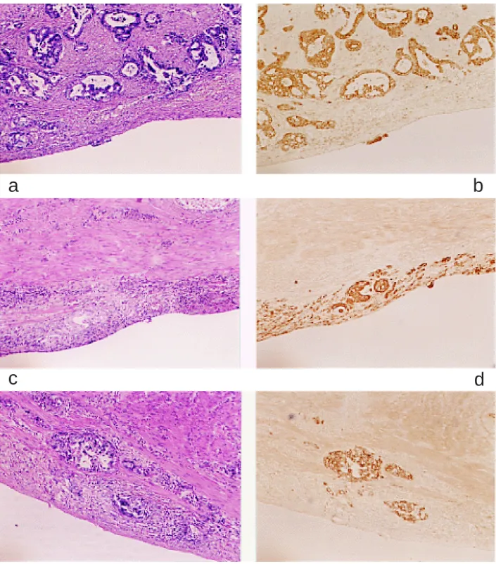
Table 1 summarizes the results of the current study in 50 gastric cancer patients with subserosal invasion in which serosal invasion was undetect-able by H&E staining. Eight patients (16%) were determined to have serosal microinvasion, includ-ing 1 patient (Patient 1) whose serosal invasion was detected by both H&E and CK stainings (Figs. 1a and b). In 7 patients (Patients 2 to 8), serosal micro-invasion was detected by CK staining, but H&E staining failed to detect it, as shown in Figs. 1c and d.
Fig. 1. Comparison of the results between H&E and CK stainings.
a and b: Example of stained primary lesion after staining with H&E (a) and CK (b) (2.5 × 10 each). Serosal microinvasion was detected by both H&E and CK stainings (Patient 1).
c and d: Example of stained primary lesion after staining with H&E (c) and CK (d) (2.5 × 10 each). Serosal microinvasion was not detected by H&E staining but by CK immunostaining.
e and f: Example of stained primary lesion after staining with H&E (e) and after staining with CK (f) (2.5 × 10 each). Serosal microinvasion was not detected by either H&E or CK staining.
c
d
a
b
0 0.2 0.4 0.6 0.8 1.0 0 20 40 60 80 100 120 140 (month) Survival time Survival rate
7 patients with se microinvasion
42 patients without se microinvasion
Fig. 2. Postoperative survivals (Kaplan-Meier method).
se, tunica serosa.
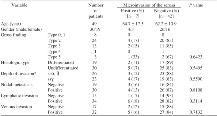
Table 2. Serosal microinvasion in the primary lesion and clinicopathologic characteristics
Variable Number Microinvasion of the serosa P value of Positive (%) Negative (%)
patients [n = 7] [n = 42] Age (year) 49 64.7 ± 17.5 62.2 ± 10.9 Gender (male/female) 30/19 4/3 26/16
Gross finding Type 0, 1 8 0 8
Type 2 24 4 (17) 20 (83)
Type 3 13 2 (15) 11 (85)
Type 4 1 0 1
Type 5 3 1 (33) 2 (67) 0.6423
Histologic type Differentiated 19 2 (11) 17 (89)
Undifferentiated 30 5 (17) 25 (83) 0.5495
Depth of invasion* ssα, β 26 3 (12) 23 (88)
ssγ 23 4 (17) 19 (83) 0.5590
Nodal metastasis Negative 19 3 (16) 16 (84)
Positive 30 4 (13) 26 (87) 0.8108
Lymphatic invasion Negative 15 1 ( 7) 14 (93)
Positive 34 6 (18) 28 (82) 0.3114
Venous invasion Negative 17 2 (12) 15 (88)
Positive 32 5 (16) 27 (84) 0.7132
Types 0 and 1, superficial, flat tumors with or without minimal elevation or depression and polypoid tumors, sharply demarcated from the surrounding mucosa, usually attached on a wide base, respectively; Type 4, diffusely infiltrating carcinomas in which ulceration is usually not a marked feature; Type 2, 3 and 5, see footnote to Table 1. * When cancerous invasion extends to the tela subserosa (ss), the growth patterns can be divided into 3 categories; ssα, expansive growth pattern with a distinct border; ssβ, intermediate pattern between ssα and ssγ; ssγ, infiltrative growth pattern with an ill-defined border.
Six of these 8 patients with serosal microinvasion had undifferentiated adenocarcinoma as a histolo-gic type of gastric cancer. The remaining 42 pa-tients were determined to have no serosal micro-invasion by either H&E or CK-staining (Figs. 1e and f).
Clinicopathologic characteristics of the 7 pa-tients with serosal microinvasion in the primary le-sions (Patient 1, whose serosal invasion was de-tected by H&E and CK stainings, was excluded) were compared with those of 42 patients without it (Table 2). Microinvasion was found to be more fre-quent in undifferentiated types of cancer and in pa-tients with lymphatic invasion by cancer. However, there were no statistical differences between the 2 groups among any variables.
Figure 2 compares the 5-year Kaplan-Meier survival curves for 7 patients with and 42 patients
without microinvasion. There was no difference in survivals between the 2 groups.
Discussion
Identification of lymph node metastasis is an im-portant factor in predicting postoperative survival of patients with gastric cancer, and H&E staining has been widely used for this purpose. However, immunohistochemical techiques with antibodies against CK can identify micrometastases in lymph nodes missed during routine H&E staining from gastric cancer (Maehara et al., 1996; Ishida et al., 1997; Maeta et al., 1998; Cai et al., 1999), colo-rectal cancer (Greenson et al., 1994; Adell et al., 1996; Sasaki et al., 1997) and breast cancer (Sedmak et al., 1989; Dowlatshahi et al., 1997). Because CK pro-teins are essential constituents of the cytoskeleton of both normal and malignant epithelial cells in lymph nodes, they serve as reliable markers for the epithelial origin of cells. Patients with these CK-positive cells in lymph nodes have a significantly decreased postoperative survival time.
Since it is known that in patients with gastric cancer, the depth of cancer invasion within the stomach wall is also an important prognostic factor, as well as nodal metastases, we reported previously that immunohistochemical staining with CK was useful for the detection of microinvasion of the muscularis propria in gastric cancer with submuco-sal invasion (Cai et al., 1999). In gastric cancer pa-tients with serosal invasion, serosally exposed can-cer cells are easily isolated in the peritoneal cavity, and these cells proliferate as a peritoneal metastasis. On cytologic examination of peritoneal lavage fluid immediately after laparotomy at the time of sur-gery, free intraperitoneal cancer cells are frequently found in such patients. Kaibara et al. (1986) re-ported that there were close relationships both be-tween the rate of detection of intraperitoneal free cancer cells and the area of serosal invasion, and be-tween the area of serosal invasion and prognosis in patients with gastric cancer with serosal invasion. Therefore, in patients with gastric cancer, the pres-ence of serosal invasion implies a very high risk for the development of postoperative peritoneal meta-stasis.
In the current study, the usefulness of CK staining was examined in order to find out whether it can detect serosal microinvasion in gastric cancer with subserosal invasion, because serosal microin-vasion may be overlooked by routine H&E staining. As a result, microinvasion was detected in 8 pa-tients, including 1 whose microinvasion was detect-ed by both CK and H&E stainings. However, sero-sal microinvasion was not a significant prognostic factor, and there were no differences in postopera-tive survivals between patients with or without microinvasion. The area of microinvasion detected by CK staining in this series may be too narrow to both introduce the isolation of free cancer cells into the peritoneal cavity and affect postoperative sur-vival.
Based on our findings above, it may be sug-gested that there is no clinical implication in detect-ing serosal microinvasion usdetect-ing CK staindetect-ing. How-ever, the important point is how to make an effort to diagnose the presence or absence of serosal in-vasion and/or microinin-vasion accurately and pre-cisely. In our series, the number of patients was too small to answer the question of whether the detec-tion of serosal microinvasion by CK staining is im-portant or not, and thus a multi-institutional study using a large number of patients is necessary. Nev-ertheless, induction of the CK staining as in this series in the pathohistological diagnosis of serosal microinvasion may be the 1st trial ever for gastric cancer. We believe that H&E and CK stainings should be seriously considered for simultaneous use in the accurate determination of both nodal metasta-sis and depth of cancer invasion.
Although CK is a reliable marker of cells from epithelial origins, Makin et al. (1984) reported that there were some exceptions to negative staining, such as normal stratified squamous esophageal epi-thelia and of positive staining of cells from non-epithelial origins, such as smooth muscle and meso-thelium. As indicated in this series, when CK im-munostaining is used for detecting serosal micro-invasion, careful attention should be paid to the un-expected exceptional positive staining of meso-thelial one-layered cells.
References
1 Adell G, Boeryd B, Franlund B, Sjodahl R, Hakansson L. Occurrence and prognostic importance of micro-metastases in regional lymph nodes in Dukes’ B colo-rectal carcinoma: an immunohistochemical study. Eur J surg 1996;162:637–642.
2 Baba H, Korenaga D, Okamura T, Saito A, Sugimachi K. Prognostic factors in gastric cancer with serosal in-vasion. Univariate and multivariate analyses. Arch Surg 1989;124:1061–1064.
3 Cai J, Ikeguchi M, Maeta M, Kaibara N. Micrometa-stasis in lymph nodes and microinvasion of the mus-cularis propria in primary lesions of submucosal gas-tric cancer. Surgery 1999;126:32–39.
4 Dowlatshahi K, Fan M, Snider HC, Habib FA. Lymph node metastases from breast cancer. Cancer 1997; 80:1188–1197.
5 Greenson JK, Isenhart CE, Rice R, Mojzisik C, Houchens D, Martin EW. Identification of occult micrometastases in pericolic lymph nodes of Dukes’ B colorectal cancer patients using monoclonal anti-bodies against cytokeratin and CC49. Cancer 1994; 73:563–569.
6 Iitsuka Y, Kaneshima S, Tanida O, Takeuchi T, Koga S. Intraperitoneal cancer cells and their viability in gastric cancer. Cancer 1979;44:1476–1480.
7 Ishida K, Katsuyama T, Sugiyama A, Kawasaki S. Immunohistochemical evaluation of lymph node mi-crometastases from gastric carcinomas. Cancer 1997;79:1069–1076.
8 Japanese Research Society for Gastric Cancer. The general rules for the gastric cancer study in surgery and pathology. Jpn J Surg 1981;11:127–145. 9 Kaibara N, Iitsuka Y, Kimura A, Kobayashi Y,
Hirooka Y, Nishidoi H, et al. Relationship between area of serosal invasion and prognosis in patients with gastric cancer. Cancer 1986;60:136-139. 10 Kaibara N, Hamazoe R, Iitsuka Y, Maeta M, Koga S.
Hyperthermic peritoneal perfusion combined with chemotherapy as prophylactic treatment of peritoneal recurrence of gastric cancer. Hepato-Gastroenterology 1989;36:75–78.
11 Koga S, Kaibara N, Iitsuka Y, Kudo H, Kimura A, Hiraoka H. Prognostic significance of intraperi-toneal free cancer cells in gastric cancer patients. J Cancer Res Clin Oncol 1984;108:236–238.
12 Maehara Y, Oshiro T, Endo K, Baba H, Oda S,
Ichiyoshi Y, et al. Clinical significance of occult micrometastasis in lymph nodes from patients with early gastric cancer who died of recurrence. Surgery 1996;119:397-402.
13 Maeta M, Cai J, Oka S, Saito H, Kondo A, Tsujitani S, et al. Cytokeratin-positive cells in lymph nodes in which metastases are undetectable by conventional histological staining in advanced gastric cancer. Yonago Acta Med 1998;41:105-112.
14 Makin C, Bobrow L, Bodmer W. Monoclonal anti-body to cytokeratin for use in routine histopatholo-gy. J Clin Pathol 1984;37:975–983.
15 Maruyama K, Gunven P, Okabayashi K, Sasako M, Kinoshita T. Lymph node metastasis of gastric cancer: general pattern in 1931 patients. Ann Surg 1989;210: 596–602.
16 Matsushita M, Hajiro K, Suzaki T, Uchida K, Okano A, Ohana M, et al. Histopathological assessment of lymph node metastasis in patients with gastric can-cer. Hepato-Gastroenterology 1995;42:861–866. 17 Nakajima T, Harashima M, Hirata M, Kajitani T.
Prognostic and therapeutic peritoneal cytology in gastric cancer. Acta Cytol 1978;22:225–229. 18 Sasaki M, Watanabe H, Jass JR, Ajioka Y, Kobayashi
M, Matsuda K, et al. Occult lymph node metastases detected by cytokeratin immunohistochemistry pre-dict recurrence in node-negative colorectal cancer. J Gastroenterol 1997;32:758–764.
19 Sedmak DD, Meineke TA, Knechtges DS. Detec-tion of metastatic breast carcinoma with monoclonal antibodies to cytokeratin. Arc Pathol Lab Med 1989; 113:786–789.
20 Siewert JR, Bottcher K, Roder JD, Busch R, Hermanek P, Meyer HJ. Prognostic relevance of systematic lymph node dissection in gastric carcinoma. Br J Surg 1993;80:1015–1018.
21 Tanida O, Kaneshima S, Iitsuka Y, Kudo Y, Kiyasu Y, Koga S. Viability of intraperitoneal free cancer cells in patients with gastric cancer. Acta Cytol 1982;26:681–687.
22 Trojani M, Mascarl ID, Bonichon F, Coindre JM, Delson G. Micrometastasis to axillary lymph nodes from carcinoma of the breast; detection by immuno-histochemistry and prognostic significance. Br J Cancer 1987;55:303–306.
Received October 15, 2003; accepted November 17, 2003 Corresponding author: Michio Maeta, MD