1. Introduction
Sediment-mixed biofilms(SMBs)are aggre-gations of microbial growth present on the
surfa-ces of most substansurfa-ces in contact with water. They are commonly found on the sedimentary surfaces of seabeds and intertidal zones(BOUDREU and JØRGENSEN, 2001; GHANNOUM and OʼTOOLE, 2004).However, the roles of SMBs in the biogeo-chemical cycle at seawater-benthic boundaries remain unclear. Biofilms have been experimen-tally grown from individual microbes isolated from natural samples using microtiter plate cul-ture(OʼTOOLEand KOLTER, 1998)and flow-cham-ber culture(WOLFAARDTet al., 1994).Mixed bio-films(containing multiple microbial species), such as those occurring on sediments, are more
Laboratory reconstruction and nearby sulfide dynamics of a
model benthic boundary layer containing sediment-mixed biofilm
Norihisa ISHIBASHI1)*, Hiroo SATOH2)and Jota KANDA1)
Abstract: Sediment-mixed biofilms of microorganisms embedded within an extracellular
poly-meric substance were reconstructed using laboratory models of seawater-benthic boundaries, and the sulfide dynamics in the boundary zone were examined. Biofilm-covered sediment was obtained from the field and cultured in a beaker of seawater with a granular formula for fish lar-vae and powdered foraminiferal limestone. In the standing culture, a floating biofilm formed on the liquid surface, and a microbiota capable of mixed biofilm formation was obtained. The mixed biofilm was cultured in a fluidization-free aerobic state, again with nutritional granules and pow-dered foraminiferal limestone. This culture formed a sediment-mixed biofilm on the bottom of the beaker. Furthermore, a seawater-benthic boundary model was developed on a hydrated sub-stratum with sediment biofilm, using a nylon mesh as the model bottom layer in the culture sys-tem. When the floating biofilm was disrupted and then dispersed on the nylon mesh, detectable biofilms formed on the surface of the model bottom. As nutrients were utilized by the microbes, sulfides accumulated beneath the model bottom. Subsequently, the sulfides passed through the model bottom and became detectable in the seawater just above the sediment-mixed biofilm, where a white-turbid layer formed. As revealed by denaturing gradient gel electrophoresis, the layer contained populations of several bacterial species.
Keywords : sediment mixed biofilm, seawater-benthic boundary, sulfide cycle, pelagic bacterial
layer
1)Graduate School of Marine Science and Technolo-gy, Tokyo University of Marine Science and Technology, 4Ȃ5Ȃ7 Konan, Minato-ku, Tokyo 108Ȃ 8477, Japan
2)6Ȃ6Ȃ9Ȃ105 Kameido, Koto-ku, Tokyo 136Ȃ0071, Ja-pan
*Corresponding author: Tel: + 81Ȃ80Ȃ1301Ȃ4588 Fax: + 81Ȃ467Ȃ24Ȃ5026 E-mail: [email protected]
1)Institute of Marine Science, Burapha University, Bangsaen, Chon Buri 20131, Thailand
2)Department of Aquatic Science, Faculty of Sci-ence, Burapha University, Bangsaen, Chon Buri 20131, Thailand
3)Atmosphere and Ocean Research Institute, The
University of Tokyo, 5Ȃ1Ȃ5, Kashiwanoha, Kashi-wa, Chiba 277Ȃ8564, Japan
*Corresponding author: Thidarat Noiraksar Tel: + 66(0)38 391671
Fax: + 66(0)38 391674
difficult to reproduce in the laboratory. In Japan, fish larvae have been cultured in large, on-land water chambers(with capacities of 10Ȃ50 m3) with powdered foraminiferal limestone as an ad-ditive since the early 1990s. Powdered foramini-feral limestone is known to increase the survival of fish larvae(SUZUKIet al., 2008),but the under-lying mechanism is unknown. In 2002, when powdered foraminiferal limestone was present at the Minami-Izu Aquaculture Center(Shizuoka, Japan),the sediment surface was covered by an SMB(IIJIMA et al., 2009). This suggests that SMBs are promoted by powdered foraminiferal limestone, which may be associated with good water quality. Based on these observations, we initiated experimental attempts to reproduce an SMB in a beaker via the addition of foraminiferal limestone.
SMBs can form on two types of substrata in sea beds and intertidal zones: water-containing types(soft bottom or aquatic sediments such as mud and sand, referred to here as “aquatic sedi-ments”)and solid types(hard bottom or solid substrata such as rocks, referred to here as “sol-id substrata”). In aquatic sediments, sol“sol-id sub-stances such as sand and mud are mixed with organic matter and infused with pore water (BOUDREU, 1997). In the seawater-benthic boun-dary zones at the bottom of shallow sea areas, abundant organic matter sedimentation accumu-lates from both land and the aquatic environ-ment and is utilized and degraded by many spe-cies of microbes(KOIKE, 2000; FENCHEL et al., 2012).Owing to the vigorous degradation of or-ganic matter, remarkable oxygen losses occur at the benthic boundary, and the high concentra-tions of sulfate ions in the anaerobic marine envi-ronment activate microbial sulfur metabolism. Consequently, a great variety of sulfur com-pounds is formed(GEMERDEN, 1993; SAMUKAWA and HIIRO, 1996; RICKARD, 2012).SMBs have been
widely investigated on solid substrata(RIDING and AWRAMIKE, 2000),but their roles in aquatic sediments are less well understood. The labora-tory study of the sulfur cycles in a sandy beach habitat using an experimental chamber suggest-ed that bacterial biofilms on sand substrata pro-duce much of the sulfur at the benthic bounda-ries between seawater and sand substrata (JØRGENSEN, 1974). The sandy beach study indi-cates that biogeochemical studies of aquatic sedi-ments would be greatly facilitated by the availa-bility of an appropriate laboratory model of aquatic sediments with SMBs.
In the present study, we developed a model experimental system that reconstructs SMBs from naturally occurring microbial assemblages in sediments. We first reproduced SMBs ob-served on solid substrata in aquaculture tanks and then, using the same method, produced SMBs from natural sediments with bacterial as-semblages collected at a variety of sites in coast-al environments. We coast-also established an experi-mental setup that can model an aquatic sediment in the laboratory and produced SMBs on the sur-face of the modeled aquatic sediment. In order to demonstrate that the experimental model could be used in biogeochemical studies of SMBs, we then analyzed the water-quality parameters(sul-fide levels and dissolved oxygen)and bacterial species at the benthic boundary of the model. Experiments were performed in the laboratory of the Minami-Izu Aquaculture Center from 2002 to 2003 and at the Tokyo University of Marine Science and Technology(Tokyo, Japan)from 2009 to 2016. While the laboratory SMBs we ob-tained may not completely reproduce or be rep-resentative of the natural SMBs in coastal envi-ronments, further improvement of the reproduc-tion technique will hopefully result in the estab-lishment of a conventional laboratory model for SMBs in natural coastal environments.
2. Materials and Methods
To reproduce the SMBs of an aquaculture fa-cility, we collected SMB-containing sediments with a spatula from the bottom of a concrete wa-ter chamber(capacity, 20 m3)in which fish lar-vae were reared at the Minami-Izu Aquaculture Center of the Japan Sea-farming Association (currently the Minami-Izu Aquaculture Center of the Fisheries Research and Education Agen-cy, Minami-Izu, Shizuoka, Japan; 34°36′43″ N, 138°50′55″ E).Moreover, seabed sediments con-taining SMBs were collected from three marine sites during the spring ebb tide at a water depth of about 0.1 m. For sampling, an open, transpar-ent acrylic cylinder(length, 20 cm; inside diame-ter, 3 cm)was thrust into the benthic layer from above and then immediately closed at both ends. The sites included a canal under the TENNOUZUBridge(Minato-ku, Tokyo, Japan; 35° 37′24″ N, 139°44′42″ E),a canal frontage site at OHISeaside Park(Ota-ku, Tokyo, Japan; 35°35′ 19″ N, 139°44′58″ E), and a shoreline off the SHICHIRIGAHAMAsand beach(Kamakura, Kanaga-wa, Japan; 35°18′9″ N, 139°31′26″ E). The two former sites occupy the innermost part of Tokyo Bay, and the latter is located along Sagami Bay. Surface substratum samples were also collected from an offshore seabed near HANEDA in Tokyo Bay(35°30′42″ N, 139°49′48″ E)at an
approxi-mate depth of 30 m using an Ekman-Birge bot-tom sampler(RIGO, Tokyo, Japan). Each sub-stratum sample was submerged in seawater and transported to the laboratory.
The formation of floating biofilms was investi-gated in standing cultures of microbes nourished with a granular fish-larvae feed[Ambrose 400; Nippon Formula Feed Manufacturing(current-ly, Feed One),Yokohama, Kanagawa, Japan; par-ticle diameter, 420Ȃ650 µm]used as a complex nutrient for microbes, hereafter referred to as nutritional granules, and powdered foraminiferal limestone as an additive(Fish Green, Green Cul-ture, Takaoka, Toyama, Japan; average diame-ter, 70 µm).The constituents of the nutritional granules and foraminiferal limestone powder are listed in Tables 1 and 2, respectively. For stand-ing culture, the SMB-containstand-ing sediment, pow-dered foraminiferal limestone, and nutritional granules were spread over the bottom of a 100Ȃcm3glass beaker or a 220Ȃcm3plastic vessel with 80 cm3of sand-filtered seawater, which was covered with PVDC film(Kureha, Tokyo, Ja-pan)to prevent evaporation.
Figure 1 shows the culture model system of
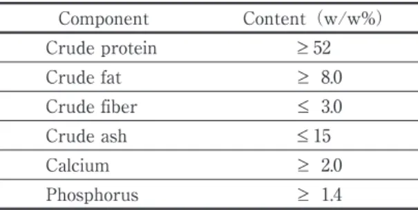
Table 1. Composition of “Ambrose 400” nutrition
granules. Component Content(w/w%) Crude protein ≥ 52 Crude fat ≥ 8.0 Crude fiber ≤ 3.0 Crude ash ≤ 15 Calcium ≥ 2.0 Phosphorus ≥ 1.4
SOURCE: NIPPONFORMULAFEEDMANUFACTURING(2009)
Table 2. Mineral element composition of “Fish
Green” foraminiferal limestone powder. Mineral element Content(g/kg)
Calcium(Ca) 302 Silicon(Si) 71.7 Magnesium(Mg) 5.94 Aluminum(Al) 5.9 Iron(Fe) 4.62 Sodium(Na) 2.7 Potassium(K) 0.74 Phosphorus(P) 0.21 Manganese(Mn) 0.2 Zinc(Zn) 0.4
the seawater-benthic boundary with the sedi-ment biofilm on hydrated substrata. To develop the culture model system, we placed a nylon mesh(pore size, 50 µm; NYTAL DIN1 10Ȃ50, Sefer, Ruschlikon, Switzerland)at the mid-height of a transparent cylindrical vessel(acrylic; in-side diameter, 9. 5 cm; height, 15 cm)with an open upper end. This system models the bottom surface of a typical aquatic sediment. The use of nylon limits the disintegration that might occur under microbial chemical activity. The space be-low the mesh was filled with seawater, modeling the pore water of aquatic sediments. This vessel was placed in a compact, glass water chamber (area, 18 cm × 31 cm; height, 24 cm).The sea-water in the sea-water chamber was maintained in a fluidization-free aerobic state. At the Minami-Izu Aquaculture Center, where an SMB covers the bottom of the aquaculture tank, the bottom layer of water is static because the air stones are
suspended at an intermediate depth in the rear-ing chamber. However, the oxygen saturation in this layer is ≥ 70%. To reproduce these condi-tions in the bottom-layer water in the aquacul-ture tank, we installed an air stone(diameter, 3 cm)in a plastic cylinder(length, 12 cm; diame-ter, 5 cm)with numerous 3Ȃmm-diameter pores to prevent water movement of the culture sys-tem. The system was aerated with an air pump (O3PROOF202, Addx, Hachioji, Japan). Test cultures were prepared with sand-filtered and mat-filtered seawater. The filter mat was a filter-ing material for aquarium water(Mat Kobo, Tokyo, Japan).The salinity of the filtered seawa-ter was not below 15.0, and the indoor light in-tensity of the laboratory was not higher than 6.8 µE・mȂ2∙ sȂ1. Culture experiments were initiat-ed by spreading the seinitiat-ed bacterial population, powdered foraminiferal limestone, and nutrition-al granules on the nylon surface.
To quantify biofilm formation, we measured the packed cell volume(PCV)and the wet weight. Biofilm samples were placed in a gradu-ated 10Ȃcm3 tube and centrifuged (H107, Kokusan Centrifuge, Tokyo, Japan)at 2000 rpm for 3 min, and the volume was defined as the PCV. To determine the wet weight of each bio-film, the floating biofilm on the liquid surface was transferred to a nylon mesh(5 × 5 cm; pore size 50 µm; NYTAL DIN1 10Ȃ50, Sefer, Ruschlikon, Switzerland),drained of the biofilm surface wa-ter by applying wawa-ter-absorbing paper from be-low the mesh, and weighed repeatedly on a pre-cision balance until the weight was nearly con-stant. The biofilm was observed both macro-scopically and micromacro-scopically(CK2, Olympus, Tokyo, Japan).
Total acid volatile sulfides(AVSs)were meas-ured using the detector tube method(HEDRO-TECHȂS330, Gastec, Ayase, Japan). AVSs are sulfide compounds that volatilize in water under
Fig. 1. Experimental model of biofilm formation at
the aquatic benthic boundary. The model bottom (nylon mesh, 50 µm)was affixed in a transpar-ent acrylic vessel(diameter, 9. 5 cm; height, 15 cm)with a closed bottom and an open mouth. The acrylic vessel was submerged in a glass chamber of dimensions 18 cm(D)× 30 cm(W) × 24 cm(H).
acidic conditions, namely, hydrogen sulfide, hy-drogen sulfide ions, sulfide ions, and sulfur from non-pyrite iron sulfide. The dominant species are H2S and HS−(DAVIDand MORSE, 2005; SUGAHARA et al., 2012).Hereafter, the total AVSs measured using this method are simply referred to as sul-fides or AVSs. The bacterial density or turbidity of the culture water was assayed by determining the absorbance at 660 nm(OD660)in a spectro-photometer(UV160, Shimadzu, Kyoto, Japan; MARDIGAN et al., 2009). The dissolved oxygen (DO)content was measured using a DO meter (YSIȂ55, Yellow Spring Instrument, Yellow Spring, OH, USA).For the culture system mod-eling the seawater-benthic boundary of an aquat-ic sediment, the sensor of the DO meter was ver-tically inserted at a non-water-agitating speed (4 cm/min)using a slow elevator(made in-house).The water samples from above the mod-el bottom surface that were used for measuring sulfide concentrations and turbidity were collect-ed with a syringe(5 cm3)joined to a silicon tube (inside diameter, 2.0 mm)fixed to the side of the sensor. The water was sampled at a depth of ap-proximately 2 cm using another syringe(capaci-ty, 1 cm3)and a syringe needle to penetrate the mesh. Oxidation-reduction potential(ORP)was measured using an ORP meter(RMȂ12P, DKK-TOA, Tokyo, Japan).
The microbial composition in the white-turbid layer was analyzed by denaturing gradient gel electrophoresis(DGGE)(MUYZER and SMALLA, 1998; ISHIIet al., 2000; NISHIJIMAet al., 2010).Sam-ples were frozen at Ȃ80 ℃ until required for DGGE analysis. DNA was extracted using MORA-EXTRACT(Kyokuto Pharmaceutical In-dustrial, Tokyo, Japan), and the bacterial 16S rDNA was amplified by a modified touchdown polymerase chain reaction(PCR)method(DON et al., 1991; MUYZER and SMALLA, 1998). A 16S rDNA sequence of approximately 200 bp was
amplified using the 341fȂGC and 534r primers. Electrophoresis was performed at 100 V for ap-proximately 12 h. The electrophoresis mixture included the PCR product, 8%(w/v)polyacry-lamide gel, and denaturant at concentrations of 25Ȃ65%. To check the DNA purity over the band width, the DNA from a band in the electrophor-esed gel was extracted and used as the template in a second PCR. The second amplification prod-uct was labeled using the Big Dye Terminator v3.1 Cycle Sequencing Kit(Applied Biosystems, Carlsbad, CA, USA),and its nucleotide sequence was determined by an ABI 3130xl Genetic Ana-lyzer System(Applied Biosystems).A homolo-gy search and simplified molecular phylogenetic analysis were performed using the Apollon 2.0 program(Techno Suruga Laboratory, Shizuoka, Japan), the International Base Sequence Data-base(Gen Bank; DDBJ; EMBL), and the Apol-lon Reference Strain Database DBȂBA9.0(Tech-no Suruga Laboratory).
3. Results
3.1 Reconstitution of sediment-mixed biofilms above the model bottom surface
In the standing culture of SMB-containing sediment collected from the bottom surface of the water chamber used for rearing fish larvae (sediment weight/base area, about 100 mg/ cm2), containing added powdered foraminiferal limestone(5 mg/cm2)and nutritional granules(10 mg/cm2),a floating membrane containing bacte-ria formed on the surface of the liquid on day 4 of cultivation at 22 ℃. Similarly, cultivation with natural coastal sediments collected from the aforementioned sites also yielded floating mem-branes. With nutritional granules of varying areal densities(5, 10, 15, or 20 mg/cm2)and powdered foraminiferal limestone(3 mg/cm2), we examined in detail the formation of the float-ing biofilm produced from the substratum
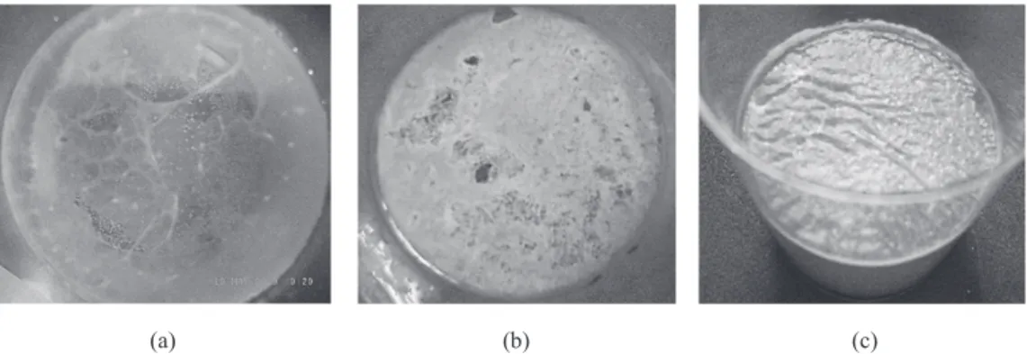
col-lected from a canal under the TENNOWZE Bridge (100 mg/cm2).On day 4, a highly fluid, sol-like membrane formed on the liquid surface(Fig. 2 (a)). On day 7, the membrane became thick, elastic, gel-like, and less fluid(0.6 cm3PCV with both the 10 mg/cm2 and 15 mg/cm2nutritional granules; Fig. 2(b)). On day 9, the gel-like
membrane formed using the 20 mg/cm2 nutri-tional granules became surface-water depleted and fragmented(Fig. 2(c)).In contrast, those formed using 5, 10, or 15 mg/cm2 nutritional granules maintained their gel-like properties with a wet surface. Figure 3 shows the relation-ship between biofilm formation and nutrient
lev-Fig. 2. Formation of floating mixed biofilm by the aquatic standing-culture method(20
℃).(a)Liq-uid sol-like biofilm(day 4 of cultivation).(b)Solid gel-like biofilm(day 7 of cultivation).(c)Gel-like colloidal biofilm deprived of surface water(day 9 of cultivation).
Fig. 3. Formation of floating mixed biofilms nourished with different amounts of nutritional
el. The wet weight of the biofilm increased as the areal density of the nutritional granules in-creased from 3 to 15 mg/cm2. However, increas-ing the areal density of the nutritional granules to 25 mg/cm2yielded no further increase in bio-film growth.
In the culture experiments with a 100Ȃcm3 beaker under fluidization-free aerobic conditions, where the culture contained a mixture of biofilm-containing sediment from the fish-larvae culture tank(weight/base area, 100 mg/cm2 ),nutrition-al granules(10 mg/cm2), and powdered fora-miniferal limestone(5 mg/cm2),the water above the sediments in the beaker became transparent, and an SMB formed on the sediment surface on day 4 of cultivation at a water temperature of 22 ℃. Under the same conditions, we cultured the floating biofilms (0.2 cm3PCV) obtained from the cultivation of the sediment-attached biofilms collected from the four marine locations. The re-sulting SMBs were similar in appearance and de-velopmental processes to those formed from the sediment collected from the aquaculture tank, in-dicating that SMBs can be reconstructed on a solid substratum using the culture conditions adopted in this study.
3. 2 Sulfide and microbial dynamics near the aquatic benthic boundary of the SMB model The above-described procedure was applied to a model aquatic sediment. To create the model aquatic sediment(Fig. 1), the floating biofilm (0.2 cm3PCV)grown from sediments collected under the TENNOUZ Bridge was disrupted and spread over the nylon bottom surface as the seed microbial population, together with nutri-tional granules(10 mg/cm2)and powdered for-aminiferal limestone(3 mg/cm2). On day 1 of cultivation, the water contained a light suspen-sion within 3 cm above and below the model bot-tom surface. After 2 days, the slightly turbid
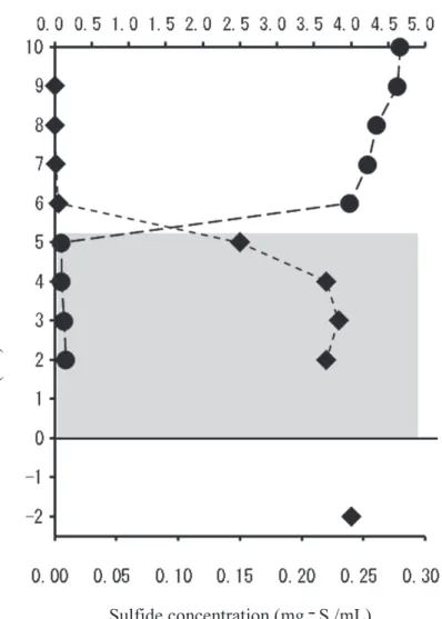
phase was distinctly separated from the trans-parent phase of the water. The boundary was observed just above the model bottom surface, on which a biofilm, presumably an SMB, had formed. On day 3 of cultivation, a dense white-turbid layer was formed above the SMB on the model bottom, remaining until day 11(Fig. 4(a)). This white-turbid layer was confirmed to be con-tain densely populated microbes under light mi-croscopy. The microbial cells(approximately 3 µm in diameter)contained green granules and were surrounded by a 30Ȃµm thick coat of trans-lucent colloidal material(Fig. 4(b)).Once this layer had been established, the DO rapidly de-creased below the upper surface of the layer. The DO level in the white-turbid layer was close to zero(≤ 0. 005 mg/dm3; Fig. 5). The sulfide concentration was nearly uniform(about 0.2 mg-S/cm3)in the white-turbid layer above the mod-el bottom surface, slightly lower in the water
be-Fig. 4. White-turbid layer above the bottom of the
modeled aquatic benthic boundary with biofilm growth after 5 days of culture at 20 ℃(a),and a microscopic image of bacteria in the white-turbid layer(b).
low the model bottom surface, rapidly decreased above the white-turbid layer, and was undetecta-ble in the transparent water above the white-turbid layer(Fig. 5).The white-turbidity(OD660)in the white-turbid layer remained essentially un-changed. Above the white-turbid layer, steep gradients(i. e., chemoclines)of DO and sulfide concentrations were observed(Fig. 5).
Forma-tions of white-turbid layers with similar patterns of sulfides and DO distribution were observed for the substrata collected from all canal and shoreline sites.
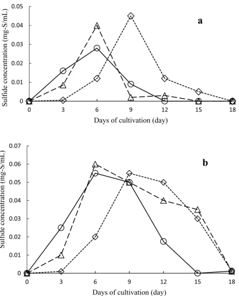
Figure 6 shows the effect of the areal density of the nutritional granules on sulfide formation. Increased amounts of nutritional granules in-creased the maximum sulfide concentrations in
Fig. 5. Vertical distributions of sulfides(AVSs)and dissolved oxygen in
the presence of the white-turbid layer in the experimental system af-ter 5 days of culture at 20 ℃. Diamonds, sulfides(AVS);circles, dis-solved oxygen(DO);shaded area: white turbid layer.
the overlying and underlying water in the mod-els containing 5, 7.5, and 10 mg/cm2 nutritional granules, as well as prolonging the time to for-mation of the white-turbid layer just above the model surface. The white-turbid layer was main-tained while sulfide was detected in the underly-ing water. In contrast, in the model containunderly-ing
20 mg/cm2nutritional granules, sulfides(AVSs) in the underlying water were detected on day 3 and peaked on day 6 but were never detected in the overlying water(Fig. 6). In addition, no white-turbid layer formed just above the model bottom surface, and the SMB that formed on the model bottom surface was white and only
slight-Fig. 6. Sulfide concentration in the water 2 cm above(a)and 2 cm below(b)
the bottom of the aquatic benthic boundary model(culture water tem-perature, 25 ℃)with biofilm growth using different amounts of nutri-tional granules(diamonds, 5 mg/cm2; triangles, 7. 5 mg/cm2; squares, 10 mg/cm2; circles, 20 mg/cm2)with a fixed volume of crushed colloidal biofilm(0.2 cm3PCV)and a constant density of powdered foraminiferal lime stone(3 mg/cm2).
ly viscous(Fig. 7).
Figure 8 shows the effect of temperature on sulfide formation in an experiment with the TENNOUZU sediment. Below the model bottom surface, the maximum sulfide concentration was nearly independent of water temperature, but above the model bottom surface, it increased with decreasing water temperature. The time required to detect the sulfides also increased with decreasing water temperature, both above
and below the model bottom surface(Fig. 8). The persistence time of the white-turbid layer increased with decreasing water temperature. Above the model bottom, the ORP of the water rapidly decreased over time; moreover, the ORP reduction rate increased with increasing water temperature. At all of the examined water tem-peratures, the ORP reached Ȃ300 mV on days 3Ȃ5. Additionally, the white-turbid layer formed when the ORP reached ≤ Ȃ100 mV above the
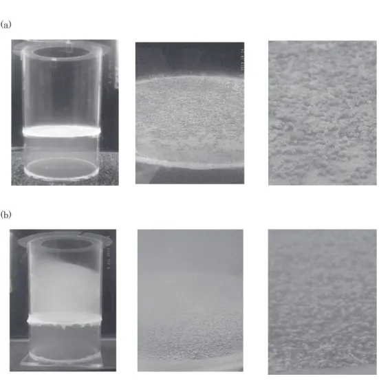
Fig. 7. Absence of white-turbid layer above the model aquatic sediment after 7 days of
cul-ture with 20 mg/cm2of nutritional granule(a),and the biofilm and the white-turbid lay-er above the model aquatic sediment aftlay-er 7 days of culture with 10 mg/cm2of nutritional granule(b).
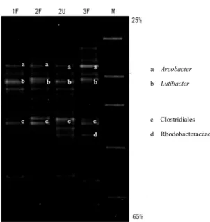
model surface and disappeared when the ORP was restored to ≥ 0 mV above the model surface. Figure 9 shows the PCR-DGGE results of an experiment with the TENNOUZUsediment indicat-ing the temporal changes in the bacterial com-munity of samples collected from the white-turbid layer above the model bottom surface.
Three distinct bands(a, b, and c; Fig. 9)ap-peared on days 1 and 2, and four distinct bands (a, b, c, and d; Fig. 9)were present on day 3 of cultivation. In the sample on day 3 when the white-turbid layer formed, the intensity of band a was markedly more intense than those of the other two bands(b and c). Sequence analysis
Fig. 8. Sulfide concentration in the water above(a)and below(b)the bottom of the
aquatic benthic boundary model(nutritional granules, 5 mg/cm2)with biofilm growth at different water temperatures. Circles, 25 ℃; triangles, 20 ℃; squares, 15 ℃. The original sediment was collected under the TENNOZUBridge.
indicated that band a was derived from Arco-bacter sp.(sequence identity, 100%),a microaer-ophilic sulfur bacterium. Band b was derived from Lutibacter sp., with 99% sequence identity to Lutibacter maritimus, a marine bacterium of the family Flavobacteriaceae isolated from a ti-dal flat sediment(CHOIand CHO, 2006),whereas band c was derived from a bacterium of the or-der Clostridiales(sequence identity, 94%), an obligate anaerobic sulfur-reducing bacterium commonly found in soil. Band d was derived
from a bacterium in the family Rhodobactera-ceae(sequence identity, 99%), a purple non-sulfur photosynthetic bacterium that utilizes S0. 4. Discussion
4.1 Reconstitution of SMBs on the model bot-tom surface
In this study, floating biofilms from sediment microorganisms were formed on liquid surfaces in standing cultures. It has long been known that Bacillus subtilis forms a floating microbial film at the air-liquid interface of a standing culture (MORIKAWAet al., 2006; KOBAYASHI, 2007).In the floating biofilm development of Shewanella onei-densis, the pellicle forms in three steps, including rapid formation of a thin pellicle, followed by evo-lution into a heterogeneous biofilm and finally in-to a thick homogeneous biofilm(ARMITANOet al., 2013). The floating biofilms in our study also showed three developmental stages: a highly flu-id, sol-like membrane stage; a thick, elastic, gel-like membrane stage; and a gel-gel-like membrane with a water-depleted surface stage, which pos-sessed a dry, gel-like consistency(Fig. 2). The growth of the floating biofilms in our experi-ments was therefore similar to that of other known biofilms (LEWANDOWSKI and BEYENAL, 2013).
As suggested by the PCR-DGGE analysis of the microbial assemblage developed using the floating biofilm as the seed population, mixed species of bacteria constituted the floating bio-films. Unlike separation methods involving ho-mogenization or sonication, in which some por-tions of a microbial assemblage might be de-stroyed(REIL, 1994),the floating biofilm of this study developed from the natural sediment com-munity under relatively mild conditions. Using this mild separation technique, we were able to produce a mixed biofilm containing a coexisting set of bacterial species.
Fig. 9. DGGE electrophoretogram showing the
mi-crobial composition of the water above the model bottom from day 1 to day 3 of cultivation(until formation of the white-turbid layer). Lanes 1F, 2F, and 3F were extracted from the water 2 cm above the model bottom on days 1, 2, and 3, re-spectively. Lane 2U was extracted from the wa-ter 5 cm above the model bottom on day 2. Lane M is a DGGE marker(Nippon Reiji, Tokyo, Ja-pan). The culture was created with disrupted biofilm (0.2 cm3 PCV), nutritional granules (10 mg/cm2), and powdered foraminiferal lime-stone(3 mg/cm2)at 25℃. The original sediment was collected under the TENNOZUBridge.
Our results further showed that, using a float-ing biofilm as the seed population, SMBs could be cultured on solid substrata and aquatic sedi-ments with nutritional granules and powdered foraminiferal limestone. Among the SMBs ob-tained in this study, we speculate that the bio-film produced from the sediments of the aquacul-ture tank should likely reflect the biofilm that was originally found in the aquaculture facility, as the reproduction conditions were essentially the same as those of the aquaculture tank. Stud-ies using this laboratory-grown biofilm would provide valuable information that could lead to the improvement of aquaculture conditions. Us-ing the same technique, we successfully pro-duced mixed biofilms from seed populations ob-tained from natural coastal sediments with SMBs. Because the growth of each species in a natural bacterial assemblage changes substan-tially during artificial cultivation, the species composition in the mixed biofilm produced in the laboratory may be different from the seed popu-lation. Nevertheless, the successful laboratory development of a mixed biofilm enabled us to ex-amine the possible role of a mixed biofilm in bio-geochemical cycles in a laboratory setup. While the mixed biofilm and the material dynamics in the laboratory setup may not completely repre-sent the processes occurring in natural benthic boundary systems, laboratory studies using this system could provide at least preliminary in-sights. Furthermore, with additional examination and improvement of the conditions used for bio-film formation in this study, it may be possible to develop a laboratory system of SMBs that could be used in biogeochemical studies of natural ma-rine environments.
4. 2 Sulfide and microbial dynamics near the aquatic benthic boundary of the SMB model Laboratory-grown culture systems that can
model common aquatic sediments in coastal en-vironments such as sand and mud should con-tribute to studies of sulfur cycles. JØRGENSEN (1974)conducted an extensive study of sulfur cycles in a sandy beach environment in a water chamber by placing sand in a chamber with sea-water and adding seagrass to model the condi-tions on a post-storm sandy beach. This was used to examine a radioactive isotope of sulfur, with sulfides accumulated below the substratum surface. Approximately 90% of the sulfides mi-grated and diffused upward from substratum surface. At the end of the experiment, the sur-face of the seagrass was covered with microbial biofilms, including those of sulfur-reducing bacte-ria. These findings suggested that mixed bio-films are responsible for sulfide accumulation be-low the sand surface(JØRGENSEN, 1974).A similar sulfur cycling process was observed in our pres-ent study. We found that the permeability of the biofilms depended on the areal density of the nu-tritional granules(Fig. 6); with an abundance (20 mg/cm2)of nutritional granules, no sulfide migration from below the substratum surface was observed. It has been generally observed that the biofilm structure is sparse under low-nu-trient conditions and dense under high-nulow-nu-trient conditions(WIMPENNYand COLASSNTI, 1997).This suggests that under abundant nutrient levels, the biofilm structure becomes very dense and blocks sulfide migration.
Unlike the sandy-beach model of JØRGENSEN (1974), our culture model system of the sea-water-benthic boundary was fluidization-free above the bottom surface. Therefore, in our study, the sulfides rose from below the substra-tum surface and accumulated in the water above the model bottom. A dense white-turbid layer formed in the same region. Using a radioactive sulfide, MATSUYAMA(1978)showed that sulfide was formed by sulfur-reducing bacteria in
strati-fied meromictic lakes. In these lakes, the deeper seawater layer is covered by a superficial fresh-water layer. However, sulfide formed not in the bottom layers of the water but on the bottom surface, with sulfide concentrations peaking im-mediately below the bottom surface. Subse-quently, the sulfides passed through the bottom surface and accumulated in the anaerobic water immediately above the surface. This sulfur cycle in the benthic boundary of the stratified water of meromictic lakes is generally consistent with the sulfur cycle in our model experiment. Sulfide production and accumulation in bottom-layer wa-ter was also observed in the so-called “blue tide” in Tokyo Bay(MARUMOand YOKOTA, 2012).MAKI et al.(2013)revealed that sulfides exist in and just above the substratum in the innermost part of Tokyo Bay during the summer. In this part of the bay, the anoxic layer is known to correspond with the sulfide-containing layer just above the bottom(OKADAet al., 2011).The distributions of DO and sulfides in the white-turbid layer just above the model bottom surface in this study were similar to those in Tokyo Bay. Similar white-turbid layers(indicating a pelagic bacteri-al layer)in the anaerobic water were bacteri-also ob-served in previous studies. In the stratified wa-ter of the Black Sea and in meromictic lakes such as Lake SUIGETSU and Lake KAIIKE, pelagic bacterial layers consisting mainly of photosyn-thetic bacteria and sulfur-containing bacteria have been observed at intermediate depths (CANFIELD et al., 2005). Furthermore, the sub-stratum surfaces of Lake Suigets and Lake Kaiike are lined with bacterial mats(MATSUYAMA and SAIJO, 1971; OGURIet al., 2002).KOIZUMIet al. (2005)diluted the microbial mat on the bottom surface of Lake KAIIKEand found that mesophilic sulfate-reducing bacteria belonging to the Delta-proteobacteria and Epsilon-Delta-proteobacteria play an important role in sulfur metabolism on the
sediment surface. The sulfur cycle in our labora-tory model thus exhibited several similarities to the sulfur cycles in these natural systems.
In the present study, mixed-species biofilms were obtained in the laboratory by simultane-ously separating the species from the substra-tum. The coexistence of these microorganisms indicates that individual microbes cooperate with each other to maintain biofilm function. Moreover, biofilms are associated with the for-mation of pelagic bacterial layers just above the bottom surface.
5. Conclusions
SMBs were reconstructed in laboratory mod-els of seawater-benthic boundaries, and the sul-fide dynamics in the boundary zone were exam-ined. Floating biofilms were formed on the liquid surfaces of vessels containing seawater, natural sediment samples, nutritional granules, and pow-dered foraminiferal limestone. The mixed micro-bial communities of these biofilms can be used as seed populations for reconstructing benthic bio-films on both solid substrata and aquatic sedi-ments in laboratory models. In our benthic boun-dary model of biofilms and hydrated substrata, anaerobic layers formed in the seawater below and above the aquatic bottom surface. A mixed biofilm then developed on the model bottom sur-face. The bacteria in this biofilm produced and accumulated sulfides in the seawater below the bottom surface. These sulfides eventually mi-grated to the anaerobic seawater above the bot-tom surface and were probably utilized by the pelagic bacterial layer. The results obtained in our model of benthic boundaries suggest that the model exhibits similarities to the natural benthic boundary environments of seawater. Acknowledgements
as-sistance. We thank Prof. MASAAKI MORIKAWA (Hokkaido University)for information and
ad-vice regarding the study of mixed biofilms.
References
ARMITANO J., V. MEJEAN and C. JOURLIN-CASTELLI
(2013): Aerotaxis governs floating biofilm for-mation in Shewanella oneidensis. Environ. Micro-biol., 15, 3108Ȃ3118.
BOUDREU, B. P.(1997): Diagenetic Models and their
Implementation. Springer, New York, 21 pp. BOUDREU, B. P. and B. B. JØRGENSEN(2001): The
Benthic Boundary Layer. Oxford University Press, New York, 404 pp.
CANFIELD, D. E., B. THAMDRUP and E. KRISTENSEN
(2005):Aquatic Geomicrobiology. Elsevier Aca-demic Press, London, 640 pp.
CHOI, D. H. and B. C. CHO(2006):Lutibacter litoralis
gen. nov., sp. nov., a marine bacterium of the family Flavobacteriaceae isolated from tidal flat sediment. Int. J. Syst. Evol. Microbiol., 56, 771Ȃ 776.
DAVID, R. and J. MORSE(2005):Acid volatile sulfide
(AVS).Mar. Chem., 97, 141Ȃ197.
DON, R. H., P. T. COX, B. J. WAINWRIGHT, K. BAKERand
J. S. MATTICK(1991): Touchdown PCR to
cir-cumvent spurious priming during gene amplifi-cation. Nucleic Acids Res., 19, 4008.
FENCHEL, T., G. M. KINGand T. H. BLACKBURN(2012):
Bacterial Biogeochemistry: The Ecophysiology of Mineral Cycling, Third edition. Elsevier-Aca-demic Press, London, 303 pp.
GHANNOUMM. and G. A. OʼToole(2004):Microbial
Bi-ofilms. ASM Press, New York, 426 pp.
GEMERDEN, H.(1993):Microbial mats: a joint venture.
Mar. Geol., 113, 3Ȃ25.
GREEN CULTURE(2000): Mineral Element
Composi-tion of Foraminiferal Limestone Powder. Green Culture, Takaoka, Japan, 2 pp(in Japanese). IIJIMA, S., K. WASHIO, R. OKAHARA and M. MORIKAWA
(2009):Biofilm formation and proteolytic activi-ties of Pseudoalteromonas bacteria that were iso-lated from fish farm sediments. Microbiol. Bio-technol., 2, 361Ȃ369.
ISHII, K., T. NAKAGAWAand M. FUKUI
(2000):Applica-tion of denaturing gradient gel electrophoresis microbial ecology. Microbes Environ., 15, 59Ȃ73 (in Japanese).
JØRGENSEN, B. B.(1974):The sulfur cycle of a marine
sediment model system. Mar. Biol., 24, 189Ȃ204. KITAHARA, F.(1994): Basis of the Interface-Colloid
Chemistry. Kodansha Scientific, Tokyo, 172 pp (in Japanese).
KOBAYASHI, K.(2007):Bacillus subtilis pellicle
forma-tion proceeds through genetically defined mor-phological changes. J. Bacteriol., 189, 4920Ȃ4931. KOIKE, I.(ed.)(2000):Analysis Method of Nitrogen
Cycle and its Actuality on Marine Benthic Boun-dary Layer. Japan Environmental Management Association for Industry, Tokyo, 195 pp(in Jap-anese).
KOIZUMI, Y., H. KOJIMAand M. FOUKUI
(2005):Poten-tial sulfur metabolisms and associated bacteria within anoxic surface sediment from saline mer-omictic Lake Kaiike. FEMS Microbiol. Ecol., 52, 297Ȃ305.
LEWANDOWSKI, Z. and H. BEYENAL
(2013):Fundamen-tals of Biofilm Research, Second edition. CRC Press, New York, 642 pp.
MAKI, H., H. KANAYA, Y. NAKAMURA and H. AZUMA
(2013): Deterioration of Sedimentary Environ-ment and AssessEnviron-ment of its Impact on Benthic Fauna in Coastal Sea Close to Urbanized Area, FY2010Ȃ2012. NIES Research Project Report No. 106, National Institute for Environmental Stud-ies, Tsukuba, 54 pp(in Japanese).
MARDIGAN, M. T., J. M. MATRINKO, P. V. DUNLAPand D.
P. CLARK(2009): Brock Biology of
Microorgan-isms, Twelfth edition. Pearson Education, San Francisco, 1061 pp.
MARUMO, K. and M. YOKOTA(2012):Review on
Aosh-io and bAosh-iological effects of hydrogen sulfide. Rep. Mar. Inst., 15, 23Ȃ40(in Japanese).
MATSUYAMA, M.(1978):Limnological aspects of
mer-omictic Lake Suigetsu: its environmental condi-tions and biological metabolism. Bull. Fac. Fish. Nagasaki Univ., 44, 1Ȃ66.
MATSUYAMA, M. and Y. SAIJO(1971):Studies on
J. Oceanogr. Soc. Japan, 27, 197Ȃ206(in Japa-nese).
MORIKAWA, M., S. KAGIHIRO, M. HARUKI, K. TAKANO, S.
BRANDA, R. KOLTER and S. KANAYA(2006):
Bio-film formation by a Bacillus subtilis strain that produces Ȃpolyglutamate. Microbiology, 152, 2801Ȃ2807.
MUZER, G. and K. SMALLA(1998):Application of
dena-turing gradient gel electrophoresis(DGGE)and temperature gradient gel electrophoresis (TGGE)in microbial ecology. ANTONIE van
LEEUWENHOEK, 73, 127Ȃ141.
NIPPON FORMULA FEED MANUFACTURING(2009):
For-mula Feed for Marine Larval Fish “Ambrose”. Nippon Formula Feed Manufacturing, Tokyo, 7 pp(in Japanese).
NISHIJIMA, M., D. LINDSAY, J. HATA, A. NAKAMURA, H.
KASAI, Y. ISE, C. FISHER, Y. FUJIWARA, M. KAWATO
and T. MARUYAMA(2010):Association of
thioau-totrophic bacteria with deep-sea sponges. Mar. Biotechnol., 12, 253Ȃ260.
OGURI, K., M. ITO, H. HIRANO, T. HISAMATSU, S. SAKAI,
M. MURAYAMA, H. KITAZATO, K. KOIZUMI, M. FUKU
and A. TAIRA(2002):Environmental
character-istics of water, sediments and microbial activi-ties at Lake Kaiike. J. Geol. Soc. Japan, 108, 23Ȃ24 (in Japanese).
OKADA, T., J. YOSHIDAand K. FURUKAWA
(2011):On-site measurement of vertical distribution of hy-drogen sulfide using micro sensor and its char-acteristics. J. Japan Soc. Civil Engineers, Ser. B3 (Ocean Engineering),67, 334Ȃ339(in Japanese). OʼTOOLE, G. A. and R. KOLTER(1998):Initiation of
bio-film formation in Pseudomonas fluorescens WCS365 proceeds via multiple, convergent sig-naling pathways: a genetic analysis. Mol. Micro-biol., 28, 449Ȃ461.
REIL, L. A.(1994):Microbial life in sedimentary
bio-films - the challenge to microbial ecologists. Mar. Ecol. Prog. Ser., 112, 303Ȃ311.
RICKARD, D.(2012):Sulfidic Sediments and
Sedimen-tary Rocks. Elsevier, Armsterdam, 801 pp. RIDING, R. E. and S. M. AWRAMIKE(2000): Microbial
Sediments. Spring-Verlag, New York, 331 pp. SAMUKAWA, K. and K. HIIRO(1996): New Sediment
Analysis and Chemical Kinetics. Gihoudou Pub. Tokyo, 233 pp(in Japanese).
SUGAHARA, S., T. YURIMOTO, K. AYUKAWA, K. KIMOTO, Y.
TIGA, M. OKUMURAand Y. SEIKE(2012):Monthly
vertical profile of dissolved sulfide in the intersti-tial water at the pen shell(Atrina pectinata) fishing ground in northeastern Ariake Bay. Jpn. J. Limnol., 73, 23Ȃ30(in Japanese).
SUZUKI, S., M. NARYUUand K. SAKAE
(2008):Improve-ment technology for healthy seed production of tiger puffer. Fish Farming Tech. Paper, 7, 18Ȃ22 (in Japanese).
WIMPENNY, J. W. T. and R. COLASSANTI(1997):A
uni-fying hypothesis for the structure of microbial biofilms based on cellular automaton models. FEMS Microbiol. Ecol., 22, 1Ȃ16.
WOLFAARDT, G. M., J. R. LAWRENC, R. D. ROBARTS, S. J.
CALDWELLand D. E. CALDWELL
(1994):Multicellu-lar organization in a degradative biofilm com-munity. Appl. Environ. Microbiol., 60, 434Ȃ446.
Received: March 16, 2017 Accepted: August 17, 2017