17
Evaluation of the Effects of Frequent Alcohol
Administration on Liver Function in Partially
Hepatectomized Rats: Serum Changes
Yurika OKAMURA
*1, Hisami ONO
*2and Akifumi ONO
*3(Accepted August 6, 2018)
Key words: liver transplantation, partial hepatectomy, alcohol, alcoholic liver disease, serum lipid level
Abstract
Alcohol toxicity effects in the liver regeneration process have been reported, but the effect of alcohol toxicity on the residual liver after partial hepatectomy (PH) has not yet been elucidated. We investigated whether or not the residual liver recovered to normal functional levels against the toxicity of alcohol after hepatectomy during liver regeneration. The partial hepatectomy group (the PH + C) showed increased cholesterol synthesis, but alcohol intake suppressed the increase. These results suggest that even if the same level of total energy is obtained, the liver activity required for its regeneration is suppressed by alcohol. The serum triglyceride concentrations of the alcohol-administered rats that underwent the sham operation were significantly elevated at 12 and 24 postoperative weeks (p<0.05). In the PH + A group, each TG level was lower than that in the A group. In addition, an indication of liver damage, serum AST, significantly increased 24 weeks after hepatectomy. In conclusion, alcohol consumption after PH causes liver damage as indicated by biochemical changes, and its long-term consumption can cause greater irreversible damage. Therefore, alcohol administration after hepatectomy obstructed the return of the liver function to its original level.
Original Paper
*1 Faculty of Medical Nutrition, Hiroshima International University, Kure, 737-0112, Japan E-Mail: [email protected]
*2 Department of Human Nutrition, Faculty of Contemporary Life Science, Chugoku Gakuen University
*3 Department of Clinical Nutrition, Faculty of Health Science and Technology, Kawasaki University of Medical Welfare
1. Introduction
In a nationwide survey of alcoholic liver disease (ALD) conducted in Japan, it was found that the prevalence of ALD has been increasing despite a gradual reduction in alcohol intake in recent years1,2). In
Japan, liver cirrhosis (LC) is the third most common cause of liver disease in patients who undergo liver transplantation (LT) due to cholestatic or viral liver disease1). Alcoholic LC (Al-LC) is an indication for LT. Of
the patients with Al-LC who undergo LT, more than 20% start drinking again after surgery. Egawa et al. reported that harmful postoperative effects on the liver were observed in nearly 70% of such re-drinkers3).
Some biochemical alterations have been reported as being caused by alcohol consumption. We quantified serum metabolite modifications, which depend on the hepatic metabolism, such as total cholesterol (T-Chol)4,5)
the alanine aminotransferase (ALT) and the aspartate aminotransferase (AST) because these enzymes classically reflect liver function that is dependent on morphofunctional integrity5,6). Furthermore, the final
liver / final body weight ratio was calculated as the liver regeneration index after hepatectomy6).
Although alcohol toxicity effects in the liver regeneration process have been reported, the effect of alcohol toxicity on the residual liver after partial hepatectomy (PH) has not yet been elucidated. This may expand our understanding in mechanisms explaining alcohol-induced inhibition of the residual liver after PH in rats. Recovery of normal liver structure and function, along with strategies to optimize compensatory liver regrowth are goals of treatment for hepatic disorders of various etiologies. These findings will lead to understanding of patients in the future as one of the management strategies after treatment. In order to clarify whether the liver recovers to its normal functional level after PH, we investigated the effects of frequent alcohol administration after PH on residual liver function using rats.
2. Methods
2.1 Animals and chemicals
Experimental male Sprague-Dawley rats (CLEA Japan, Inc.) with a mean initial body weight of 200 ± 210 g were used throughout the study. The rats were housed at 22 °C under a 12-h light-dark cycle and allowed to consume standard rat pellet chow (Oriental Yeast Co., Ltd., Osaka, Japan) and water ad libitum. The rats were randomly divided into two surgical groups: the sham operation group and PH group. After a certain body weight (300 ± 10 g) had been achieved, two-thirds PH was performed according to the technique devised by Higgins and Anderson7). It has been clarified that hepatic regeneration when resected
2/3 contributes to the same degree of hypertrophy and division of hepatocytes8). The day before surgery,
both surgical groups were held without food. The sham operation group underwent abdominal surgery under anesthesia. The PH was carried out under pentobarbital anesthesia and involved the removal of the medial and left lateral lobes of the liver. Both surgical groups were held without food for 48 hours after the operation before being reared individually and allowed ad libitum access to rat pellet feed. The rats were divided into four groups (see below), including two alcohol-administered groups and two non-alcohol groups, and alcohol administration was started from the first week after surgery. Based on the results of preliminary experiments, the alcohol-administered group had a 50-mL/L ethanol solution added to their drink containers every day from 8 pm to 8 am the next morning.
The rats were divided into the following groups: (1) the control rats (C group: n=6), which received water ad libitum after the sham operation; (2) the alcohol rats (A group: n=7), which received the alcohol solution after the sham operation; (3) the PH + control rats (PH+C group: n=6), which received water ad libitum after PH; and (4) the PH + alcohol rats (PH+A group: n=6), which received the alcohol solution after PH. The body weight, food intake, and alcohol consumption were measured at the same time each day. Alcohol-derived energy intake was calculated using the effective energy level of alcohol (7.1 kcal/g9,10)) by
determining the amount of pure alcohol consumed each week from the average amount of alcoholic drinks consumed per week. The total amount of energy intake (kcal/g/day) was calculated by adding the amount of energy (kcal/g/day) derived from food intake to the alcohol-derived energy intake.
The animal experiment protocols were approved by the animal experimentation committee of Kawasaki University of Medical Welfare, and the study was conducted in accordance with the animal experimentation guidelines of Kawasaki University of Medical Welfare (10-012).
2.2 Biochemical assays
Blood was collected from the tail vein of each rat under pentobarbital-induced anesthesia after 18 hours of fasting at 12 and 24 postoperative weeks. The collected blood was centrifuged (Himac CT 4D; Hitachi, Ltd.), and then serum was collected and stored at -40 °C until it was analyzed. Regarding the tested parameters, triglycerides (TG) and total cholesterol (T-Chol) were measured using enzymatic methods (Total cholesterol E-Test Wako and Triglyceride E-Test Wako, Wako Pure Chemical, Osaka, Japan). Aspartate
aminotransferase (AST) and alanine aminotransferase (ALT) activity were also measured using an assay kit for each (Alanine Aminotransferase Activity Assay Kit Wako Pure Chemical, Osaka, Japan).
2.3 Statistical analysis
The experimental data are expressed as the mean ± standard error (S.E.). For the statistical analyses, we used the statistical software IBM SPSS Statistics 17 (IBM Japan, Ltd., Tokyo). To test the significance of inter-group differences, Tukey’s multiple range test was performed after conducting one-way analysis of variance. Differences between groups were considered significant at p<0.05.
3. Results
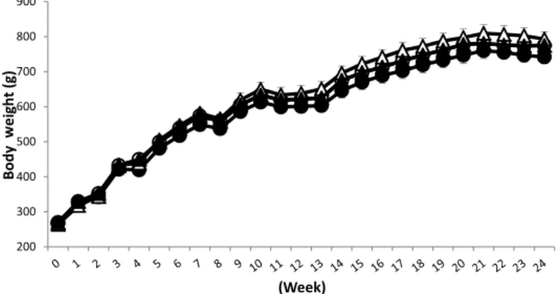
Body weight and food intake were measured at the same time during the experiment. The changes in body weight during the experimental period are shown in Figure 1. The body weight changes observed in each group did not differ significantly. However, food intake tended to be lower in the A and PH+A groups (the alcohol-administered groups) than in the C and PH+C groups (the non-alcohol groups) , and there was no difference in food intake between the A and PH+A groups (Figure 2). Also no changes in the amount of drinking were seen between the two groups (Figure 3). The mean alcohol consumption in these two groups
Figure 1 The effect of alcohol administration on body weight before and after partial hepatectomy ○, control (C) group; ●, alcohol (A) group; △, partial hepatectomy+control (PH+C) group; ▲, partial hepatectomy+alcohol (PH+A) group
Data represent mean ± S.E.
Figure 2 The effect of alcohol on food intake in sham-operated and partially hepatectomized rats ○, control (C) group; ●, alcohol (A) group; △, partial hepatectomy+control (PH+C) group; ▲, partial hepatectomy+alcohol (PH+A) group
was calculated to be 2.0 g/kg body weight per day. When the energy derived from alcohol (7.1 kcal/g) was added to the energy derived from food, it was found that the total daily energy intake of the A and PH+A groups were equivalent to those of the C and PH+C groups (Figure 4).
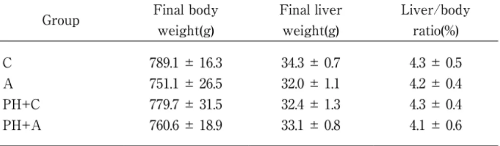
The data serum T-Chol concentrations are shown in Figure 5. While the level tended to increase by PH operation, alcohol intake reduced it. The serum T-Chol concentration in the PH + C group significantly increased at 12 weeks after pH compared with the alcohol administered group (p<0.05). The serum TG concentrations are shown in Figure 6. At 12 and 24 postoperative weeks, only the A group demonstrated significantly higher levels than the other groups (p<0.05). Alcohol increased the TG level significantly both at 12 and 24 postoperative weeks (p<0.05), while the PH operation suppressed the alcohol effect. The serum AST levels are shown in Figure 7. There was no significant difference among the four groups at 12 postoperative weeks. On the contrary, at 24 postoperative weeks, only the PH+A group exhibited significantly higher levels than the C group (p<0.05). On the other hand, the serum ALT levels showed no difference among the four groups at 12 and 24 postoperative weeks (Figure 8). Final body weight, final liver weight and liver/body ratio in the four groups were shown in Table 1. Each value showed no difference among the four groups, suggesting that the operated on livers returned to control levels after 24 weeks. 4. Discussion
Hepatocytes have a reserve capacity and a special regenerative ability. The proliferation stops when the liver volume reaches an appropriate size11). The liver’s ability to renew itself is essential for survival
following liver damage caused by toxic substances, viral infections, metabolic or immune dysfunction, or the surgical removal of parts of the liver12,13). Here, we examined the effect of frequent alcohol administration on
residual liver function in partially hepatectmized rats.
In groups A and PH+A, food consumption tended to be lower than in the other two groups. It was presumed that the resultant energy shortage was compensated by the consumption of alcohol, so no weight difference was observed between the alcohol-administered groups (the A and PH+A groups) and the non-alcohol groups. In humans, daily non-alcohol intake should not exceed 30 g, which is consistent with our finding, even if it is regarded as energy intake for weight maintenance14-16). However, if we routinely ingest a large
amount of alcohol (96 g/day), it can lead to weight gain17). In addition, the energy utilization efficiency of
alcohol varies depending on conditions, such as alcohol intake14-16), alcohol metabolising ability14-16), dietary
content17), physical condition16), etc. The liver has the ability to metabolize alcohol absorbed by the stomach
and small intestine. Alcohol is metabolized to acetaldehyde by alcohol dehydrogenase (ADH), following to harmless acetic acid by acetaldehyde dehydrogenase type 2. Finally it is converted to carbon dioxide and water, which are discharged from the body. In addition, enzymes, such as the microsomal ethanol oxidizing system (MEOS18)) and catalase, help metabolise alcohol. There are various types of enzymes in the MEOS18),
and they play an important role in alcohol metabolism. Cytochrome P450 2E1 is typical of such enzymes19).
ADH acts at low alcohol concentrations in blood, whereas the activity of the MEOS18) increases with the
blood alcohol level. The risk of liver cell damage has been reported to increase with the activity of the MEOS18) and increased alcohol tolerance20). We did not examine MEOS activity in this study, and so we
need to consider how particular MEOS18) enzymes, such as CYP2E1, influence the effects of alcohol in future
studies.
The cholesterol concentration of blood is affected by a feedback mechanism involving cholesterol synthesis21), which is mainly achieved in the liver22). Madrigal-Santillán E et al5). examined the effect of
Geranium Schiedeanum against EtOH toxicity during the hepatic regeneration process after partial hepatectomy. The concentration of serum cholesterol in the PH-EtOH group (34 mg/dL) was statistically significant compared with those obtained in the sham (57 mg/dL; p<0.05) and the PH (41 mg/dL; p<0.05) groups5). These results agreed withthe findings. Although the partial hepatectomy group (the PH+C)
showed an increase in cholesterol synthesis, alcohol intake suppressed the production (Figure 5). These results suggest that liver activity necessary for its regeneration is suppressed by alcohol, even if the same
Figure 3 Change in the amount of alcohol drinking in sham-operated and partially hepatectomized rats ●, alcohol (A) group; ▲, partial hepatectomy+alcohol (PH+A) group
Data represent mean ± S.E.
Figure 4 Food- and alcohol-derived energy intake in sham-operated and partially hepatectomized rats C, control group; A, alcohol group; PH+C, partial hepatectomy+control group; PH+A, partial hepatectomy+alcohol group
Data represent mean ± S.E.
Figure 5 Serum total cholesterol (T-Chol) levels of rats at 12 and 24 weeks after partial hepatectomy C, control group; A, alcohol group; PH+C, partial hepatectomy+control group; PH+A, partial hepatectomy+alcohol group
Figure 6 Serum triglyceride (TG) levels of rats at 12 and 24 weeks after partial hepatectomy C, control group; A, alcohol group; PH+C, partial hepatectomy+control group; PH+A, partial hepatectomy+alcohol group
Data represent mean ± S.E. values; *p<0.05
Figure 7 Serum aspartate aminotransferase (AST) levels of rats at 12 and 24 weeks after partial hepatectomy C, control group; A, alcohol group; PH+C, partial hepatectomy+control group; PH+A, partial hepatectomy+alcohol group
Data represent mean ± S.E. values; *p<0.05
Figure 8 Serum alanine aminotransferase (ALT) levels of rats at 12 and 24 weeks after partial hepatectomy C, control group; A, alcohol group; PH+C, partial hepatectomy+control group; PH+A, partial hepatectomy+alcohol group
levels of total energy are acquired. This study suggested a cholesterol synthesis inhibitory action by alcohol consumption in the residual liver after liver recovery.
In general, the serum TG level increases when alcohol is taken. NADH is produced during ethanol metabolism by ADH, which results in excessive amounts of NADH accumulation in mitochondria. This increase in the mitochondrial NADH concentration inhibits gluconeogenesis and the tricarboxylic acid cycle, whereas it promotes ketone body and fatty acid synthesis from acetyl-CoA. In addition, since NADH suppresses NAD-dependent dehydrogenase reactions, the level of glycerol triphosphate, which is another substrate for TG production, increases23). Fatty acids are β-oxidized in mitochondria, but chronic
ethanol intake downregulates carnitine palmitoyltransferase activity and reduces fatty acid uptake into mitochondria, which suppresses fatty acid β-oxidation24). In addition, fatty acid-metabolizing enzyme activity
is induced in peroxisomes via peroxisome proliferator-activated receptor (PPAR) α, which helps to regulate fatty acid concentrations. However, acetaldehyde, which is a metabolite of ethanol, suppresses PPARα expression and reduces the β-oxidation of fatty acids25). Therefore, the β-oxidation of fatty acids is inhibited
by alcohol metabolism24,25), and so dietary fatty acids cannot be used by the body. Therefore, TG accumulate,
and liver cells become fatty26). It was reported that acute treatment with ethanol in rats induced hepatic
steatosis accompanied by an increase in the production of neutral fats (TG). Thus, the serum levels of the TG may not only reflect the production of the liver, but also the equilibrium between the production and utilization of neutral fats4). The TG level in this experiment increased significantly in the A group (Figure
6). However, this increase by alcohol intake was suppressed by hepatectomy. These findings suggest that a large quantity of TG by drinking may be unable to flow out, resulting in a fat liver. Alternatively, the alcohol dose did not yet demonstrate the inhibiting effect of chylomicrons. The liver of PH+A group rats looked like fatty liver at 24 postoperative weeks (data not shown).
We determined serum AST and ALT levels as indicators of liver damage. There was no difference of ALT in the four groups. On the other hand, a significant increase of AST was observed in the PH+A group at 24 postoperative weeks. This increase was not obtained in the PH+C group, suggesting that simultaneous stress of alcohol and hepatectomy may inflict dangerous damage upon the PH+A group rats. The enzymatic rise in the serum in the remaining liver when alcohol was administered daily for 24 weeks can most likely be attributed to damage to the structural integrity of the liver cells. Ramírez-Farías et al6) reported an increase in the serum levels of the transaminases (AST and ALT) at one week after the
administration of ethanol to hepatectomized rats. This is presumed to be an alcohol toxic effect in the liver regeneration process. And our study suggested a long-term alcohol toxicity effect on either of regeneration processes.
On the other hand, compensatory liver regeneration proliferation after hepatectomy in this experiment returned to the control level, and there was no difference among the 4 groups. Both hepatic resection and alcohol influence was not observed among four the groups.
In summary, hepatic recovery was observed after hepatectomy. Cholesterol synthesis was driven by the operation, while the synthesis was suppressed by alcohol intake. Alcohol derived increase in TG
Table 1 Final weight, final liver weight and liver/body ratio in the groups
Group Final body
weight(g) Final liver weight(g) Liver/body ratio(%) C A PH+C PH+A 789.1 ± 16.3 751.1 ± 26.5 779.7 ± 31.5 760.6 ± 18.9 34.3 ± 0.7 32.0 ± 1.1 32.4 ± 1.3 33.1 ± 0.8 4.3 ± 0.5 4.2 ± 0.4 4.3 ± 0.4 4.1 ± 0.6
concentration was reduced by hepatectomy, suggesting that fat accumulation in the liver may be promoted by alcohol intake after the treatment. The risk of liver injury due to the influence of long-term drinking on the residual liver after PH was also suggested. In conclusion, the results obtained herein provide solid evidence that alcohol consumption after PH gives rise to liver damage, demonstrated by the biochemical alterations, and their prolonged consumption can cause greater, irreversible damage. Therefore, it was concluded that in alcohol administration after hepatectomy, the function in the remaining liver did not return to the original level. However, the detailed mechanism of alcohol-induced fatty liver promoting action in the residual liver by the PH rat has not been elucidated. We need to continue discussing this topic in the future.
References
1. Horie Y, Ebinuma H and Kanai T : Current status of alcoholic liver diseases in Japan. Nihon Shokakibyo
Gakkai Zasshi, 112(9), 1630-1640, 2015. (In Japanese, translated by the author of this article)
2. Horie Y, Ishii H, Yamagishi Y, Ebinuma H, Kikuchi M, Umeda R, Saito H, Kato S and Hibi T : Current status of alcoholic liver cirrhosis and factors involved in the profression. Kanzo, 50(9), 507-513, 2009. (In Japanese with English abstract)
3. Egawa H, Ueda Y, Kawagishi N, Yagi T, Kimura H and Ichida T : Significance of pretransplant abstinence on harmful alcohol relapse after liver transplantation for alcoholic cirrhosis in Japan. Hepatology Research,
44(14), 428-436, 2014.
4. Hernández-Muñoz R, Santamaría A, García-Sáinz JA, Piña E and Chagoya de Sánchez V : On the mechanism of ethanol-induced fatty liver and its reversibility by adenosine. Archives of Biochemistry and
Biophysics, 190(1), 155-162, 1978.
5. Madrigal-Santillán E, Bautista M, Gayosso-De-Lucio JA, Reyes-Rosales Y, Posadas-Mondragón A, Morales-González Á, Soriano-Ursúa MA, García-Machorro J, Madrigal-Bujaidar E, Álvarez-Morales-González I and Morales-González JA : Hepatoprotective effect of Geranium schiedeanum against ethanol toxicity during liver regeneration. World Journal of Gastroenterology, 21(25), 7718-7729, 2015.
6. Ramírez-Farías C, Madrigal-Santillán E, Gutiérrez-Salinas J, Rodríguez-Sánchez N, Martínez-Cruz M, Valle-Jones I, Gramlich-Martínez I, Hernández-Ceruelos A and Morales-Gonzaléz JA : Protective effect of some vitamins against the toxic action of ethanol on liver regeneration induced by partial hepatectomy in rats. World Journal of Gastroenterology, 14, 899-907, 2008.
7. Higgins GM and Anderson RM : Experimental pathology of liver: Restoration of liver of white rat following partial surgical removal. Archives of Pathology, 12, 186-202, 1931.
8. Ozaki M : The roles of Jak/STAT3 and PI3-K/Akt pathways in liver injury and regeneration. Biochemistry,
80(5), 399-408, 2008. (In Japanese, translated by the author of this article)
9. Ministry of Education, Culture, Sports, Science and Technology Council Resource Survey Subcommittee :
Standard tables of food composition in Japan 2010. National Printing Bureau, Tokyo, 2010. (In Japanese,
translated by the author of this article)
10. Atwater WO and Benedict FG : An experimental inquiry regarding the nutritive value of alcohol. Obesity
Research, 1(3), 228-244, 1993.
11. Fausto N : Liver regeneration. Journal of Hepatology, 32(1 Suppl), 19-31, 2000.
12. Bucher NL and Swaffield MN : The rate of incorporation of labelled thymidine into thedeoxyribonucleic acid of regenerating rat liver in relation to the amount of liverexcised. Cancer Research, 24, 1611-1625, 1964.
13. Michalopoulos GK and DeFrances MC : Liver regeneration. Science 276, 60-66, 1997.
14. Rumpler WV, Rhodes DG and Baer DJ : Energy value of moderate alcohol consumption by humans.
American Journal of Clinical Nutrition, 64, 108-114, 1996.
15. Cordain L, Bryan ED and Melby CL : Influence of moderate daily wine consumption on body weight regulation and metabolism in healthy free-living males. Journal of American College of Nutrition, 16(2),
134-139, 1997.
16. Clevidence BA, Taylar PR and Compbell WS : Lean and heavy women may not use energy from alcohol with equal efficiency. The Journal of Nutrition, 125(10), 2536-2540, 1955.
17. Suter PM, Schutz Y and Jequier E : The effect of ethanol on fat storage in healthy subjects. The New
England Journal of Medicine, 326(15), 983-987, 1992.
18. Lieber CS and DeCarli LM : Hepatic microsomal ethanol-oxidizing system: In vitro characteristics and adaptive properties in vivo. The Journal of Biological Chemistry, 245, 2505-2512, 1970.
19. Lasker JM, Rancy J and Kubota S : Purification and characterization of human liver cytochrome P-450-ALC. Biochemical and Biophysical Research Communications, 148, 232-238, 1987.
20. Badger TM, Huang J, Ronis M and Lumpkin CK : Induction of cytochrome P450 2E1 during chronic ethanol exposure occurs via transcription of the CYP2E1 gene when blood alcohol concentrations are high. Biochemical and Biophysical Research Communications, 190(3), 780-785, 1993.
21. Di Buono M, Jones PJ, Beaumier L and Wykes LJ : Comparison of deuterium incorporation and mass isotopomer distribution analysis for measurement of human cholesterol biosynthesis. Journal of Lipid
Research, 41(9), 1516-1523, 2000.
22. McNamara DJ, Kolb R, Parker TS, Batwin H, Samuel P, Brown CD and Ahrens EH Jr : Heterogeneity of cholesterol homeostasis in man: Response to changes in dietary fat quality and cholesterol quantity. The
Journal of Clinical Investigation, 79(6), 1729-1739, 1987.
23. Pownall HJ : Dietary ethanol is associated with reduced lipolysis of intestinally derived lipoproteins.
Journal of Lipid Research, 35(12), 2105-2113, 1994.
24. Tijburg LB, Maquedano A, Bijleveld C, Guzman M and Geelen MJ : Effects of ethanol feeding on hepatic lipid synthesis. Archives of Biochemistry and Biophysics, 267(2), 568-579, 1988.
25. Crabb DW, Galli A, Fischer M and You M : Molecular mechanisms of alcoholic fatty liver: Role of peroxisome proliferator-activated receptor alpha. Alcohol, 34(1), 35-38, 2004.
26. You M, Matsumoto M, Pacold CM, Cho WK and Crabb DW : The role of AMP-activated protein kinase in the action of ethanol in the liver. Gastroenterology, 127(6), 1798-1808, 2004.