A physiological study of brain activity
-a
report on the special・class children's EEG
and key reaction time
Ikuko OiKAWA
I. Introduction
In the education of the mentally retarded, teachers and parents have to know their
children's physical and mental ability or developmental levels as compared with normal children of the same age, in order to bring up the children's potential ability. EEG is an excellent way to provide information roughly about the developmental level of the・ brain or to predict brain damage by a subclinical seizure discharge. Comparative EEG characteristics of
problem juveniles", mentally retarded2'3.4'5'6'8)orother kinds ef exceptional childrenlo'11'12) have been studied for a long time. As a result some kind of anticonvulsant treatment has been recommended especially for the epileptic, which appears often in these exceptional children. The problem of exceptional children is now a question of social welfare which has
to be solved。
Almost every pupil studying in a special-class has his own etiology, combined with each handicap. Proper guidance may be provided for each pupil through an association of parents or teachers who make careful daily observations plus clinical diagnosis confirmed by EEG. ぺA^iththis point of view, pupils' EEGs in the special-class were studied as compared with those in the normal class in collaboration with their‘teachers. It showed a higher proportion of EEG abnormality than that of the control group, as from the serious patient, who has a severe EEG abnormality and needs integral medical treatment, to one with subclinical seizure discharges. It is necessary to take the EEG into account in order to prevent further
progress of mental retardation or to understand the pupils' condition. This is a report of the actual circumstances about the pupils' EEG in the speciaトclasses in Kochi. At the same time their key reaction times as induced by photic stimulation were studied as a means to provide information about their brain activity. The present report includes a quantitative data concerned with it.
11. Experimental subjects and methods
The present experimental subjects were composed of school children in the special-classes of A and B primary schools : fourteen children from 7 t0 12 years of age from group A, nineteen children from g t0 13 years of age from group B respectively. The A primary school is located in the poorest district in Kochi city. In its eχperimental subiectSj 5 were children of families helped by the Livelihood Protection Law. This group included 4 children with the I.Q. of 60-70, 10 with the I.Q. of 70-90. Ten out of fourteen (71%)had some
etiologies of brain defect in their infancies or positive hereditary factor. The rest had no etiology of brain damage but had common environment of noninterference, domestic trouble and poverty. 0 n the other hand the B primary school is a representative of enro川ng pupils
78 高知大学学術研究報告 第15巻 自然科学 n 第11号
一一一一一一一一-of good homes. Experimental subjects in B included 7 children with the I.Q. of 50-70, 8 children with the I.Q. of 70−90 and 4 unknown. There were 15 out of 19 (79%)who had some etiology of brain damage or positive hereditary factor. The remainders were composed of those grown up in the uninterference families or orphans who had no information about their infancies. ’
Control subjects were 55 school children at about the same ages, from 8 t0 10 years old. Nine (16%)of them were found to have some etiology of brain defects (Table 1).
A nine channel EEG machine was・used in the present eχperiment with time constant 0. 3 sec. Monopolar recording referred to the ears ・was used with Gibbs' way of electrode distribution : on the frontal, parietal, occipital and tempora卜areas symmetrically with both cerebral hemispheres。
The recording of EEG were performed in a quiet state, awake with eyes closed. Hyperven-tilation for three minutes and photic stimulation were used as methods for activation of EEG abnormality. Gibbs' classification of EEG was referred to define its normality. Positive effects of hyperventilation were those build-ups during or after hyperventilation and those developed bursts of high voltage B waves in the frontal area which appeared only after activation. In photic stimulation, EEG abnormality during or after flicker stimulation, which correspond to Gibbs' photoactivation and photoconvulsive, were observed.
Flicker apparatus) PS-101 type, Sanei Co. was used for photic stimulation in which frequencies・ 5 − 16 Flickers/sec. were applied. For the measurement of key reaction time induced by single photic stimulation, the same methods were applied as those used in the former report.9)
Table 1. Past histories of special-class children at the A and B primary school with those of the controL common primary school children.
Medical history Past history Encephalitis Cerebral palsy Difficultbirth Febrile disease . Jaundice Bruise
Positive hereditary factor
Positive hereditary factor 十Fever or Bruise
Psychological history
Noninterference, Domestic trouble
A 1 2 2 2 1 2 4 B 2 1 6 1 1 2 り 乙 1 Control 1 1 / 0 1
Negative medical historywith brain defector unknown
3 46Total
14 19 55Rate of those with positive medical history to the whole 10/14・(71%) =15/19(79%)
9/55
(16%)
Febrile disease : autointoxicatioriiacute pneumoniaj scarletfever, etc. in the infancy. Difficultbirth : perinatal injury, premature birth etc.
Bruise : fall from the upstair, trafficaccident etc.
A ical study of brain activity.(I. Oikawa) 79
III. Experimental results :
(a) EEGs of the control
In 55controls, there ・were 48 (87 %) normal and 7(13%)・abnormal EEGs (Table 2). EEG abnormalities were as follows : CD moderately slow Q6 c/s) for age (9 years old) (Fig. 1(1)) (2) slightly abnormal fast (F-1)ぐFig. 1(2)) (3) diffuse paroxysmal slow (Fig. l(3)(a), Fig. l(4)(a)). Six of 48 normal control EEGs were different frと)mothers in the following points : (1) Appearance of many intermediate fast waves (15-20 c/s) in the frontal area. (2)Asymmetry(20−30%)in the amplitude of the occipital basic wave. (3) Sporadic high voltage slow waves associated with slight asymmetry in the occipital basic waves. (4) Dominant fast waves (30 c/s) and bursts of high voltage 0 waves in the frontal area (Fig. 1(5))。
As will be discussed later, these EEG abno耳nalitiesin the control express a fact that EEG abnormality in normal children is not always correlative with an organic or functional
brain defect, especially for children. Diffuse paroxysmal slow waves, which appeared in the normal children, indicates their nature of subclinical epilepsy which sometimes subsides
without producing symptome or becomes clinically eχpressed at a・later date. After all, in 87 % the common primary school children had normal EEG。
In control children there were about 21 %of activation by hyperventilation in those
who showed normal EEG, including slight, medium, big build・ups or bursts of d waves
L. F. R.F. L. p. R 。 0 , L.T. R.T.
二
Fig. 1(1) Normal boy> age g years・awake. Moderately slow waves (6 c/Sj)in all areas. Vertical
and horizontal lines in the right bottom, Fig. 1-3 mean 50μv and l sec. respectively.
Abbrevi- ations on the left in each Fig. indicate recording parts on the skull : the frontal (F. ), parietal
(P.)。occipital Co.) and temporal (T. ) areas of the left (L. ) or right (R. ) cerebral hemisphere
80 高知大学学術研究報告 第15巻 自然科学 n 第11号 L. F. L. p. R. P. ・ ’ L. T. R. T.
_・ |
Fig. 1(2) Normal girl, age g years-awake. Slightly abnormal fast waves (F−1)
A physiological of brain activity. (I. Oikawa)
EKG
|
81、
IFlicker ' 1 1 1 1 1
Fig. l(3)(b) Same subject as in Fig. 1(3)(a). Photoactivation at the end of 14 c/s flicker stimulation
for 10 sec.
L.0・
R.O. み
R.T
|
82 高知大学学術研究報告 第15巻 自然科学 n 第11号 -R. Pi R。0. 、 L.T、
こ
Fig. l(4)(b) Same subject as in Fig. l(4)(a). Build-up in the end of hyperventilation for 3 rain.
L. p. R. p. L 。 0 , R.O. __, │’ R.T. ●
Fig. l(r,) Normal boy, age 8 years-awal<e. Fast waves (30 c/s) and high voltag, bursts of. e
A physiologica】 of brain activity. (I. Oikawa) 85
・Table 2 : EEGs of control children :(17 of 8 years> 25 of g years and 13 of 10 years of age)
EEG
finding
Age . constituent N0.lj?i§7venti-Flicker.
stimulation
8 yr. 9 yr. 10 yr- SM
∂ B SM
び B ● = - 4 R Z NormalIntermediate fast waves (15-20 c/s) in the frontal.
ASymmetry(20−30%)in the amplitude of occioital basic waves.
Sporadic high voltage slow waves in the occipitalwith amplitude asymmetry. Fast waves (30 c/s) with bursts of θin the frontal. \ 12 2 18 2 1 12 1 42 1 2 1 2 5 1 1 1 2 2 5 3 1 1
Total
48 (87%)Positive
rate: 10/48
(21%)
Positive
rate: 12/48
(25%)
7 Moderately slow waves (6 c/s basic E wave of g years of age)
J l
Slightly abnormal fast waves (F−1) べ Diffuse paroxysmal slow waves 3
1 1 2 1 1 5 1 1 2 1
Total ・ 7(13%)
Positive
rate: 4/7
rate: 1/7
Positive
Total of normal and abnorma】EEGs.・
17 25 . 13 55 7 2 1 4 2 5 4 2Total rate:
Total rate:
Of 55 control children! 7 showed EEG abnormalities, 6 included in these with normal EEC had background activitiessuggestive mentally retarded type. Effects of hyperventilation and flicker stimulation included small-(S), medium-(M)> bigCB)-builcl-ups and bursts of 0 waves (S). Same abbreviations were used in Table 3A and 3B.
in the frontal area. Its rate in total control was
25 %
(Table
2,Fig. l(4)b).
EEG
abnormality
activated by photic stimulation was
about 25 %
including from slight
to big activations, photoconvulsive・ and
bursts of d waves in
the frontal 尽rea which
appeared only after stimulation (Fig. l(3>b, Table
2).
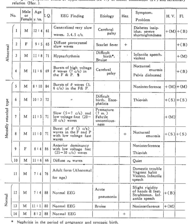
(b) Special-class children's EEGs in the A primary schooけTable 3A) 一 Four out of fourteen at A showed abnormal EEGs : (1) generalized very slow waves (No. 1. Table 3A) (2) diffuse paroxysmal slow waves (No. 2, Table 3A) (3) hypsarhythmia (No. 3, Table 3A) (4) bursts of high voltage d waves in the frontal and parietal・ areas (No. 4, Table 3A) (Fig. 2ci)∼(4)). Five showed EEGs of mentally retarded type as follows ;(1) bursts of d (5, 6 c/s) waves in the frontal and parietal areas (No. 5, 6, Table 3A)(2) mixture of slow (5, 6 c/s)andlow voltage fast(25−30 c/s) waves (No. 7, 8, Table 3A, Fig. 2(8)) (3) anterior dominancy with low voltage fast waves (25−30 c/s) (No. 9, Table
3A). (4) diffuse a waves i.e. ocφpital dominant very regular high voltage αlwave (N0. 10, Table 3A,Fig. 2(10)). These were already assumed as EEG characteristics of mentally
84 高知大学学術研究報告 第15巻 自然科学 n 第11号
Table 3A: EEG findings of special-class children in the A primary school accompanied with etioloeiesjsyraptomsj effectsof hypervendlation (H. V.) or flicker stimulation (Fl. ) and hereditary relation (Her. ). N0. Male O「Female Age y.:m.
I.Q.
EEG
Finding
Etiology
Her.
Symptom!
Problem
H. V. Fl. −a Q 呂 笥 ど 1M
12:4 61 Generalized very slow waves, 3,4,5 c/s. Cerebral palsy Diabetes insip・ idus, severe shortsightedness 十(M) 十(B) 2 F 9:5 65 Diffuse paroχvsmalslow waves ゛ Scarlet fever + 十(B)
3
M
11:8 71Hypsarhythmia
・birth*,‘Difficult Bruise Infantile speech, violent 十(M) 4 1M
11:6 69Bursts of high voltage 0 waves f7 c/s) in the F & p.§ Cerebral palsy Nocturnal enuresis Pelvis dislocated 十(B)
ト
M
8:10 84 Bursts of 0 waves (5,6 c/s) in the F& P. Noninterference 十(M) 十(M)
} 1 吻 4 - J Q Q = { J 6
M
10:3 72 // Difficult birth, Ence-phalitisThievish
十(S) 十(S) 7M
11:5 71 Slow(5−7 c/s) and low voltage fast(20−30 c/s) waves Premature、 (7 m.) Febri】e unconsious-ness ・ 十(M) 8
M
11:0 71 Burst of θ(5 c/s) waves in the F and P with low voltage fast waves十 Nocturnal
enures IS 十(S) 十(S)
9 F 8:4 86 Anterior dominancy
with low voltage fast (25−30 c/s) waves
Noninterference
Th ievish
いo
M
11:6 66Diffuseびjwaves
Quiet11
M
7:4 76Adult form (Abnormal
for age)
≒塵cご1)1e
昌rrt,lnfantile
−∃ Q 2 /12
M
7:4 88Normal EEG
Acute
pneumonia
Slight rigidity of hands & feet,
Strabismus, Inf-antile speech 十(B)
一
一
13 ' M 11:1 80Normal EEG
Bruise
Noninterference 十(M)い4
M
8:2 88Normal EEG
+* Nephritis in the period of pregnancy and syncopic birth.
§F, P or O:Abbreviations of the frontaU parietal and occipitalareas in Table 3A and 3B.
retarded.*''' Moreover, No.
11, Tab】e 3A was a prob!em
boy of seven years old and was
included in the abnormal
because of its adu!tform (Fig. 2(u)).
The
rate of abnormal
and mentally retarded type EEGs
to the who】eEEGs
in A
was
78%(11
out of 14), about 6 times that of control. Of those with
abnormal
and mentally
retarded type EEGs
6 had
etiologies of some
kinds of brain deficiencies;2 had positive
A
L.T.
j
85
Fig. 2(1) Special・class boy. age 12years-awake. Generalized very s】ow waves. Numbers in parentheses
attached to Fig. 2 and 3 indicate individuals of those in Table 3A and 3B respectively.
L.O. u v
I..T.
R.T.
二
86 高知大学学術研究報告 第15巻 自然科学 H゛第11号
L.O.
L.T.
R 。 ‘ T . -」
Fig. 2(3) Special-class boyj age 11 years. Hpsarhythmia.
R 。 0 .
一 ニ
→
Fig. 2(4) Special-class boy・age 11 years. High voltage θ・waves (7 c/s) in the frontal and parietalareas.
A study of in activity. (I. OiK人wA) R。0・ L.T. j l 丿 I R.T. 々 ` 87
Fig. 2(8) Special-class boy, age 11 years・awake. Burst of e waves (5 c/s) in the frontal and parietal areas with low voltage fast waves (25 c/s) in the frontal area.
R.F, .. .
L.P,
R.P.
w/Wyvi-v- ●●
___ ___1
88 高知大学学術研究翰悟.j第i,5巻 自然科学 立 第11号 L . p . ダ ・ \ , R , P 。 , ゝ . ● ・ I S ゛ . . ● y L ● , 0 ● , ● , / ‘ ; I . ヽ ● 〆 ● I 「 ・ p R . O . ’ ● 心 ・ 岫 『 L . T . ゛ , _ご
Fig. 2(m Special-class boy> age 7 years-awake. Adult form EEC unusual for his age.
hereditary factor and 4 had no etiology of brain damage with negative hereditary factor. Positive activation effects were induced in 50 %by hyperventilation and in 50 %by photic stimulation which showed an easiness of its activation rather than the control.
(c) Special・class children's EEGs in the B primary school (Table 3B)
Abnonnal EEGs were showed in 3 out of 19 children at B : (1) petit mal (spike and wave) type (No. 1, Table 3B) (2) asymmetry (No. 2, Table 3B) (3) diffuse paroxysmal slow waves (No. 3, Table 3B) (Fig. 3(1)∼(3)).
Furthermore, 9 expressed mentally retarded type or border line・ EEGs. Their characteristics were as follows:(I) anterior dominancy with bursts 'of d waves (6, 7 c/s) (No. 4, 5, 6, Table 3B Fig. 3(4)) (2) bursts of 0 waves in all areas (No. 7, Table 3B) (3) mixture of slow (6 c/s) and intermediate fast (18-22 c/.s) waves (No。8, Table 3B,Fig. 3(8))(4) bursts of intermediate fast waves (18−20 c/s) in the frontal area (No. 9, Table 3B,Fig.
3(9)) (5) slightly abnormal fast (F-1) (Noバ0, Table 3B> Fig. 3(10)) (6) diffuse a waves ゛l
[No. 11, 12, Tab]e 3B)。 犬 The seven remainders were normal EEGs. ・
The rate of abnormal, mentally retarded or border line type EEGs to the whole EEGs was 63 % (12 t0 19), about 5 times that of control. In those with abnormal, mentally retarded or border line type EEGs, 10 of 12 had 雨olびgies of some kinds of brain damage, 3 had positive hereditary factor. Thus there were large numbers of EEG abnormalities
Table 3B
A ical study of brain、 activity.(I. Oikawa) 89・
EEG finding of special-elass children・in the B primary school with etiologies,
effects of hyperventilation (H. V. ) or flicker stimulation (Fl. ) and hereditary
symptoms, effects of hyperventilation relation (Her. ). N0. Male O「Female Age y.:m.
I.Q.
EEC
Finding
'EtiologyHer.
Symptom,Problem H. V. Fl.1 § i
/
1 F 9:11 85 Petit mal (spike and
wave) type Encephalitis
Nocturnal . enuresis. Speech disorder 2 F・ 10:2 50
かに雪言路首
autointoxi-cationI大穴庁hlf)
? -十(B〉レ
M
12:6 89Diffuse paroxysmal
slow waves
Bruise
+Itch
十(M)に
M
10:2 ‘65 ‘Frontal domihancy, Burst of ∂wa、.'es C6, l c/s)in the F &P Febrile disease + Seizure! Speeeh disorder 十(M)十(M〉 モ | M I 泡 5M
11 : 11 70 Frontal dominancy> Bursts of d waves (7c/s)in the F &P
Jaundice
Head-ache, impulsive 十(M) 十(M) 6
M
11:7 74 77 ノ♂ 十(S) 十(S) 7M
13:8 Bursts o{ 0 waves(6, 7 c/s) in all areas autoln・● ●toxication Speech disorder 十(M)
8 F 13:1 50
Mixture of slow (6 c/s) and intermediate fast waves Encephalitis ?
?
9 F 9:9 78 Bursts of intermediate fast waves (18−20c/s) in the Fpneumonia
Clumsiness, hyperactive ? ? 10 F 12:5 68認雲1y≒-16t
+ Introversive 11 F 12:7 65 Diffuseαwaves with bursts of 6 waves (6 c/s) in the F Di伍cult. birth 十(M)い2
M
11:2 53 Di仔useαwaves colitis 十(∂)レ
F 10:7 70Normal EEG
pneumonia
Inferior physi-cal strength, stammer 十(M) − E 私 ・ 2 14M
13:2 65Normal EEG(manyl8
c/s in the F &P)
Autointoχi-cation Chronic hives, stammer15
M
12:9 76Normal EEG
Bruise
Head-ache.
restless
16 F 11:9 ノノ 〆7
Nocturnal
enure-sis wear glasses
after Neuritis
optics
十(M) 17 F 12:5 ♂Z 7♂昌こげ゜白eづ
18 F 10 : 10 // X/ Tonsilar hyper・ trophy 十(B)い
F 12:6 // // 十90 高知大学学術研究報告 第15巻 自然科学 n 第11号 R.P. ● 。’∧、 L,0, 1 ‘:./ / j ν.. ・f ・ 祠)ノ y / ’ト ・‥., ゴ L.T. R.T.
Fig. 3(1) Spec祐1一山ss girl・ age g years・awake.∧Petit mal (spike-wave) type.
L.F. j 。 L, p. R. p. R。0. , L.T.
二
R.T.Fig. 3(2) Special-class girl, age 10 years-awake. Asymmetry【iso-e】ectric in the !eft
A physiological study of brain acti (LO限八w人) 91
R.T-. ノ
Fig. 3!3) Special-c】ass boy・ age 12 years-awake. Diffuse paroxysmal s】ow waves in
a quiet condition.
L.T.
二
92 高知大学学術研究報告 第15巻 自・然科学 n 第,11号
R。0, ’
・lこ
Fig. 3(8) Special-class girl, age 13 years-awake. Mixture of slow and intermediate fast waves.
L. T.
__‥ j
R.T. ●’
Fig. 3(9) Special-class girl, age g years-awake. Intermediate "fast waves (18-22 c/s) remarkable
A physiological study of brain activity.(I. Oikawa)
L.P.
9ろ
L. T.
R.T
Fig.・ 3(10) Special-c】ass girl, age 12 years・awake. Slightly abnormal fast waves (F−1).
caused by secondary brain obstacles in their infancies. effects by hyperventilation or photic stimulation. They undecided ones interfered by EMG.
There were 7 positive activation
corresponded to ii %
exclusive of
(d) Brain activities of special-class chi】dren ― information obtained by key reaction time。 There was almost paral】el relationship between EEG abnormality and l. Q. inferiority in special・class children. Then whether there is any difference in the activity of neuronal tract inducing motor reaction between the special・c】assand the common primary school children was studied quantitatively by measuring key reaction time. By applying a method
described in previous report・ key reaction times induced by single flicker or head light were obtained with eyes closed or open (Table 4 (a), 4 (b), 5)。
As controls; 38 of 8,9 and 10 year old children were used. As indicated in Table 4 (a). 4 (b) there was a difference of speed, to some extent, according to their ages when a 5 % level 0f significance was used. With their eyes open children of 10 years 01dcould respond faster to the head light than those of g years old. Also children of 10 years old were quicker to respond to single flicker than those of 8 years 01d with their eyes closed. In the key reaction time there was a tendency of the older child to respond faster than the younger
child。
Ho■weverア inthe present experiment, one group of 8,9 and`10 year old children was used to compare with the special-class children. Even though the latter was composed of
94 高知大学学術研究報告.第15巻 自然科学 n 第U号 ”’  ̄ ̄ ̄ ̄ ̄ ̄7 ’ レ
older children than the control, as shown in Table 5,t・hey were slower to respond to the head light than the control. They could respond to the supramaximal stimulation of flicker as quickly as the control, but were slow, 1.3-1.5 times the control, to respond to mild stimulation of head light.
Table 4(a): Mean values of key reaction time (msec. ) in controls! 8,9 and 10 years of age.
Age
Y.
n.
Eyes Open
Eyes Closed
Flicker
Head Light、
Flicker
Head Light
10 9 8 8−10 8 19 11 38 298土163 279土 69 323土 64 296士 94 476士 93 603士126 625±205 582士155 ・ . 229士 71 302士127 354士 99 301土116 579士156 646士180 719士307 653・士223
Table 4(b): t for testing difference of the mean value of key reaction time with ages in the control.
Comparison
Level of
significance
5%
Eyes Open ・
Eyes Closed
Flicker
Hed Light
Flicker
Head
Light
8y': 9y' 8:10 9:10 士2.048 土2.110 士2.060 1,328 0.463 - 0.434 0.364 1.886 2.562一一 1. 160 3.005 -1.524 0.855 1. 167 0.918
With a level of significance; 5 % there was a significantdifferenとein those of g and 10 years of age in which the key reaction was induced by head light with their eyes open. Also, a significant difference in 8 and 10years of age was observed when the kとy reaction was induced by a flicker with their eyes closed.
Table 5 : Key reaction time (msec. ) of special-classchildren in A and B primary school with those of controls. A> B and control were composed of number of children, 8-13 years of age indicated. When experimental values were compared with controls using level of significance, 5%(±1.95996), those induced by head light with both eyes open or closed had significant differences judging from their t values.
Control
A
B
301士116 324士130 371土252 t - 0.611 - 1.483 653土223 850士165 t −3.002 - 4.66 296士94 387土363 n ゛317士149 t - 1.435 - 0. 666 582士155 789±191 t - 3.996 - 1.972 963土2411) 737土453 1 ; l1) Total number was 17 without one, 10 year old.
No. in each age
Total
No.
8 9 10 11・ 12 13Control
A
B
11 3 19 1 3 8 3 4 6 ・4 1 5 2 38 14 18A physiological study of brain activity.(I. O限awa) 95
IV. Discussion
1)On the normal and abnormal EEGs
It is a question how many abnormal EEGs would be found in the control primary school children as compared with those in special-class children. In the present study, 7 of 55 controls had the following kinds of EEG abnormalities : (1) moderately slow (2) slightly abnormal fast (F-1) (3) diffuse paroxysmal slow. Moderately slow activity is a minimal form of EEG abnormality, usually reversible and creating no presumption of structural damage. Also slightly abnormal fast waves do not correlate highly with any symptomatology. Number of persons with moderately slow or slightly abnormal fast waves was both l in 55 control subjects (1. 8 96), which was a low proportion. Moreover, diffuse paroxysmal slow activity is rarely an exclusive abnormality in childhood. It is not highly epileptic unless it is associated with epileptic or epileptiform symptoms. The rate of appearance in controls was 9.196, which was rather high contrasted with about 4.2 % for ages up to 9 years old2).Long・term follow-up studies accompanied serial EEGs of children, who showed diffuse paroxysmal slow activity, may be able to predict a seizure and to avoid a production
of symptoms provided that suitable medical) nutritional ・or environmental precautions were taken。
In controls, the remaining 48showed normal EEGs in which 6 had background activities
suggestive of mentally retarded type. On the other hand, 10 out of 33 special-class children had normal EEGs (Table 6). Generallvj normal EEGs are a presumptive but not positive evidence of normality. In undifferentiated mental retardation, Gibbs found normal EEGs about 50 96 for 5−9 year 01d children and 39% for 10 − 19 year old children. In the present study, the number of normal EEGs in undifferentiated type of special-class children was 5 out of 12 (42 %') (Table 6)。
Anyhow, normal EEG would not guarantee・ normal brain function, for EEG does not indicate all types of brain function ゛nor does it view all parts of the brain. However, it suggests that there is no acute or progressive damage in the accessible cortex, no highly active epileptic process and no disturbance of those types of neuronal metabolism that are
concerned in electrogenesis。
2)On the hyperventilation and photic stimulation
A big build・up activated by hyperventilation is not definitely abnormal at any age. In general, the younger the child, the greater will be the build-up. ThuSi big build-up was observed in 50 %of 6-10 year old normal controls and in 87 96 of epileptics of the same ages1). Below 10years of age. therefore, it is normal. It is not very significant by itself and is classified as slightly abnormal in adults. It may reinforce a judgment of abnormality which is based on other features of EEC In the present study. 25 % of control showed a sign of build・up as compared with 43 −44 %of those in the children of special-class. The build・up is considered to be a result of failure in homeostatic mechanism, which ordinari】y 'abiusts the arterial carbon dioχide tension in the brain. Present results suggest that the
special-class children's competence of the homeostatic mechanisms was feeble and sensitive
to acapnia in the cerebral neurons。
% 高知大学学術研究轍告 第15巻 自然科学 H 第11号
-activity or slow waves occur, has not been clear】y established in its diagnostic value. The
photoconvulsive, in which seizure discharges appear even after discontinuation of photic stimulation, commonly associates with clinical manifestat・ons of an epileptic character. It is also able, however, to induce in a small percentage of normal persons. They, therefore,
were concerned as a reference data attached to children's EEC In the present result, the・ children in the special-class showed 1.7 times positive effects of control with photic Sti-mulation. ,. ●
3) EEG abnormality in the special-class children
EEG abnormality observed in the special-class children ’w卵 classified into two groups : (a) EEGs accompanied by epileptic or other disease and (b) mentally retarded type or
border line EEGs. They included EEGs with the following characteristics :
(a) 1. Petit mal type 2. Asymmetry 3. Diffuse paroxysmal slow wave 4. Generalized slow wave* 5. Hypsarhythmia.
(b) 1. Bursts of 0 waves (in all areasi in the frontal and parietal areas with or without frontal dominancy).
2. Miχture of slow, and low voltage fast(25−30 c/s) waves. 3. Mixture of slow and intermediate fast (18-22 c/s) waves.
4. Intermediate fast (18−20 c/s) waves in the frontal area. ,
5. Frontal dominancy with low voltage fast (25-30 c/s) waves in all areas. 6. Slightly abnormal fast (F-1) wave. , 7. Diffuse a waves.
It is already well known that, hypsarhythmia, epileptic EEC especially appears often in the children with mental retardation and others in (a)ヽalso tend to accompany mental
retardation. Medical treatment may be given to the children with epileptic EEGs, petit mal, diffuse paroxysmal slow activity and hypsarhythmia. A child with EEG asymmetry caused after cerebral palsy should be rehabilitated to prevent further invasion of paralysis.
Kasamatsu6)and Izawa et a13)have reported characteristics of EEG in the mentally retarded as follows : (1) asymmetry (2) intermediate fast waves (18−22 c/s) (3) anterior
dominancy (4) bursts of high voltage 0 waves (5) mixture o・ffast and irregular slow waves. After analytical study Katada also reported that dominant components of EEG in the mentally retarded exist in the lower frequency range than that in the nonnal.“The lower range component becomes n!ore dominant on from the occipital to the frontal region”8).EEG characteristics written in (b) roughly coincided with these observations. 0n the other hand, diffuse a wave has been observed often in the mentally retarded. In the present study this pattern was found in the children with l. Q. inferiorities associated with quiet temper in 11 − 12 years of age> i.e. in rather grown-up stage electroencepharographically.
After an・ children with EEGs of mentally retarded type may be directed to develop their own potentialities m order to join m social life.
* A child (No. 1, Table 3A) suffered from Diabetes insipidus, which suggests a EEG disorder caused by a infectious disease of hypothalamus. |
A 97
■4) ()n the brain activity of special-clss children
Parallel with EEG recording, key reaction time induced by photic stimulation was measured as a study of their physiological activity. This is a time from a moment of illumination untilla response of pushing a key made after a sensation of light is received by impulses passing through the occipital visual cortex. This is a simple reaction, rather . close to a reflex phenomenon, which connects two conditions “feel the light”then “push the key"。transmitted through brain activity. There were no differences observed in the key reaction times induced by supramaximal stimulation between experimental subjects and controls. This means both have equal activities to respond reflexively. Delays in key reaction for a weak photic stimulation observed in the experimental subjects suggests a delay in central nervous activity which would affect a skill 0f physical exercise.
5)On the etiology of EEG abnormality
In the medical histories of special-class children, there were several etiologies which might cause cerebral obstacles or EEG abnormalities : difficult birth. prematurity, nephritis during the period of pregnancy, cerebral palsy> encephalitis, febrile disease (scarlet fever, pneumonia) etc. in A, and difficult birth, encephalitis! fever caused by autointoxication,
pneumonia, jaundice, colitis, bruise etc. in B. These facts suggest that the disturbances around birth, cerebral disease and infant's disease with fever tend to accompany mental retardation secondarily.
Table 6 : EEGs and their medical histories.
Groups with DOS. med. history
`ぶミiゴヘ、へEtiology
Finding `犬
Perinatal disturbances
Braini'
disease Febrile2)disease
Jaundice Bruise
Febrile disease or Bruise 十 Pos. Her. ≪ Total
Abnormal
1 3 1 2 7 Mentally retarded orborder line type 2 2 2 1 2 9
Normal
3 2 5Total 3 3 6 1 2 4 21
Groups with undifferentiated med. history
犬
Finding 犬
3) Pos. Her.≧ぷヤ
Noninterference
Total
Abnormal
次次ご詰(│゜「
2 5 7Normal
2 3 5 Total 4 8 12The rate of abnormal, retarded or borderline total EEGs, mentally EEGs to 23/33 : 70 % 1 ) 2 ) 3 )
Brain disease : Encephalitis and cerebral palsy.
Febrile disease : Pneumonia, autointoχication>scarlet feverj colitis etc. Pos. Her.:Positive hereditary factor.
98 高知大学学術研究報告 第15巻 `自然科学 11 第11号
Table 6 indicates a summary of EEGs and their etio】ogies. Experimental subjects with positive medical history were 21 0f 33 in total (64%).The remainders were composed of those with negative medical history of cerebral obstacles, unfavorable environment and positive hereditary factor. Those with negative medical history accompanied with EEGs of
mentally retarded or border line type had commonly poor, noninterference homes. This might mean normal cerebral development occurs in those with certain nutrition and careful environment. Those with positive hereditary factor had also poor, noninterference homes in which effects of hereditary and environmental factor could not be discriminated with each
other. In conclusion, most of EEG abnormalities observed in the special-class children occurred secondarily from their‘medical history during the embryo or the infant. However, lack of suitable precautions, educational 0r developmental. might be another important
factor of EEG abnormality. .
V. Summary
Thirty-three EEGs of special-class children (7 to 13 years of age) at A and B primary Schoo】sin Kochi city were compared with 55 0f those of common primary school children
(8 to 10 years of age). The following facts were observed :
1. Abnormal, mentally retarded or border line EEGs in the‘special・class children occupied 70 % of all, 5 −6 times that of control. Percentage of eχperimental subjects with positive medical history of cerebral obstacles within them corresponded t0 70 96. The rest was com-posed of those with positive hereditary factor, unfavorable environment or negative medical ●history. in which a lack of educational or developmental precautions was a common factor. and which suggested its effect upon the cerebral development.
2. Their main etiologies were encephalitis; cerebral palsy, febri】e disease, obstacles around birth and bruise.
3. EEG abnormalities included petit mal type; asymmetry, generalized slow waves, diffuse paroχysmal slow waves and hypsarhythmia. EEGs of mentally retarded or border line type were observed as follows:
(1) Bursts of 0 waves (in all areas, in the frontal and parietal areas with or without anterior dominancy). に
(2) Miχture of slow and low voltage fast waves. (3) Miχture of slow and intermediate fast waves. (4) Intermediate fast waves in the frontal area.
(5) Mixture or low voltage fast waves in the all areas with anterior dominancy. (6) Slightly abnormal fast wave (F-1).
(7) Diffuseαwaves. \・
Children would l)e taught in consideration of their EEG findings : especially children with subclinical seizure discharges would need to take care of their symptoms.
4. Hyperventilation and photic stimulation produced more build-up, photoactivation and photoconvulsive in the experimental subjects than controls.
5. Key reaction times induced by photic stimulation were measured as an index of brain activity. Compared with the control, there were no di仔erencesin the key reaction
A physiological study of brain activi (I. OlK八w八) 99
times induced almost, reflexi vely by supramaximal flicker stimulation. However, there were generally delays in these induced by weak photic stimulation which might suggest delays in the central nervous activity.
Acknowledgements
The
auther wishes
to eχpress her sincerest appreciation to American
Association of
University Women
for a study grant in 1963 -64. which
made
this work
possible. The
kind cooperation of teacherS) parents and children in the attached primary school) Kochi
University or the Shovva primary school are heartily appreciated. My
grateful thanks are
also due to Dr. Mikinao
Mimoto
for his generous guidance ; to Dr.
Takaharu
Yamada
for
his efficientadvice; to Mrs.
Yoshi Ujihara
for her help
especially in providing
infor-mation about special-classchildren; to Mrs.
Michie
Abo,
Mrs.
Yasuko
Moriguchi,
Miss
Noriko
Matsui and Miss Sumiko
Ichikawa
for their assistance in recording EEC
References :
L・ Gibbsj F. A. and Gibbs, E.L.:Atlas of electroencephalography, Addison Wesley Press,
Inc. I (1958).
2. GibbS) F. A. and GibbS) E. L.:ibid, in (1964).
3. Izawaj S. . Hirai) T. > Kurisu. E. . Mabuchi, M. & [wata> T.:EEG and mental development
-1. EEGs of mental defectives -P roc. Vllth Ann. Meet. Jao. EEG So(こ 8,(1958).
ミ4. Izawa, S. , Hirai, T. , Kamicle, H. , Mabuchi, M. & Kurisu, E.: EEG and mental
develop- ment C3) Proc。IXth Ann. Meet. Jap. EEG Soc. 49, (1960).
5. Kasamatsu) A. , Hirai, T. . Izawai S. & Mabuchi, M.: On the hypersynchronous d rhythm
in normal infancy. Proc. Xth Ann. Meet. Jap. EEG Soc. 9, (1961).
6.笠松 章・伊沢秀而:知能及び精神薄弱と脳波.精神医学最近の進歩第am (1960).
7.梶谷 喬・大田原俊輔・志茂 実・向井幸生・高畠美人:問題行動児の脳波学的研究.小児の精神と神 経.第4巻第3.号(19,64).
8. Katada, A.:Analytical studies on the EEG of mentally retarded children by the multi・channel
magnetic recording. 東京教育大学部紀要11 (1965).
9. Oikawa, I.:A physiological study of brain activity.一An investigation concerned with the
latency of a ―block and key reaction time. (Proceedings) Bull. Fac. Education, Kochi Univ. No.
18, (1966).
10.大田原俊輔・梶谷 喬・三崎 功:小児てんかんにおける知能障害.小児科診療,第25巻第7号 (1962).
11.下田又季雄:広義異常児の医学的研究一脳波の立場からー“異常児”診断と治療社(1958). 12.高畠芙人:脳性小児麻扉の脳波に関する研究.臨床脳波第7巻,第2号(1965).