Force-dependent allostery of the
α-catenin
actin-binding domain controls adherens junction
dynamics and functions
Noboru Ishiyama
1
, Ritu Sarpal
2
, Megan N. Wood
3
, Samantha K. Barrick
4
, Tadateru Nishikawa
1
,
Hanako Hayashi
5
, Anna B. Kobb
6
, Annette S. Flozak
3
, Alex Yemelyanov
3
, Rodrigo Fernandez-Gonzalez
2,6
,
Shigenobu Yonemura
5,7
, Deborah E. Leckband
4,8
, Cara J. Gottardi
3,9
, Ulrich Tepass
2
& Mitsuhiko Ikura
1,10
α-catenin is a key mechanosensor that forms force-dependent interactions with F-actin,
thereby coupling the cadherin-catenin complex to the actin cytoskeleton at adherens
junc-tions (AJs). However, the molecular mechanisms by which
α-catenin engages F-actin under
tension remained elusive. Here we show that the
α1-helix of the α-catenin actin-binding
domain (
αcat-ABD) is a mechanosensing motif that regulates tension-dependent F-actin
binding and bundling.
αcat-ABD containing an α1-helix-unfolding mutation (H1) shows
enhanced binding to F-actin in vitro. Although full-length
α-catenin-H1 can generate epithelial
monolayers that resist mechanical disruption, it fails to support normal AJ regulation in vivo.
Structural and simulation analyses suggest that
α1-helix allosterically controls the
actin-binding residue V796 dynamics. Crystal structures of
αcat-ABD-H1 homodimer suggest that
α-catenin can facilitate actin bundling while it remains bound to E-cadherin. We propose that
force-dependent allosteric regulation of
αcat-ABD promotes dynamic interactions with
F-actin involved in F-actin bundling, cadherin clustering, and AJ remodeling during tissue
morphogenesis.
DOI: 10.1038/s41467-018-07481-7
OPEN
1Princess Margaret Cancer Centre, University Health Network, Toronto, ON M5G 1L7, Canada.2Department of Cell and Systems Biology, University of Toronto, Toronto, ON M5S 3G5, Canada.3Department of Medicine, Northwestern University Feinberg School of Medicine, Chicago, IL 60611, USA. 4Department of Chemistry, University of Illinois, Urbana, IL 61801, USA.5RIKEN Center for Life Science Technologies, Kobe, Hyogo 650-0047, Japan. 6Institute of Biomaterials and Biomedical Engineering, University of Toronto, Toronto, ON M5S 3G9, Canada.7Department of Cell Biology, Tokushima University Graduate School of Medical Science, Tokushima 770-8503, Japan.8Department of Chemical and Biomolecular Engineering, University of Illinois, Urbana, IL 61801, USA.9Department of Cellular and Molecular Biology, Northwestern University Feinberg School of Medicine, Chicago, IL 60611, USA. 10Department of Medical Biophysics, University of Toronto, Toronto, ON M5G 1L7, Canada. Correspondence and requests for materials should be addressed to N.I. (email:[email protected]) or to M.I. (email:[email protected])
123456789
T
he mechanical coupling of intercellular adhesion proteins
to the cytoskeleton plays a key role in balancing the
integrity and plasticity of epithelial tissues. Mechanical
tension generated by cortical actomyosin is transmitted through
the epithelial sheet by adherens junctions (AJs), allowing
con-tractile forces to change cell and tissue shape
1,2. The
cadherin-catenin cell adhesion complex is the major building block of AJs,
and has a crucial function in the dynamic behaviors of epithelial
cells, such as cell polarization and cell rearrangements
3,4. The
enormous versatility of cadherin-mediated cell adhesion in tissue
morphogenesis and homeostasis requires catenin-dependent
regulation of the dynamic cadherin-actin interface in response
to variable tension.
α-catenin is an actin-binding and actin-bundling protein
responsible for connecting the cadherin-catenin complex to
fila-mentous actin (F-actin) at AJs
5–8. It plays critical roles in
development and tissue homeostasis across the metazoans
9–12,
and
α-catenin gene mutations have been linked to a variety of
physiological abnormalities
13–15, including tumor metastasis
16.
The
α-catenin family includes three paralogs expressed in
amniotes, E (epithelial), N (neuronal), and T (testis and heart), as
well as a single homolog expressed in invertebrates, such as
Drosophila
17. Monomeric
α-catenin binds to cadherin-bound
β-catenin and anchors the cell adhesion complex to the actin
cytoskeleton
7,18,19.
α-catenin dissociated from β-catenin can
homodimerize to promote actin bundling
5, but the underlying
mechanism and function of
α-catenin dimers in cell adhesion
have been controversial
20,21and remain to be clarified.
The structure of
α-catenin (100 kDa) consists of three distinct
domains. The N-terminal (N) domain (30 kDa) facilitates
β-catenin binding and homodimerization in a mutually exclusive
manner
22,23. The central mechanosensitive modulatory (M)
domain (40 kDa) contains a cryptic binding site for another
F-actin-binding protein vinculin
6,24–27. The C-terminal
actin-binding domain (ABD) (28 kDa), which is connected to the rest
by a
flexible P-linker region
28(2 kDa), directly binds to F-actin,
and closely resembles the vinculin ABD (vin-ABD)
27,29. Unlike
vinculin that forms an autoinhibitory head-to-tail interaction
30,
the unhindered
αcat-ABD
23,27forms a catch bond with F-actin
that stabilizes the interaction under tension
8. However, the
molecular basis of this catch bond is unknown, and the
physio-logical significance of its distinctive mechanical properties has not
yet been demonstrated.
Here we reveal that a force-dependent conformational change
in the
αcat-ABD allosterically regulates direct F-actin binding.
Several lines of evidence suggest that
α1-helix unfolding changes
the conformational dynamics of the actin-binding site.
Further-more, the
αcat-ABD in an activated state homodimerizes to
facilitate actin bundling. Our data suggest that manipulation of
the ABD-dependent mechanosensory function of
α-catenin
severely interferes with AJ remodeling in mammalian cells and
Drosophila embryos. Surprisingly, not only loss but also gain of
F-actin binding propensity dramatically compromises
α-catenin
function in morphogenesis. Based on these results, we propose a
new mechanism of the force-dependent, dynamic cadherin-actin
linkage regulated by the ABD of
α-catenin.
Results
Force-dependent unfolding of
αcat-ABD enhances actin
binding. The direct interaction between
α-catenin and F-actin
was demonstrated to be a catch bond
8, an interaction that is
stabilized by increased force
31,32. Since the C-terminal tail
(resi-dues 865-906) of
α-catenin is postulated to be part of the interface
between the
αcat-ABD and F-actin
33–35, we hypothesized that a
regulatory motif resides within or near the N terminus of ABD.
We monitored the disassembly and reformation of AJs in
α-catenin-deficient R2/7 epithelial cells
36,37expressing various
αE-catenin deletion mutants (Supplementary Fig. 1a; Supplementary
Table 1). We found that the deletion of residues 663-696 from the
ABD was associated with an unusual accumulation of
cadherin-catenin-F-actin complexes in the cytoplasm after trypsinization of
cell monolayers (Supplementary Fig. 1b, c), and delayed
refor-mation of AJs with a unique square wave-like arrangement
(Supplementary Fig. 2a). Cells with these deformed junctions
showed diminished tight junction barrier function compared to
full-length
αE-catenin (αEcatFL)-expressing cells (Supplementary
Fig. 2b). In addition, the
αEcat-ABD residues 663-906 expressed
in R2/7 cells colocalized with actin-rich regions at the cell
per-iphery (Fig.
1
a), whereas an N-terminally truncated form of ABD
(ABD*; residues 697-906) prominently accumulated along stress
fibers and actin rods (Fig.
1
a), consisting of tightly packed actin
bundles (Supplementary Fig. 2c). These results suggest the
αE-catenin residues 663-696 regulate the association of
αcat-ABD
with different actin assemblies (Fig.
1
a), and are critical for the
normal function of
αcat-ABD in forming AJs and, consequently,
epithelial differentiation.
Comparison of crystal structures of
αcat-ABDs
27,38with the
vin-ABD
30revealed several highly conserved motifs of
α-catenin
potentially involved in its unique actin-binding mechanism: an
N-terminal
α1-helix (αE-catenin residues 669-675), a β-hairpin
(βH; residues 799-810), and a C-terminal tail (Fig.
1
b, c and
Supplementary Fig. 3). Considering that the
α1-helix is part of the
ABD truncation (residues 663-696) that resulted in abnormal
F-actin association and a failure to form normal AJs in R2/7 cells
(Fig.
1
a and Supplementary Fig. 1b, 2a‒c), we sought to explore
the potential role of
α1-helix in the regulation of force-dependent
αcat-ABD-F-actin interaction. We performed equilibrium and
constant-force steered molecular dynamics (SMD) simulations of
the
αN-catenin ABD (αNcat-ABD) to gain insights into how
α1-helix may respond to increasing mechanical tension at the
cadherin-actin interface. To help discuss equivalent residues
between
αN-catenin and αE-catenin with different residue
numbering (e.g., V795 of
αN-catenin is equivalent to V796 of
αE-catenin), henceforth the αN-catenin residues will be denoted
by using the equivalent
αE-catenin residue numbers accompanied
by a subscripted
‘N’ (e.g., V795 as V796
N) for clarity. The SMD
simulations showed
α1-helix unfolding after a constant pulling
force was applied on
αNcat-ABD for 60 ns (Fig.
1
d,
Supplemen-tary Fig. 4a, b and SupplemenSupplemen-tary Movie 1). Interestingly, shortly
before
α1-helix unfolded (at ~45 ns), the side chain of V796
Nturned over from a cryptic position to an exposed position
(Fig.
1
e and Supplementary Movie 2).
αN-catenin residues V796
Nand I792
Nare equivalent to the vinculin actin-binding site
residues, V1001 and I997
39(Fig.
1
c). These results suggest that
the conformational
flexibility of α1-helix and the dynamics of
V796
Nare mechanically coupled within the
αNcat-ABD. This
mechanism would be consistent with catch bond formation, if the
conformation change of
α1-helix exposes V796
Nand enhances
the bond strength between
αcat-ABD and F-actin.
To assess whether the
α1-helix affects the α-catenin-F-actin
interaction, we performed in vitro actin cosedimentation assays
with three ABD variants of
αE- and αN-catenin (αE-catenin
residue numbers are shown): a wild type form of ABD (ABD-WT;
residues 652-906), an ABD with a structure-guided helix-1
mutation (H1) designed to unfold
α1-helix (ABD-H1;
RAIM670-673GSGS) (Fig.
1
c), and an ABD with a partially deleted
α1-helix
(ABD-Δα1; residues 671-906)
33. The structural integrity of
αcat-ABD was not affected by these mutations (Supplementary Fig. 4c‒
e). We observed a nearly two-fold increase in the cosedimented
amount of either ABD-H1 or ABD-Δα1 compared to ABD-WT
(Fig.
1
f). These results indicate that the
α1-helix attenuates the
a
αN-catenin (4K1O) αE-catenin (4IGG:B) Vinculin (1ST6) α1 α2 α3 α4 α5 α6 α1 α2 α3 α4 α5 α6 βH CT α1 α2 α3 α4 α5 βH CT CT β1 β2 mαE-catenin hαE-catenin 663 mαE-catenin 663 mαN-catenin 662 mαT-catenin 655 dα-catenin 676 vinculin 864 α1 α2 G ARA M QLPQEQK KI LIA QS I A A G ARA M QLPQEQK KI LIA QS I A A G ARA M QLPQEEK KI LIA QS I A A G RA M QLPE EK KI ... KTD K T A E AR M M EEDK KI ISGICT EA RK T Q PEE PLPEGEVPPPRPPP KDEEF 684 684 683 673 697 885 GSGS H1 TT α5 β1 β2 α6785 Q I S VKA S LYCH LN C K EVQNLGGELVV G D V 785 Q I S VKA S LYCH LN C K EVQNLGGELVV G D V 784 Q I S VKA S LYCH LN C K EVQNLGGELIV G D L 774 Q I S VKA S Y H L C EIQNLGGELIV DF S K Q AL 798 Q I S VKA S LYCH Q K DVQN GELIV G D I T IS L 990 Q I S VKA S L I ETIST K L T TMLG...RTN DE 813 813 812 802 826 1015 S P S P S P S P S P S P WT (652–906) H1 (652–906) Δα1 (671–906) αEcat-ABD WT (651–905) H1 (651–905) Δα1 (670–905) αNcat-ABD αcat-ABD (WT: 28 kDa) Actin (42 kDa) 0 ns 60 ns 120 ns α5 F729N M673N A798N A815N L676N V796N R670N G734N P735N L736N K737N V796N* α4 α6 α3 α2 α1 β2 β1 α5 α4 α6 α3 α2 α1 β2 β1 α1 ABD FLAG ABD* ABD* 663 906 697 906 FLAG
b
c
d
e
f
ABD FLAG F-actin ABD* ABD* αNcat-ABD-Δa1 αNcat-ABD-H1 αNcat-ABD-WT αEcat-ABD-WTαEcat-ABD-H1αEcat-ABD-Δa1 0.0 0.2 0.4 0.6 0.8 1.0 Bound /total
***
***
ns***
***
αcat-ABD-F-actin interaction, and alterations in α1-helix
sig-nificantly enhance the F-actin-binding activity of both
αEcat-ABD and
αNcat-ABD.
α1-helix unfolding induces weak αcat-ABD homodimerization.
To examine the structural details of
αcat-ABD with enhanced
F-actin binding, we determined crystal structures of
αNcat-ABD-H1 (Fig.
2
a, Supplementary Fig. 5, and Supplementary Table 2).
The
αNcat-ABD-H1 structure closely resembles the overall fold of
αNcat-ABD-WT (PDB ID: 4K1O)
27(Supplementary Fig. 6a),
except for the
α1-helix residues. However, unlike the monomeric
αNcat-ABD-WT structure, αNcat-ABD-H1 crystallized as a
homodimer connected by two
βH motifs (Fig.
2
a and
Supple-mentary Fig. 6b). The dimer interface involves L807
Nof the
βH,
which mimics M673
Nof
α1-helix interacting with the
hydro-phobic patch in the
αNcat-ABD-WT structure (Fig.
2
b and
Supplementary Fig. 6c). Moreover, the observation of
αNcat-ABD-H1 dimerization, which occludes 3100 Å
2of
solvent-accessible surface (Fig.
2
a), in two distinct crystal forms
(Sup-plementary Fig. 6b) provides a basis for further examining the
physiological relevance of this ABD-dimer interface. Our NMR
analysis
of
αNcat-ABD-H1 in solution showed that
concentration-dependent chemical shift perturbations (CSPs)
mostly occurred in the
βH motif (Fig.
2
c and Supplementary
Fig. 7a, b). In addition, we observed the increased propensity for
αEcat-ABD-H1 to dimerize, albeit very weakly, in a
concentration-dependent manner compared to
αEcat-ABD-WT
by size-exclusion chromatography-coupled multiangle light
scattering (SEC-MALS) (Fig.
2
d and Supplementary Fig. 7c).
These results further support that the unfolded
α1-helix
propa-gates the weak dimerization of
αcat-ABD through the
βH-dependent interface.
One functional implication for
α-catenin dimerization is actin
bundling
5, which has been presumed to occur through N-domain
dimerization of
αcatFL
20,27,38. However, the ability of
α-catenin
to homodimerize through the ABD suggests an alternative
actin-bundling mechanism. Indeed, actin actin-bundling assays showed that
both isolated
αEcat-ABD-WT and αEcat-ABD-H1 proteins are
capable of actin bundling (Fig.
2
e). We next examined the
involvement of
α1-helix and βH motifs in ABD-dependent actin
bundling. A
βH-deletion mutant (αEcat-ABD-ΔβH) and a
construct carrying both the H1 and
βH-deletion mutations
(αEcat-ABD-H1ΔβH) were well folded (Supplementary Fig. 4d,
e), but cosedimented markedly less with F-actin at a high
centrifugal force (100,000×g), indicating that the
ΔβH mutation
inadvertently affected F-actin binding of
αEcat-ABD (Fig.
2
e).
Nevertheless,
αEcat-ABD-ΔβH displayed residual actin bundling,
whereas
αEcat-ABD-H1ΔβH was unable to bundle F-actin
(Fig.
2
e). These results suggest that actin bundling can be
facilitated by ABD dimerization through the
βH-dependent
interface, as well as through an unknown mechanism involving
the
α1-helix in our assays. In addition, our NMR transferred cross
saturation (TCS) experiments with
15N/
2H-labeled
αNcat-ABD-WT and unlabeled F-actin indicated that the ABD directly
interacts with F-actin through
α5- and α6-helices, likely involving
I792
Nand V796
N, and, unexpectedly, through
α3- and α4-helices
on the opposite side of ABD (Fig.
2
f and Supplementary Fig. 7d).
This
finding may point to a secondary contact site involved in
actin bundling (Fig.
2
f). Collectively, these results support the
view that
α-catenin facilitates actin bundling through ABD
homodimerization.
ABD mutations compromise AJ remodeling in cells and
embryos. Our
finding that α-catenin can dimerize and mediate
actin bundling independent of the N domain implicates the
AJ-associated pool of
α-catenin in reorganization of the actin
cytoskeleton. To determine how alterations of
α1-helix and βH
would affect cadherin-mediated cell-cell adhesion, we tested the
function of
α-catenin mutants in R2/7 cells and Drosophila
embryos. First, we examined R2/7 cells stably expressing
αEcatFL
fused with monomeric GFP (Supplementary Fig. 8a). Cells
expressing
αEcatFL or αEcat-H1 showed the typical cobblestone
appearance of well-adhered epithelial cells with consistent
colo-calization of
α-catenin and actin at AJs (Supplementary Fig. 8b).
In contrast, cells expressing
αEcat-ΔβH, αEcat-H1ΔβH, or a
construct that lacks ABD entirely (αEcat-ΔABD) did not form
cohesive cell monolayers and showed increased presence of
α-catenin in protrusions (Supplementary Fig. 8b). Similarly, both
αEcatFL or αEcat-H1 cells formed three-dimensional spheroids
on ultra-low-attachment plates, whereas cells expressing other
mutants remained in a semi-aggregated state (Supplementary
Fig. 8c).
To
find out how the H1 and ΔβH mutations affect the cell-cell
adhesive strength, we performed an epithelial sheet disruption
assay
40.
αEcatFL or αEcat-H1 cell monolayers lifted as a
continuous sheet from the culture plate upon dispase treatment
prior to mechanical disruption (Fig.
3
a), but
ΔβH,
αEcat-H1ΔβH, and αEcat-ΔABD cell sheets disintegrated into
numer-ous pieces (Supplementary Fig. 8d). Subsequent mechanical
disruption of cell monolayers caused
αEcatFL monolayers to
fragment, whereas
αEcat-H1 monolayers remained mostly intact
(Fig.
3
a, b). These observations indicate that monolayers formed
by
αEcat-H1 cells have increased resistance towards mechanical
stress compared to
αEcatFL cells.
Next, we challenged R2/7 cells in scratch wound assays. In
contrast to unchallenged cells,
αEcatFL cells at the wound front
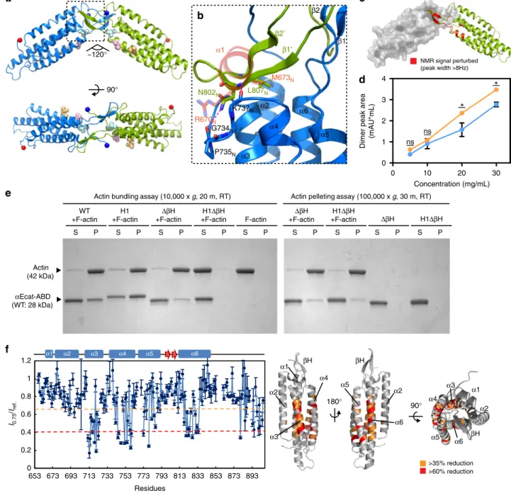
Fig. 1 Force-induced unfolding ofα1-helix enhances the F-actin-binding activity of the αcat-ABD. a R2/7 cells transiently expressing ABD (residues 663-906) or ABD* (residues 697-663-906).αcat-ABD/ABD*-FLAG and actin were labeled with the anti-DDDDK antibody and phalloidin, respectively. Scale bar, 10 μm. b Comparison of the ABD crystal structures of αN-catenin, αE-catenin and vinculin. The αcat-ABD contains three distinct structural motifs: α1-helix (α1; red circle),β-hairpin (βH; magenta circle), and C-terminal tail (CT; black circle). PDB ID codes are indicated in parentheses. c Multiple sequence alignment ofα-catenin and vinculin primary sequences. The α1-helix and βH sequences are highly conserved among three paralogs of α-catenin (E, N and T; h, human; m, mouse), as well as in Drosophilaα-catenin (dα-catenin). The H1 mutation (RAIM670-673GSGS) is indicated. Conservation of three actin-binding site residues inα-catenin, as well as the vinculin actin-binding site residues, I997 and V1001, are marked by purple dots. d Snapshots of the structure of αNcat-ABD at select time points during a constant-force SMD simulations (100-pN pulling force for 120 ns) (Supplementary Movie 1). Cartoon representation showsα1-helix (blue) starts to unfold at ~60 ns. e A close-up view of α1-helix and V796Nin theαNcat-ABD crystal structure. During constant-force SMDsimulations, V796Nin a cryptic state is exposed (V796N*; magenta) at 45 ns, shortly beforeα1 unfolding occurred at 60 ns (d). Two conserved α1-helix
residues, R670Nand M673N, engage in critical interactions with thefive-helix bundle of ABD to attenuate the ABD-F-actin interaction. f Actin
cosedimentation assays comparing WT, H1 andΔα1 variants of αEcat-ABD and αNcat-ABD. Whereas less than half of total ABD-WT (0.37-0.45) cosedimented with F-actin for bothαE-catenin and αN-catenin, alterations in α1-helix, either by deletion or unfolding via the H1 mutation, significantly increased the amount of mutantαcat-ABD proteins cosedimented with F-actin (0.71–0.81). Supernatant (S) and pellet (P) fractions are indicated. Data are presented as mean ± standard error of the mean (SEM) (N= 3). Significance by ANOVA: ***P < 0.001
displayed punctate AJs connected to actin cables aligned along
the wound edge, whereas
αEcat-H1 cells formed less organized
punctate AJs and actin assemblies (Fig.
3
c and Supplementary
Fig. 9a). High-resolution live-imaging revealed that
αEcat-H1 AJs
were less organized towards the wound front, resulting in
unproductive cell-cell tugging events that appeared to interfere
with forward sheet migration (Supplementary Movie 3). In fact,
αEcat-H1, αEcat-ΔβH, and αEcat-H1ΔβH cells were all inferior to
αEcatFL cells in wound closure, and no better than αEcat-ΔABD
cells (Fig.
3
d, e and Supplementary Fig. 9b). By tracking cells
individually, we found that
αEcat mutant cells moved with similar
speeds as
αEcatFL cells, but less persistently, contributing to
0 10 20 30 0 1 2 3 4
*
*
ns ns Concentration (mg/mL)Dimer peak area
(mAU*mL) I0.75 /Iref. Residues 0 0.2 0.4 0.6 0.8 1 1.2 653 673 693 713 733 753 773 793 813 833 853 873 893 α2 α3 α4 α5 α6 α1 β β 90° ~120° α1 α2 α3 α4 α5 α6 β1 β2 G734N P735N K737N N802N L807N R670N M673N β1’ β2’
b
a
NMR signal perturbed (peak width >8Hz)c
d
Actin (42 kDa) αEcat-ABD (WT: 28 kDa) S WT +F-actinActin bundling assay (10,000 x g, 20 m, RT) H1 +F-actin ΔβH +F-actin H1ΔβH +F-actin F-actin
Actin pelleting assay (100,000 x g, 30 m, RT)
ΔβH H1ΔβH ΔβH +F-actin H1ΔβH +F-actin P S P S P S P S P S P S P S P S P
e
f
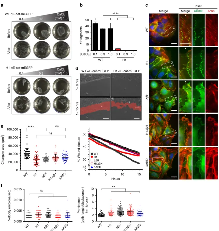
180° >35% reduction >60% reduction α2 α2 α1 α3 α4 α5 α6 βH βH 90° α2 βH α1 α3 α4 α5 α6Fig. 2 Crystal structure ofαNcat-ABD-H1 reveals a novel ABD dimer interface. a Crystal structure of the αNcat-ABD-H1 dimer in form A (two protomers shown as blue and green). The N and C termini of ABD are indicated by blue and red spheres, respectively. Three actin-binding site residues, L785N, I792N
and V796N, are shown as light blue, pink and orange spheres.b A close-up view of the ABD dimer interface. The dashed-line box in a is rotated by ~90°
CCW. TheβH motif from one protomer covers the hydrophobic patch exposed by α1-helix unfolding in the adjacent protomer (the α1-helix of αNcat-ABD-WT is shown in red).c Concentration-dependent CSPs ofαNcat-ABD-H1 are localized to the βH residues. Residues with CSP greater than 8 Hz are indicated on theαNcat-ABD-H1 structure in red. d SEC-MALS analysis of αEcat-ABD. The integrated dimer peak area was plotted against the αEcat-ABD concentration forαEcat-ABD-WT (blue) and αEcat-ABD-H1 (orange). Data are presented as mean ± SEM (N = 3). Significance by ANOVA: *P < 0.05. e In vitro actin cosedimentation assays ofαEcat-ABD variants, WT, H1, ΔβH, and H1ΔβH. Actin bundling was analyzed by sedimentation at low RCF (10,000×g). The F-actin-bound ABD was sedimented at high RCF (100,000×g).f TCS experiments with unlabeled F-actin and15N/2H-labeled αNcat-ABD-WT. Plots of the reduction ratios of the backbone amide signal intensities observed with and without presaturation. Residues with >60% and >35% signal reduction are indicated on theαNcat-ABD-WT structure (right). The affected residues are mostly located in the last four α-helices (α3-α6)
overall reduced epithelial sheet migration (Fig.
3
f, Supplementary
Fig. 9b and Supplementary Movie 4). In addition, differences
between
αEcatFL and αEcat-H1 cell trajectories were
indepen-dently validated using a particle image velocimetry (PIV)-based
tracking method (Supplementary 9c). These observations suggest
that
α-catenin with a defective α1-helix can support AJs in static
epithelia but fails to support dynamic AJ rearrangements and cell
movements.
c
e
d
t = 0 hrs t = 15 hrs WT αE-cat-mEGFP H1 αE-cat-mEGFP 0 20,000 40,000 60,000 80,000 100,000 Changein area ( μ m 2)****
ns ns WT H1 ΔβH H1Δβ H ΔABD WT H1 ΔβH H1Δβ H ΔABD WT H1 Δβ H H1Δβ H ΔABD WT H1 ΔβH H1ΔβH ΔABD 0 5 10 15 0 15 30 40 50 Hours % Wound closurea
0 0.1 0.3 1.0 0.1 0.3 1.0 10 20 30 40 50 # Fragments****
WT H1 [CaCl2] Before After CaCl2 (mM) 1.0 0.1 0.3 Before Afterb
Δ ABD αEcat Actin Merge WT H1 Δβ H H1 Δβ H Inset Mergef
0.000 0.005 0.010 0.015 Velocity (microns/sec) ns 0 2 4 6 8 10 Persistence (path length/displacement in microns)**
WT αE-cat-mEGFP H1 αE-cat-mEGFP 0.1 0.3*
*
**
*
*
*
*
*
*
*
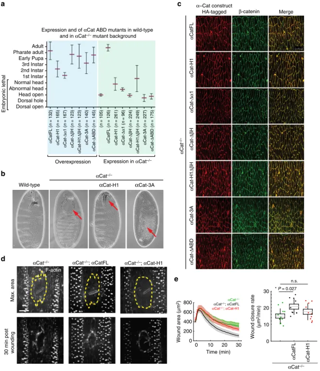
CaCl2 (mM) 1.0Fig. 3α1-helix and βH are critical for the formation of multicellular structures and wound healing. a Epithelial sheet disruption assay of R2/7 cells expressingα-catenin variants. Representative αEcat monolayers before and after mechanical stress treatment are shown. b Plots showing total cell monolayer fragments after mechanical stress treatment. Mechanical disruption causedαEcatFL cell monolayers to fragment, whereas αEcat-H1 monolayers remained intact with only few fragments forming at a low calcium concentration. Data are presented as mean ± standard deviation (SD) (N= 3). Significance by ANOVA; ****P < 0.0001. c Confocal images of R2/7 cells expressing αEcat variants at the wound fronts. Close-up views of inset boxes are shown. Scale bar, 20μm. d Scratch wound healing assays with R2/7 cells expressing αEcat-WT or αEcat-H1. The areas of wound healing after 15 hrs are shown in red. Scale bar= 50 μm. e Plots showing changes in total wound closure area and the wound closure percentage over time. Data are presented as mean ± SD (>35fields of view (FOV); > 5 biological replicates (BR)). Significance by ANOVA; ****P < 0.0001. f Plots showing changes in the persistence, but not the velocity, ofαEcat mutant cells at the wound front compared to αEcatFL cells. Data are presented as mean ± SD (>35 FOV; > 5 BR). Significance by ANOVA; **P < 0.01
To further assess
α1-helix and βH functions in tissue
organization we generated mutants in Drosophila
α-Catenin
(α-Cat) and tested their function in transgenic animals. Both
α1-helix and
βH regions are conserved in α-Cat (Fig.
1
c), and
previous work showed that the ABD of Drosophila
α-Cat
(αCat-ABD) is essential for cell adhesion
7. Moreover,
αCat-ABD-H1
showed enhanced actin binding and bundling activity compared
to
αCat-ABD (Supplementary Fig. 10a) similar as mammalian
proteins (Fig.
2
e). Zygotic null mutants for
α-Cat (αCat
−/−) show
embryonic lethality and severe defects in head morphogenesis
9(Fig.
4
a, b). Expression of full-length
α-Cat (αCatFL) did rescue
αCat
−/−mutants to adulthood. In contrast,
αCat-H1, αCat-Δα1,
αCat-ΔβH, and αCat-H1ΔβH did not rescue the embryonic
lethality of
αCat
−/−mutants similar to
αCat-ΔABD (Fig.
4
a).
Expression of
αCat-H1, αCat-Δα1, and αCat-H1ΔβH led to some
improvements in head morphogenesis, and a small number of
animals expressing
αCat-H1 or αCat-H1ΔβH survived to larval
stages (Fig.
4
a). Immunoblot analysis (Supplementary Fig. 10b)
and tissue staining in an
αCat
−/−mutant background (Fig.
4
c)
showed that our constructs are expressed at levels similar to
endogenous
α-Cat and are effectively recruited to AJs. Efficient
recruitment of
α-Cat proteins to AJs in a wildtype background
indicated that mutant proteins are not outcompeted by
endogenous
α-Cat (Supplementary Fig. 10c). We noted that
overexpression of
αCat-H1 or αCat-Δα1 had a toxic effect on
survival with most animals dying as larvae, whereas
over-expression of other
α-Cat constructs led to pupal lethality or adult
survival (Fig.
4
a). The failure of
α-Cat proteins with a
compromised
α1-helix to substantively rescue αCat
−/−mutants
was surprising as those variants are likely capable of coupling
cadherin to the actin cytoskeleton to promote intercellular
adhesion. On the other hand, enhanced F-actin binding of
αCat-ABD-H1 (Supplementary Fig. 10a) could explain the
observed toxicity upon overexpression of these constructs. Our
findings indicate that the function of the α1-helix in attenuating
interactions between
α-catenin and F-actin is instrumental for AJ
function in developing epithelia.
We further examined the role of
α1-helix in wound repair,
which is driven by the polarized assembly of actin at the interface
between wounded and adjacent cells in the Drosophila embryonic
epidermis
41. Polarization of actin (and the non-muscle myosin II)
in the cells adjacent to the wound results in the assembly of a
supracellular contractile cable around the perimeter of the wound
that drives tissue repair
42. The quantified wound closure
dynamics revealed that
αCat
−/−embryos expressing
αCatFL
repaired damage to their epidermis faster than
αCat
−/−embryos,
whereas
αCat-H1 expression did not significantly accelerate
wound closure in an
αCat
−/−epidermis. (Fig.
4
d, e). These results
are consistent with our whole animal rescue experiments, as well
as our scratch wound healing assays (Fig.
3
d‒f), and collectively
suggest that a compromised
α1-helix severely interferes with
α-catenin function in tissue morphogenesis.
Actin-binding site residues are essential for
α-cat function.
Considerable in vitro evidence suggests that
α-catenin can
directly interact with F-actin
5,7,8,20,27,29,33,35,43,44. A previously
determined low resolution (18 Å) cryo-EM map of an
αcat-ABD-F-actin complex precluded any detailed analysis of the complex
interface
29. Nonetheless, it suggested that the
αcat-ABD interacts
with two actin monomers adjacently aligned on the long axis of
F-actin. A similar arrangement was observed in a recently
determined 8.5-Å cryo-EM structure of a vin-ABD-F-actin
complex, which revealed that the last two
α-helices of vin-ABD
interact with F-actin
45. Considering the relatively high sequence
identity shared between
αcat- and vin-ABDs (~30%)
27, we
generated an atomic model of the
αNcat-ABD-H1-F-actin
com-plex based on the vin-ABD-F-actin structure. In this model,
α5-and
α6-helices of the αNcat-ABD-H1 interact with two axially
arranged actin monomers of F-actin (Fig.
5
a). In particular, the
α5-helix contains the highly conserved residues, I792
Nand V796
N(Fig.
1
c). I792
Nof
αN-catenin assumes an exposed position
clo-sely resembling the vinculin actin-binding site residue I997
30. In
contrast, the conformation of V796 remains ambiguous, partly
due to poorly defined electron density of this region in the 3.7-Å
crystal structure of human
αE-catenin
38, and a cryptic position of
V796
Nin the
αNcat-ABD structure
27(Supplementary Fig. 11a)
compared to the fully exposed V1001 of vinculin
30.
To better characterize the
αE-catenin actin-binding site, we
elucidated a crystal structure of
αEcat-ABD-WT at 2.2-Å
resolution (Fig.
5
b, Supplementary Fig. 5, and Supplementary
Table 2). The electron density map of
α5-helix clearly shows that
V796 adopts a conformation that exposes its side chain on the
ABD surface, along with two additional hydrophobic residues
L785 and I792 (Supplementary Fig. 5g and 11b). Our site-directed
mutagenesis and actin cosedimentation assays with the
αEcat-ABD variants support critical roles of these hydrophobic residues
in F-actin-binding: Ala substitutions of L785, I792 and V796,
individually or together as 3A, led to a range of reduction (75, 36,
47, and 78%, respectively) in the amount of ABD cosedimenting
with F-actin compared to
αEcat-ABD-WT (Fig.
5
c). The effects of
I792A and V796A were greater in the H1 background (reduction
of 70% and 73%, respectively), confirming that alterations of
these residues significantly reduce F-actin binding by
αEcat-ABD-H1 (Fig.
5
c). In contrast, Ala substitution of V714, which is
located on the
α3-helix surface, resulted in no reduction (Fig.
5
c).
Also, none of the above mutations appear to interfere with the
ability of
αEcat-ABD to bundle F-actin (Supplementary Fig. 12).
The equally significant reduction observed with either the L785A
mutation alone or 3A suggests that L785 plays a central role in
establishing the critical hydrophobic interface between F-actin
and
αcat-ABD. The measurable reduction in F-actin binding with
I792A or V796A suggests that I792 and V796 are likely involved
in further stabilizing this interface, and any changes to these
residues could modulate the F-actin-binding activity of
αcat-ABD. These results confirm that the hydrophobic residues on the
α5-helix surface constitute an important binding surface for
F-actin interaction.
Next we tested the in vivo importance of this interaction by
expressing an
αCat-3A (L798A + I805A + V809A) mutant in
Drosophila. All three key hydrophobic residues identified in
mammalian
αE-catenin or αN-catenin are conserved in
Droso-phila
α-Cat (Fig.
1
c).
αCat-3A was recruited normally to the
cadherin-catenin complex (Fig.
4
c and Supplementary Fig. 10c)
but failed to show a rescue of the
αCat
−/−mutant phenotype; a
fraction of embryos showed more severe defects than
αCat
−/−mutants, consistent with a mild dominant-negative effect of
αCat-3A expression (Fig.
4
a, b). We conclude that direct interaction
between
α-catenin and F-actin is essential for AJ assembly and
function during development.
Allosteric coupling between
α1-helix and V796 dynamics. Our
observations of the cryptic (attenuated) and exposed (activated)
conformations of V796 (Supplementary Fig. 11a, b), despite the
nearly identical primary sequences of
αEcat- and αNcat-ABDs
(87% identity; Supplementary Fig. 3), indicate that this residue
resides within a conformationally dynamic region. Consistent
with this idea, the
αEcat-ABD-WT structure contains an internal
cavity that could accommodate V796 in the cryptic state similar
to V796
Nin the
αNcat-ABD-WT structure (Fig.
5
b,
Supple-mentary Fig. 11c). This internal cavity is partly formed by the side
c
α Cat-Δ ABD α Cat-3A α Cat-H1 Δβ H α Cat-Δβ H α Cat-H1 Merge β-catenin α–Cat construct HA-tagged α CatFL α Cat –/–d
αCat–/– F-actin Max. area30 min post wounding
αCat–/–; αCatFL αCat–/–; αCat-H1
a
Expression and of αCat ABD mutants in wild-type and in αCat–/– mutant background
α Cat-Δ ABD ( n = 175) α Cat-3A ( n = 227) α Cat-H1 Δβ H ( n = 249) α Cat-Δβ H ( n = 224) α Cat-Δα 1 ( n = 96) α Cat-H1 ( n = 261) α Cat-Δ ABD ( n = 145) α Cat-3A ( n = 140) α Cat-H1 Δβ H ( n = 123) α Cat-Δβ H ( n = 123) α Cat-Δα 1 ( n = 167) α Cat-H1 ( n = 165)
Overexpression Expression in αCat–/–
α CatFL ( n = 126) (n = 105) α CatFL ( n = 132) Adult Pharate adult Early Pupa 3rd Instar 2nd Instar 1st Instar Embryonic lethal Normal head Abnormal head Head open Dorsal open Dorsal hole
b
αCat–/–Wild-type αCat-H1 αCat-3A
e
0 200 400 600 800 Time (min) 0 αCat–/– αCat–/–; αCatFL αCat–/–; αCat-H1 30 20 10 0 α CatFL α Cat-H1 * P = 0.027 n.s. α Cat-Δα 1 Wound area ( μ m 2) 10 20 30 αCat–/–Wound closure rate
(μ
m
2/min)
Fig. 4αCat ABD mutants fail to rescue αCat function in Drosophila. a Phenotypic consequences of the overexpression of αCat ABD mutants and rescue activity ofαCat ABD mutants expressed in αCat−/−zygotic null mutants. Overexpression: all mutant constructs showed significantly reduced survival (P < 0.0001) compared toαCatFL overexpression. Rescue experiments: Mutant constructs showed a significant rescue (αCatFL, H1, Δα1, αCat-H1ΔβH [P < 0.0001]) or enhancement (αCat-3A [P < 0.0001], αCat-ΔABD [P = 0.0071]) of the αCat−/−zygotic mutant phenotype. Expression of αCat-ΔβH did not significantly modify the αCat−/−mutant phenotype. Data are presented as mean ± SD.b Cuticles of wild-type embryo, ofαCat−/−mutant showing failure in head morphogenesis (‘head open’; arrow), of αCat−/−mutant expressingαCat-H1 showing a defective head skeleton (‘abnormal head’; arrow), and ofαCat−/−mutant expressingαCat-3A showing dorsal hole (arrow) in addition to an open head. c HA-tagged αCat variants were expressed with Act5c-Gal4 da-Gal4 in the epidermis of Drosophila embryos mutant forα-Cat (αCat−/−) at stage 15. AJs marked byβ-catenin. d Epidermal wounds in αCat−/−,αCat−/−mutant expressingαCatFL, and αCat−/−mutant expressingαCat-H1. F-actin was labeled with GFP::UtrophinABD. Top panels show time of maximum wound area (yellow lines outline the wounds) and bottom panels show epidermis 30 min after wounding. Anterior left, dorsal up. Scale bar, 10μm. e Wound area over time (left) and wound closure rate (right) for αCat−/−(red, n= 12 wounds), αCat−/−mutants expressingαCatFL (cyan, n = 10 wounds), andαCat−/−mutants expressingαCat-H1 (green, n = 10 wounds). αCatFL, αCat−/−embryos repaired damage to their epidermis significantly faster thanαCat−/−embryos (P= 0.027), whereas αCat-H1 αCat−/−embryos did not show a significant difference to αCat−/−embryos. The box plot shows the mean (gray line), SEM (box), and SD (black lines)
chain of M673 from
α1-helix (Fig.
1
e), hence raising the
possi-bility that
α1-helix unfolding allosterically affects the
F-actin-binding site by changing the conformational dynamics of V796.
To determine the influence of α1-helix on the actin-binding
site of
α-catenin, we developed a new bio-layer interferometry
(BLI) approach to measure the kinetics of the
αcat-ABD-F-actin
interaction. We immobilized F-actin onto the streptavidin-coated
optical sensor with biotinylated LifeAct actin-binding peptides
(LAbio)
46, and measured subsequent association and dissociation
of
αcat-ABD (Fig.
6
a and Supplementary Fig. 13a). We
determined that concentration-dependent F-actin binding curves
of
αEcat-ABD-WT fit well with a 2:1 hetero-ligand:receptor
model with two K
Dvalues, K
D1= 2.0 μM and K
D2= 0.3 μM
(Fig.
6
b, Table
1
, and Supplementary Fig. 13b). This model
supports
αEcat-ABD-WT in an equilibrium between the
attenuated and activated actin-binding states, respectively. The
lower K
D2value is consistent with the positive cooperativity of
F-actin binding by
αEcat-ABD as previously reported
8,29. In
contrast, the BLI data of
αEcat-ABD-H1 fit well with a 1:1
ligand:receptor model with the single K
Dvalue of 0.58
μM
(Fig.
6
c, Table
1
and Supplementary Fig. 13b), reflecting a
predominantly activated state of
αEcat-ABD-H1. The effects of
mutations in the
α-catenin actin-binding site, as well as ΔβH
mutation, resulted in decreased affinity (Table
1
and
Supplemen-tary Fig. 14a, b) that are consistent with our actin
cosedimenta-tion assay results (Figs.
2
e and
5
c). Our data support the
conclusion that the unfolded
α1-helix contributed to an apparent
equilibrium shift towards an activated state of
αcat-ABD.
Comparison of the
15N/
1H TROSY NMR spectra of
αNcat-ABD-WT
47and
αNcat-ABD-H1 showed that a region
(αN-catenin residues 794–814) containing V796
Nand
βH was one of
three regions affected by the H1 mutation (Supplementary
Fig. 15a), likely indicating an altered conformation in this
region (Supplementary Fig. 15b). We further confirmed by
NMR relaxation and MD simulations studies that the unfolded
α1-helix increased molecular motions in the V796
N/βH region
(Supplementary Fig. 15c, d). In addition, we performed
chemical shift (CS)-based Rosetta comparative modeling
(CM)
48to show that V796
Nof
αNcat-ABD-WT remained in
the cryptic state, whereas
αNcat-ABD-H1 displayed a large
conformational change that exposed V796
Non the surface
(Fig.
6
d), resembling V796 in the crystal structure of
αEcat-ABD-WT (Fig.
5
b). Our extended equilibrium MD calculations
of
αcat-ABDs support that the unfolded α1-helix accelerates the
conformational change to favor the exposed state of V796
(Fig.
6
e, f). As the exposure of V796
Nprecedes complete
unwinding of
α1-helix during the constant-force simulation
(Supplementary Movie 2), we expect that
α1-helix unfolding
‘locks’ V796
Nin the activated state. Taken together, our
observations indicate that allosteric coupling between
α1-helix
and the actin-binding residue V796 is central to the
force-induced association of
α-catenin with F-actin.
V796 V796N αN-ABD-WT αE-ABD-WT α1 α2 α3 α4 α5 α6 β1 β2 α1 α2 α3 α4 α5 α6 β1 β2 I792N I792 M673N M673 L785N L785 αNcat-ABD-H1 α2 α3 α4 α5 α6 βH L785 L785N I792 I792N V796 V796N V714 V714N L785N I792N V796N V714N
a
b
c
S P S P S P S P Actin (42 kDa)ABD L785A I792A V796A 3A V714A
S P S P
S P S P S P S P
H1 H1L785A H1I792A H1V796A
ns ns
***
***
***
ns ns** ***
ABD L785A V796A 3A V714A H1H1L785A H1I792A H1V796A
***
αEcat-ABD (28 kDa) Actin (42 kDa) αEcat-ABD (28 kDa) 1.0 0.9 0.8 Bound/total 0.0 0.1 0.2 0.3 0.4 0.5 0.6 0.7 I792AFig. 5 Identification of the critical actin-binding site residues in α-catenin. a Model of the αcat-ABD (red) bound to two axially adjacent actin monomers (dark and light teal) within F-actin, based on the vin-ABD/F-actin cryo-EM structure (PDB ID: 3JBI).b Comparison of high-resolution crystal structures of αEcat-ABD-WT (red) and αNcat-ABD-WT (blue). The overall structure of αEcat-ABD-WT closely resembles αNcat-ABD-WT, as two ABD structures can be superposed with RMSD of 0.53 Å over 156 residues. A close-up view (right) shows thatαEcat-ABD-WT contains a cavity (pink molecular envelope), which could accommodate V796 in a cryptic state similar to V796Nin theαNcat-ABD-WT structure. c Actin cosedimentation assays of αEcat-ABD
variants: WT, L785A, I792A, V796A, 3A, V714A, H1, H1L785A, H1I792A, and H1V796A. Data are presented as mean ± SEM (N= 3). Significance by ANOVA: **P < 0.01, ***P < 0.001
Discussion
We show that the unique molecular features of
αcat-ABD,
α1-helix, V796, and
βH, confer mechanosensitivity to α-catenin and
its ability to dynamically regulate and reorganize actin
filaments
directly associated with cadherin-catenin complexes at
inter-cellular junctions. The importance of
α-catenin to directly
associate with F-actin in a mechanosensitive manner is
under-scored by experiments showing that
αcat-H1 with enhanced
F-actin binding was equally inferior to
αcatFL function as mutants
with diminished F-actin binding (e.g., 3A) during mammalian
and Drosophila wound healing, and Drosophila development
(Figs.
3
and
4
). Although a high-resolution structure of the
αcat-ABD-F-actin complex remains to be solved, we have shown that
the critical actin-binding site residues, L785, I792, and V796, are
located away from the
αcat-ABD mechanosensory motif,
α1-helix, thus raising the possibility that the N-terminal region of
ABD acts allosterically to regulate F-actin binding. Based on these
observations, we propose that the coupled conformational states
of
α1-helix and V796 provide the structural basis of
force-dependent
allosteric
regulation
of
the
α-catenin-F-actin
interaction.
In the proposed mechanism, the ABD of
α-catenin in the
attenuated state can weakly associate with F-actin, whereas its
interaction with F-actin under force would trigger
α1-helix
unfolding and the exposure of V796 to form a catch bond
interaction between the cadherin-catenin complex and F-actin at
nascent contacts
8(Fig.
7
). As nascent contacts grow, multiple
α-catenin molecules will bind to F-actin in a cooperative manner
8,29to promote the formation of cadherin-catenin complex clusters
(Fig.
7
). Although
αcat-H1 or other constructs without the
α1-helix can support AJ formation in R2/7 cells (Supplementary
Fig. 1b and 8b), these do not restore normal
α-catenin function
(Supplementary Fig. 2a, b) and may reflect a lack of extensive
junctional remodeling in these cells. A similar discrepancy
between confluent R2/7 cells, wound-healing assays, and in vivo
performance was noted for
αEcat-NM
I(residues 1-402): this
ABD-deficient construct forms AJs in R2/7 cells through the
recruitment of vinculin
6, but does not support normal
α-catenin
function during wound closure
6, and a corresponding Drosophila
construct (αCat-NM1) showed no rescue of αCat
−/−embryos (R.
S. and U.T., unpublished).
Cadherin clustering and AJ maturation likely require
trans-interactions and cis-trans-interactions of cadherin ectodomains, as well
as an active process involving intracellular coupling of the
cadherin-catenin complex to actin networks
49. Our
αNcat-ABD-H1 crystal structures revealed an unexpected ABD
homo-dimerization (Fig.
2
a), which can facilitate F-actin bundling
in vitro (Fig.
2
e). It involves the
βH motif forming an extensive
dimer interface with the hydrophobic patch uncovered by
α1-helix unfolding (Fig.
2
b and Supplementary Fig. 6c). Considering
the very weak
αcat-ABD dimerization (Fig.
2
c and Supplementary
Fig. 7c), which is marginally increased by the H1 mutation in
solution (Fig.
2
d), it is possible that tension-induced unfolding of
α1-helix allosterically changes the conformational dynamics of
V796N-WT V796N-H1 I792N-WT I792N-H1 CS-Rosetta:WT αNcat-ABD-WT CS-Rosetta:H1 0 ns 54 ns 187 ns 0 ns 54 ns 189 ns 0 ns 16 ns 43 ns V796N I792N H788N V796N I792N H788N V796 I792 H788 Cryptic Exposed Strepavidin -coated optical sensor Biotinylated LifeAct peptide F-actin α-catenin ABD 4 μM 2 μM 1.5 μM 1 μM 0.5 μM 3 μM αEcat-ABD-WT 2 μM 1 μM 0.5 μM 0.25 μM 0.125 μM αEcat-ABD-H1 αE V796-I792 Response (nm) Response (nm) 0.12 0.1 0.08 0.06 0.04 0.02 0 0.12 0.2 0.18 0.16 0.14 0.1 0.08 0.06 0.04 0.02 0 Time (s) 0 10 20 30 40 Time (s) 0 10 20 30 40 0 50 100 150 Time (ns) Distance (Å) 10 5 αN V796N-I792N H1 V796N-I792N KD1 = 2.0 μM KD2 = 0.3 μM KD = 0.58 μM
a
b
c
d
e
f
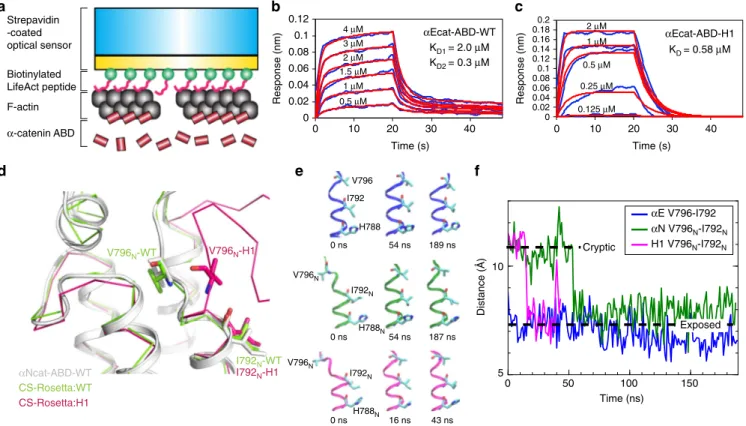
Fig. 6 Unfolding ofα1-helix affects the conformational dynamics of V796. a A scheme of BLI experiment for a kinetic analysis of direct interaction between αEcat-ABD and F-actin. The streptavidin-coated optical sensor with LAbio peptides immobilizes F-actin through high avidity, thereby restricting the movement of attached F-actin to minimize the occurrence ofαcat-ABD-induced actin bundling. b BLI responses curves of the αEcat-ABD-WT. The KD
values were obtained byfitting concentration-dependent F-actin binding curves (blue) to a 2:1 heterogeneous binding model (red curves). c BLI response curves of theαEcat-ABD-H1. The KDvalue was obtained byfitting concentration-dependent F-actin binding curves (blue) to a 1:1 binding model (red
curves).d CS-Rosetta-CM models ofαNcat-ABD-WT and αNcat-ABD-H1 based on NMR CS data and the αNcat-ABD-WT crystal structure as the template.e Conformational states of V796 during the equilibrium MD simulations ofαEcat-ABD-WT (blue), αNcat-ABD-WT (green) and αNcat-ABD-H1 (magenta). Snapshots of the region ofα5-helix containing V796 at specified time points are shown. f Evolution of distance between the β-carbon atoms of V796 and I792 during the equilibrium MD simulations. Dotted lines mark the approximate inter-residue distances when V796 is in the cryptic and exposed positions
the actin-binding site without affecting dimerization.
None-theless, AJ-localized
α-catenin cooperatively binding to F-actin
would likely increase the propensity of
αcat-ABD to dimerize and
promote F-actin bundling. Uncovering this ABD dimerization
interface motivated us to propose a new monomer-dimer model
for
α-catenin at the cadherin-actin interface (Fig.
7
). Although
both in vitro
8and in vivo
7studies consistently concluded that
monomeric
α-catenin forms the essential link between the
cadherin-β-catenin complex and F-actin, the current model fails
to account for the capacity of
α-catenin to bundle F-actin
5at AJs.
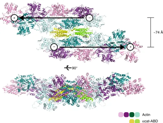
The ABD-dependent dimerization as demonstrated here allows
actin
filaments to be tightly bundled in an antiparallel fashion
(Fig.
8
) and places the
α3-α4 surface of ABD in close proximity
with F-actin, which is consistent with our NMR saturation
transfer data (Fig.
2
f). In addition, the ABD dimerization allows
F-actin bundling to occur while the N domain of
α-catenin
remains associated with cadherin-bound
β-catenin (Fig.
7
).
Hence, our proposed model differs from the previous
monomer-dimer model of
α-catenin by arguing that (i) the
E-cadherin/β-catenin/α-catenin/F-actin complex regulates the cadherin-actin
linkage without disrupting the
β-catenin-α-catenin interaction;
(ii) that
α-catenin as a component of the complex can bundle
F-actin, and (iii) that
α-catenin controls actin binding through
force-dependent allosteric regulation of the actin-binding site
within the ABD. The versatility of
α-catenin to modulate the
attachment of the cadherin-catenin complex to F-actin from
transient interaction to stable actin bundling, and to the dynamic
cortical actin network
50, will likely involve additional dynamic
connections provided by the recruitment of other F-actin-binding
proteins, such as vinculin, afadin, ZO-1 and EPLIN, to
inter-cellular junctions
6,27,51–53.
We have employed an integrative structure/function approach
to show that the structural motifs of
αcat-ABD involved in the
regulation of tension-sensitive actin binding are essential for
normal tissue morphogenesis and wound healing. Although the
occurrence of actin bundling involving ABD-linked
α-catenin
dimers at intercellular junctions remains to be tested, our model
reconciles previous observations of
α-catenin as a critical
mechanosensor engaged in reorganization of AJs by facilitating
dynamic F-actin association
8,21, and actin bundling through
homodimerization
5,20.
Moreover,
the
significance of this
mechanism lies in the ability of
α-catenin to modulate
cadherin-mediated cell adhesion through force-dependent F-actin binding
and actin remodeling without dissociation from the
cadherin-β-catenin complex.
Methods
Protein expression and purification. The cDNA corresponding to the actin-binding domain (ABD) of mouseαE-catenin (652-906), mouse αN-catenin (651-905) and all related mutants (e.g., theαE-catenin H1 mutation RAIM670-673GSGS) were amplified by PCR and individually subcloned into the pGEX4T1
vector (GE Healthcare). Thefly αcat-ABD (659-917) cDNA was amplified by PCR and subcloned into a modified pET-SUMO vector. Site-directed mutagenesis was performed using the Quikchange protocol (Stratagene) to produce all single-/ multiple-residue and deletion mutants. Recombinant proteins were expressed as N-terminal glutathione S-transferase (GST) fusion proteins in Escherichia coli BL21-CodonPlus cells. Cells were grown to an O.D.600 of 0.8 at 37 °C and the recom-binant protein expression was induced with 0.5 mM isopropyl
β-D-1-thiogalactopyranoside for 16 h at 16 °C. Cells harvested by centrifugation were resuspended in the lysis buffer (50 mM Tris-HCl, pH 8.0, 300 mM NaCl, 10 mM β-mercaptoethanol, 1 mM Tris(2-carboxyethyl)phosphine (TCEP)), sonicated on ice, and subjected to centrifugation to isolate soluble proteins. GST-fusion proteins were isolated using the glutathione-sepharose resin (GE Healthcare). His-SUMO fusion proteins were isolated using the Ni2+-NTA resin (ThermoFisher Scientific). GST-fusion and His-SUMO proteins were cleaved by thrombin or SUMO protease (Ulp-1), respectively. The cleaved proteins were further purified by size-exclusion chromatography using Superdex 75 (GE Healthcare) in the running buffer (50 mM Tris-HCl, pH 8.0, 300 mM NaCl, 1 mM TCEP). The purified proteins were exchanged into protein storage buffer (50 mM Tris-HCl, pH 8.0, 100 mM NaCl, 1 mM TCEP).
Size-exclusion chromatography-multiangle light scattering. Purified protein (5 mg/mL, 100μL injection volume) was subjected to size-exclusion chromatography (SEC) using a Superdex-200 Increase 10/300 GL column (GE Healthcare) equili-brated in SEC-MALS buffer (20 mM Tris-HCl pH 7.0, 100 mM NaCl) at aflow rate of 0.5 mL/min. Multi-angle light scattering (MALS) measurements were performed in-line with SEC by using a three-angle (45°, 90°, and 135°) miniDawn light-scattering instrument and an Optilab rEX differential refractometer (Wyatt Technologies). Molecular weight was calculated by using the ASTRA software (Wyatt Technologies). Dimer peak area integration was performed by using ImageJ54. Statistical analysis was performed by Two-way ANOVA followed by
Bonferroni’s comparison test.
Actin cosedimentation assay. Monomeric rabbit skeletal muscle actin was pur-ified from rabbit muscle acetone powder55(Pel-Freez Biologicals). Purified globular
actin (G-actin) was diluted to 20μM in a fresh Buffer-G (2 mM Tris-HCl, pH 8.0, 0.2 mM ATP, 0.5 mM DTT, 0.1 mM CaCl2), and subsequently polymerized in
Buffer-F (5 mM Tris-HCl, pH 8.0, 50 mM KCl, 2 mM MgCl2, 1 mM ATP, 0.2 mM
CaCl2, 0.5 mM DTT) for 1 h at RT. Theαcat-ABD samples were subjected to buffer
exchange into Buffer-F. Samples of F-actin and ABD were mixed (the protein mixture contains 5μM ABD and 5 μM actin in 50 μL) in Ultra-Clear Centrifuge Tubes (Beckman Coulter) and incubated for 1 h at RT. F-actin with bound protein samples were cosedimented by centrifugation using a Beckman Coulter Airfuge with a chilled A-100/30 rotor at 28 psi (≥100,000×g) for 20 min at RT. To assess actin bundling, F-actin with bound protein samples were cosedimented by cen-trifugation using a benchtop microcentrifuge at low relative centrifugal force (RCF; 10,000×g) for 30 m at 4 °C. Supernatant and pellet fractions were analyzed by SDS-PAGE with coomassie blue stain. Gel band intensity was measured by using ImageJ54. Statistical analysis of three or more groups was performed by One-way
ANOVA followed by Tukey’s multiple comparison test. Statistical analysis of two groups was performed by Two-way ANOVA followed by Bonferroni’s comparison test.
Crystallization and data collection. Crystals of theαN-catenin ABD-H1 were grown at 277 K by vapor diffusion. For crystallization theαNcat-ABD-H1 sample was exchanged into Buffer-P (20 mM K/Na phosphate, pH 6.0, 150 mM NaCl, 1 mM TCEP), and the protein solution (30 mg/mL) was mixed with an equal volume of the reservoir solution, which consists of either solution A (2.0 M (NH4)2SO4, 10
mM CoCl2) for form A crystals, or solution B (100 mM Na acetate/acetic acid, pH
4.5, 0.8 M NaH2PO4, 1.2 M K2HPO4) for form B crystals. Similarly, crystals of the
αEcat-ABD-WT in Buffer-P (30 mg/mL) were grown at 277 K by vapor diffusion with the reservoir solution consisting of 0.2 M KBr, 2.2 M (NH4)2SO4and 3% (w/v)
Table 1 BLI data for
αEcat-ABD variants binding to F-actin
αEcat-ABD Fitting model KD1(μM) KD2(μM) kon1(1/Ms) kon2(1/Ms) koff1(1/s) Koff2(1/s) KD1/KD2(%)b
WT 2:1 HLa 2.0 0.3 2.34 × 10⁵ 3.19 × 10⁴ 4.65 × 10⁻¹ 9.64 × 10⁻³ 82/18 H1 1:1 0.58 – 4.55 × 10⁵ – 2.64 × 10⁻¹ – – L785A 2:1 HLa 27.8 4.8 2.49 × 10⁴ 2.21 × 10³ 6.90 × 10⁻¹ 1.07 × 10⁻² 71/29 I792A 2:1 HLa 4.0 0.6 2.34 × 10⁵ 1.45 × 10⁴ 9.45 × 10⁻¹ 8.81 × 10⁻³ 76/24 V796A 2:1 HLa 3.7 1.8 2.11 × 10⁵ 6.91 × 10³ 7.73 × 10⁻¹ 1.25 × 10⁻² 63/37 3A 2:1 HLa 38.4 6.1 2.08 × 10⁴ 1.40 × 10³ 7.99 × 10⁻¹ 8.49 × 10⁻³ 65/35 V714A 2:1 HLa 2.6 0.7 2.21 × 10⁵ 2.01 × 10⁴ 5.68 × 10⁻¹ 1.35 × 10⁻² 77/23
a2:1 heterogeneous ligand (HL) model provides two sets of kinetics parameters (kon1, koff1, KD1) and (kon2, koff2, KD2) bThe percentage of two kinetic interactions in the total binding was determined based on Rmaxvalues
D-Galactose. Crystallization ofαEcat-ABD-H1 was unfruitful. Crystals were briefly soaked in crystallization solution containing 25% glycerol for data collection at 100 K. Diffraction data were collected at the Canadian Light Source-Canadian Mac-romolecular Crystallography Facility (CMCF) beamline 08ID-1 (Saskatoon, Canada) and processed with HKL200056. Br-SAD data were collected with
αEcat-ABD crystals. Statistics pertaining to the diffraction data are presented in Sup-plementary Table 2.
Crystal structure determination and refinement. Crystal structures of the αNcat-ABD-H1 in forms A and B were determined at 2.2 and 2.8 Å resolution, respectively. The structure solution was solved by molecular replacement using PHASER57with theαNcat-ABD-WT crystal structure (PDB ID: 4K1O) as a search
model. Successive rounds of manual model building and refinement were per-formed by using Coot58and PHENIX59to refine the models of αNcat-ABD-H1.
The crystal structure of theαEcat-ABD-WT was initially determined at 2.3 Å resolution by the single-wavelength anomalous dispersion method, and further refined at 2.2 Å by using PHENIX. Refinement statistics are presented in Supple-mentary Table 2. Molecular graphics representations were prepared using PyMOL (http://www.pymol.org/).
NMR spectroscopy. The NMR experiments of15N/13C labeledαNcat-ABD-WT andαNcat-ABD-H1 were performed on Bruker AVANCE II 800 MHz (Bruker Biospin) spectrometer equipped with a cryogenic triple-resonance z-gradient probe. Labeled proteins were expressed in E. coli BL21-CodonPlus with M9 minimal media containing15N-ammonium chloride and13C-glucose for 15 h at 288 K. The purification of labeled ABD proteins was performed in a similar manner as described above. The backbone assignment ofαNcat-ABD-H1 was processed using standard1H-15N experiments.15N relaxation data were acquired at 288 K in the presence and absence of a 3 s1H saturation period prior to15N excitation using the15N-1H heteronuclear NOE pulse sequence60. NMR spectra
were processed using NMRPipe61and resonance assignment was carried out using
NMRView62. Errors in peak intensity values were estimated from the
signal-to-noise ratio of each spectrum.
The transferred cross saturation (TCS) experiments were performed at 293 K to detect the resonances ofαNcat-ABD-WT in the free state after binding to F-actin in solution. The15N/2H-labeledαNcat-ABD-WT was mixed with unlabeled F-actin at the molar ratio of 1:0.1 (ABD:G-F-actin) in the modified actin
polymerization buffer (20 mM Tris-HCl, pH 7.0, 150 mM NaCl, 0.2 mM ATP, 0.1 mM CaCl2, 1 mM TCEP, 50 mM KCl, 2 mM MgCl2) containing 12% H2O to avoid
the dipole coupling between the amides63. Control TCS experiments were carried
out without F-actin to assess the effects of the residual aliphatic protons in the ABD.
CS-Rosetta-CM. NMR chemical shift (CS)-guided structure modeling of the αNcat-ABD-WT and αNcat-ABD-H1 (28 kDa) was performed by employing the CS-Rosetta-CM approach48with NMR chemical shift data (αNcat-ABD-WT and
αNcat-ABD-H1) and the αNcat-ABD-WT crystal structure (PDB ID: 4K1O) as the template. This approach enables CS-Rosetta modeling to be effective for proteins larger than 15 kDa. The POMONA server (https://spin.niddk.nih.gov/bax/ nmrserver/pomona/) was used to prepare the Rosetta inputfiles.
Biolayer interferometry. To determine a dissociation constant for the αcat-ABD-F-actin interaction, we devised a biolayer interferometry approach which uses label/modification-free F-actin and minimizes any occurrence of actin bundling. All BLI experiments were performed at 26 °C using Octet384 (Fortebio). All proteins used in BLI experiments were buffer exchanged into the assay buffer (2 mM Tris-HCl, pH8.0, 0.2 mM ATP, 0.5 mM DTT, 0.1 mM CaCl2, 50 mM KCl, 2
mM MgCl2, 0.1% BSA, 0.02% Tween-20). F-actin was polymerized for 1 h at RT,
and subsequently diluted to 1μM for the assay. We first load the optical surface of the Streptavidin (SA) biosensors with the widely-used F-actin-binding peptide LifeAct46containing a C-terminal biotinylation (LAbio) at the concentration of 2
μg/mL. The SA sensors coated with the N-terminally biotinylated LA (bioLA) did not produce any response signals in the presence of F-actin (Supplementary
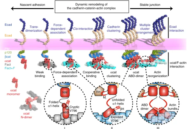
Unfolded α1-helix βH Exposed V796 V MI Vin Folded α1-helix NM α1 VCryptic V796 ABD V V Actin bundle ABD dimer Ecad Ecad p120 βcat αcat Fact Fact+F Force-dependent association Cadherin clustering Multiple cluster arrangement Cooperative binding αcat clustering αcat ABD-dimer Actin reorganization PM αcat monomer αcat N-dimer αcat/F-actin interaction Ecad interaction Weak binding Trans-dimerization
Nascent adhesion Dynamic remodeling of Stable junction the cadherin-catenin-actin complex
Force-dependent association
I II III
Cis-interaction
Fig. 7 Dynamic remodeling of the cadherin-catenin-actin complex. A model ofα-catenin-dependent cadherin-actin linkage, cadherin clustering and F-actin bundling involved in the regulation of cadherin-mediated cell-cell adhesion facilitating nascent and stable junctions. The ABD ofα-catenin bound to the cadherin-β-catenin complex is in an attenuated state with the folded α1-helix and cryptic V796 to form weak interactions with F-actin (I). α-catenin dissociated fromβ-catenin can exist as a monomer and an N-terminally linked homodimer (N-Dimer). When the cadherin-catenin complex encounters F-actin under force (Fact+ F), αcat-ABD exposes V796 on the surface while the α1-helix unfolds to form a catch bond with F-actin (II). The force propagates throughα-catenin to unfold the MIregion, which facilitates the recruitment of vinculin (Vin) to AJs. Strong F-actin binding promotes cooperative binding of
ABD. Asα-catenin clusters together on F-actin, ABD dimerization between two ABD-coated actin filaments promotes actin bundling and lateral clustering of cadherin-catenin complexes at AJs (III)