Bilateral segmental lung transplantation for children:
Transplantation using split adult living-donor lower lobe
Takahiro Oto, MD,
a,bYukiko Hikasa, MD,
c,dAkikazu Hagiyama, PT,
eand Motomu Kobayashi, MD,
cOkayama, Hyogo, and Tokyo, Japan
Video clip is available online.
The shortage of pediatric brain-dead donors is a significant limiting factor for lung transplantations in small children.1,2 In Japan, living-donor lobar lung transplantation is a widely used alternative procedure, accounting for 33% of lung transplantations in children.3 However, even lower lobe grafts might be too large to fit into a small child’s chest cav-ity.2,4,5A bilateral segmental lung transplantation using an adult living-donor lower lobe was performed on 3 children who were considered unlikely to survive until a suitable pe-diatric brain-dead donor was allocated to them.
CLINICAL SUMMARY
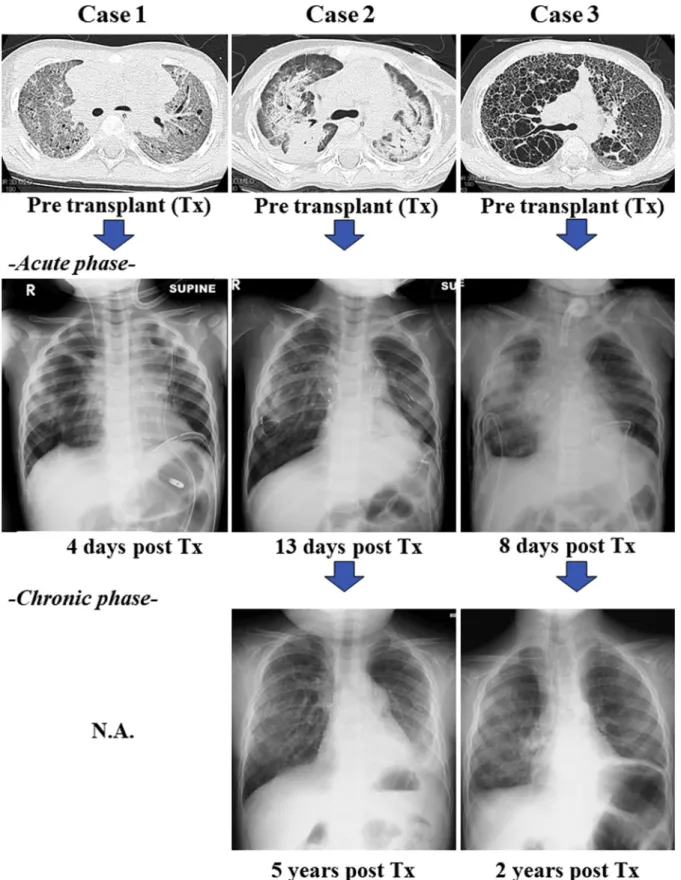
Between August 2014 and December 2018, 3 children age 1, 2, and 4 years with idiopathic pulmonary fibrosis un-derwent bilateral segmental transplantation. All 3 children were mechanically ventilated with 80% to 100% oxygen concentration before transplantation (Figure 1). Character-istics of the children and their donors are shown listed in
Table 1. The children’s parents provided informed consent. The donor operation commenced before the recipient operation. Splitting of the lower lobe into superior and basal segments was completed in vivo using electrocautery (Figure 2). The intersegmental veins were identified and preserved during division of the lung parenchyma (Video 1). The segmental grafts were reinflated, and hemostasis and sealing of air leaks from the dissected surface of the graft were secured.
The recipient operation began with hilum dissection and the initiation of cardiopulmonary bypass. The donor was then heparinized, and the bronchus and pulmonary vascula-ture was cut at the level of the lower lobe branches. Ante-grade followed by retroAnte-grade pulmonary arterial flushing was performed on a back table. The donor graft was then divided into superior and basal segments by cutting the pul-monary artery, vein, and bronchus.
In each case, the superior and basal segments were trans-planted into the recipient’s left and right chest cavities, respectively. The superior segment was transplanted without changing its direction, and the segmental vein was anastomosed to the recipient’s lower pulmonary vein. The basal segments were rotated 180 degrees horizontally and 90 degrees vertically, and then the segmental vein was attached to the recipient’s upper pulmonary vein. Two patients required delayed chest closure due to size mismatch between the graft and the recipient’s chest cavity (Table 1). The chest X-rays in acute and chronic phase are shown in Figure 1. Two patients recovered without requiring oxygen inhalation; however, 1 patient died due to legionellosis at 3 months after transplantation. The pul-monary arterial pressure of patient 2 gradually increased
A wide communication can be seen between the right side of the aorta and the right lung.
CENTRAL MESSAGE
In this case report, we had the
chance to revisit syphilitic aortitis
in the modern antibiotic era.
Despite its rarity, cases of
syphi-litic aortitis have been
increas-ingly reported recently.
See Commentary on page XXX.
From theaTransplant Center, Okayama University Hospital, Okayama;bThoracic Surgery, Himeji Daiichi Hos-pital, Hyogo;cAnesthesiology and Resuscitology, Okayama University Hospital, Okayama;dAnesthesiology, National Cancer Center, Tokyo; andePhysical Medicine and Rehabilitation, Okayama University Hospital, Okayama, Japan.
Disclosures: The authors reported no conflicts of interest.
The Journal policy requires editors and reviewers to disclose conflicts of interest and to decline handling or re-viewing manuscripts for which they may have a conflict of interest. The editors and reviewers of this article have no conflicts of interest.
Received for publication June 3, 2020; revisions received June 3, 2020; accepted for publication June 17, 2020. Address for reprints: Takahiro Oto, MD, 2-5-1, Shikata-cho, Kita-ku, Okayama 700-8558, Japan (E-mail:
[email protected]). JTCVS Techniques 2020;-:1-4 2666-2507
CopyrightÓ 2020 The Authors. Published by Elsevier Inc. on behalf of The American Association for Thoracic Surgery. This is an open access article under the CC BY-NC-ND license ( http://creativecommons.org/licenses/by-nc-nd/4.0/).
https://doi.org/10.1016/j.xjtc.2020.06.022
JTCVS TechniquescVolume-, Number - 1
SURGICAL TECHNIQUE
FIGURE 1. Acute and chronic phases of computed tomography scan and chest X-ray. Tx, Transplant; N.A., not applicable.
2 JTCVS Techniquesc- 2020
(right, 64/35/46/14 mm Hg; left, 77/38/52/16 mm Hg; sys-tolic/diastolic/mean/mean pulmonary capillary wedge pressure, respectively) with a normal cardiac index
(4.17), pulmonary vascular resistance (8.82 mm Hg/L/ min), left ventricular end-diastolic pressure (15 mm Hg), and mean aortic pressure (56 mm Hg), 68 months after
TABLE 1. Characteristics of recipients and donors and outcomes of segmental transplant
Characteristic Patient 1 Patient 2 Patient 3
Recipient age, y 1 2 4
Height/weight 79 cm/10 kg 92 cm/12 kg 79 cm/8.8 kg
Preoperative pathology NA NSIP with DAD UIP
Genetic abnormality SFTPCI73Thetero No No
Donor Mother Mother Father
Donor genetic abnormality No NA NA
Donor graft Right lower lobe Left lower lobe Right lower lobe
Graft implanted Right: basal/left: superior Right: basal/left: superior Right: basal/left: superior
Donor/recipient size matching by CT volumetry,%
213.5 200.6 188.6
Immunosuppression Tacþ MMF þ steroid Tacþ MMF þ steroid Tacþ MMF þ steroid
Extubation, d 4 13 8*
Follow-up period, mo 3 68 29
Growth of patient, cm NA 105.8 (þ13.8) 80.5 (þ1.5)
Current status Died Alive Alive
Cause of death Legionellosis NA NA
NA, Not applicable; NSIP, non-specific interstitial pneumonia; DAD, diffuse alveolar damage; UIP, usual interstitial pneumonia; SFTPC, surfactant protein C; CT, computed tomography; Tac, tacrolimus; MMF, mycophenolate mofetil. *Patient weaned from mechanical ventilation 8 days after transplantation and retained spontaneous breathing through a tracheostomy created during the preoperative period.
FIGURE 2. The donor’s lower lobe was split into 2 segmental grafts in vivo, and graft preservation, including cold flushing, was performed ex vivo. The divided basal segments were rotated at 180 degrees vertically and 90 degrees horizontally and then placed into the recipient’s right chest cavity. The re-cipient’s left lower and right upper pulmonary veins were used for venous anastomosis.
JTCVS TechniquescVolume-, Number - 3
transplantation. Therefore, the patient is now being consid-ered for a retransplantation.
DISCUSSION
A previous report described living-donor middle lobe transplantation in toddlers, stating that the “middle lobe should have single arterial, venous, and bronchial branches; otherwise, the requirement for a patch repair might increase the potential surgical risks of the donor operation.”4In the 3 current cases, middle lobe transplants were considered; however, anatomic problems, including 2 middle lobe ar-teries, were discovered in 2 donors, and 2 middle lobe veins were discovered in 1 donor during the evaluation process. We consider a size mismatch between the pediatric recipi-ent’s chest cavity and the adult donor graft of<250% to
be acceptable in a bilateral transplantation2,4,5; however, the indication may change depending on the stiffness of the recipient’s chest.
Although urgent transplantation using an adult living donor is possible, the patient is not likely to survive until a pediatric brain-dead donor is allocated. Bilateral trans-plantation is possible using a living-donor lower lobe.
The procedure does have some limitations. Retransplan-tation may be required in the near future as the recipient grows. In addition, a segmental graft carries a greater risk of bleeding and air leak compared with a whole lung or lobe.
Our experience with these cases demonstrates that bilat-eral segmental lung transplantation using a split adult living-donor lower lobe could bridge these children to suit-able pediatric brain-dead transplants.
References
1.Sato M, Okada Y, Oto T, Minami M, Shiraishi T, Nagayasu T, et al. Registry of the Japanese Society of Lung and Heart-Lung Transplantation: official Japanese lung transplantation report, 2014. Gen Thorac Cardiovasc Surg. 2014;62:594-601. 2.Oto T. Living donor lobar lung transplantation. Kirklin J, ISHLT monograph
series, Pediatric Lung Transplantation. Volume 7, Birmingham, AL: University of Alabama at Birmingham Printing; 2013:220-7.
3.The Japanese Society of Lung and Heart-Lung Transplantation. Registry Report of Japanese Lung Transplantation -2019. Jpn J Transplant. 2019; 54:105-10.
4.Oto T, Miyoshi K, Sugimoto S, Yamane M. Living related donor middle lobe lung transplant in a pediatric patient. J Thorac Cardiovasc Surg. 2015;149:42-4. 5.Oto T, Date H, Hayama M, Ando A, Shimizu N. Peripheral lung volume reduction
improved early graft function in severe size mismatched living donor lobar lung transplantation. Transplant Proc. 2005;37:4515-21.
VIDEO 1. Donor and recipient operations. Video available at:https:// www.jtcvs.org/article/S2666-2507(20)30300-X/fulltext.
4 JTCVS Techniquesc- 2020