INTRODUCTION
NC/Nga (NC) mice have been shown to develop human atopic-like skin lesions with elevated se-rum IgE level when kept in conventional conditions (1, 2). NC mice have been used as model animals for evaluating the effects of anti-inflammatory drugs and food-derived components on the development of atopic dermatitis. In addition to allergic response, NC mice have been shown to possess unique im-munological phenotypes, including a defect in toll-like receptor 9-mediated activation, deletion poly-morphysm of the FcγIIB gene promoter region, and a defect in oral tolerance (3-5). In this study, we found that NC mice have and impaired antigen (Ag)-specific T cell response after ovalbumin (OVA)
immunization. Therefore, we focused on the func-tion of antigen (Ag)-presenting cells in NC mice
MATERIALS AND METHODS
Mice and immunizationSpecific pathogen-free female NC (H-2d) (6) ,
BALB/c (H-2d) and DBA/2 (H-2d) mice were
pur-chased from SLC (Hamamatsu, Japan). DO11.10 mice were purchased from Jackson Lab. (Bar Harbor, ME, USA). All studies were performed in accordance with the ethical guidelines for animal experimentation by the Institution of Health Bi-oscience, the University of Tokushima and were ap-proved by the institutional review board of the ani-mal ethics committee.
Mice were intraperitoneally immunized with 10μg of OVA (Sigma Chemical Co., MO, USA) absorbed in 1 mg of Aluminium Hydroxide Gel Adjuvant (HCI Biosector, Denmark) 2 times at a 2-week interval.
ORIGINAL
Decreased expression of MHC class II and cathepsin E in
dendritic cells might contribute to impaired induction
of antigen-specific T cell response in NC/Nga mice
Tohru Sakai, Emi Shuto, Tomoyo Taki, Honami Imamura, Miku Kioka,
Akiko Nakamoto, Seiji Mizoguchi, Shouko Amano, Yukari Kawano,
Mariko Nakamoto, Rie Tsutumi, and Toshio Hosaka
Department of Public Health and Applied Nutrition, Institution of Health Bioscience, the University of Tokushima Graduate School, Tokushima, Japan
Abstract : NC/Nga (NC) mice are an animal model for human atopic dermatitis. We found that induction of antigen (Ag)-specific T cell response is diminished in ovalbumin (OVA)-immunized NC mice. Ability of Ag presentation in NC mouse dendritic cells (DCs) was sig-nificantly weaker than that in BALB/c and DBA/2 mouse DCs. Expression levels of MHC class II molecules and cathepsin E in NC mouse DCs were significantly lower that those in BALB/c and DBA/2 mouse DCs. These results indicate that low expression levels of MHC class II and cathepsin E might contribute to the defect in induction of Ag-specific T cells in NC mice. J. Med. Invest. 59 : 95-101, February, 2012
Keywords : cathepsin E, dendritic cells, MHC class II, NC/Nga mice
Received for publication October 28, 2011 ; accepted November 21, 2011.
Address correspondence and reprint requests to Tohru Sakai, Department of Public Health and Applied Nutrition, Institution of Health Bioscience, the University of Tokushima Graduate School, Kuramoto cho, Tokushima 770 8503, Japan and Fax : + 81 88 -633 - 9427.
Proliferation assay
Splenocytes from OVA - immunized mice were cultured in the medium supplemented with 10% fe-tal bovine serum, 50μmol/L 2-mercaptoethanol, 100μg/mL streptomycin, and 100 U/mL penicillin. Five
!
105splenocytes were stimulated with 200μg/mL OVA, 1,000μg/mL OVA or with plate-bound anti-CD3 mAb (coated overnight at 1μg/mL) in a 96-well flat-bottom plate at 37"!under 5% CO2for
72 h. For the last 8 h of culture, 1 μCi of [3H]
thymidine deoxyribose (TdR) was added to the wells, and the amount of [3H]TdR incorporated was
measured by a scintillation counter (Aloka, Tokyo, Japan).
Ag-presenting ability
CD11c+cells were purified from 3 to 4 mice
us-ing CD11c microbeads and a MACS LS column (Miltenyi Biotec Inc., CA, USA) according to the manufacturer’s instructions. CD4+T cells were
pu-rified from DO11.10 transgenic mice, which express OVA-specific T cell receptor, using a CD4+T cell
isolation kit (Miltenyi Biotec). CD4+T cells (2
!
105cells) from a DO11.10 mouse spleen were cultured in a 96-well plate with CD11 c+cells (104cells) in
the presence of 200μg/ml OVA for 48 hr at 37"!/ 5% CO2. Cultures were pulsed with 1μCi of [3H]TdR
for the last 8 hr of culture. Incorporated [3H]TdR
was determined by a scintillation counter.
Flow cytometry
Expression of MHC class I, MHC class II and CD80 molecules was analyzed by flow cytometric analysis. Purified CD11c+cells were incubated with
FITC-conjugated anti-mouse H-2KdmAb (clone
SF1-1.1), PE-conjugated anti-mouse I-A mAb (clone M5/114/15.2) or FITC-conjugated anti-CD80 mAb (clone 16-10A1) or 30 min at 4"!. After washing the cells, stained cells were analyzed with a FACSCalibur and CellQuest software (Becton Dickinson, CA, USA).
Quantitative PCR analysis
To assess gene expressions, mRNA levels of genes involved in MHC class II expression were de-termined by reverse transcription of total RNA fol-lowed by PCR analysis. Total RNA was isolated from CD11c+ cells using a RNeasy Micro kit (Qiagen
Science, MD, USA). First-strand cDNA was reverse-transcribed at 42"!for 60 min and at 95"!for 5 min from 2μg of the extracted total RNA with M-MLV
reverse transcriptase (Invitrogen, CA, USA) and a random primer. We performed real-time PCR us-ing specific primers and SYBR green dye (Takara Bio, Japan) in a Light Cycler real-time PCR system (Roche Diagnostics, Germany) according to the manufacturer’s instructions. The primers used were 5’ - CAGATACCATCAACTGCGACCA - 3’ (sence) and 5’-TCTGCTCCAATGTGCTCTATGAAG-3’ (an-tisense) for CTIIT, 5’-AGTCAGTGGAGGCCAGC-TTA-3’ (sense) and 5’-CCAGGAGGCACCAACTG-TAT-3’ (antisence) for NF-Yα, 5’-GCTGACGCTG-GAGAACCTAC-3’ (sense) and 5’-CGATGGGAG-CTGAAGGTAGA-3’ (antisense) for RFX5, 5’-CAC-AGACGGCGTTTATGAGA-3’ (sense) and 5’-TGC-TCCACCTTGCAGTCATA-3 (antisense) for H-2d
, 5’-CTGACCCTGAAGTACCCCATTGAACA-3’ (sense) and 5’-CTGGGGTGTTGAAGGTCTCAAACATG-3 (antisense) forβ-actin’. The relative abundance of target transcripts was normalized to the constitutive expression ofβ-actin.
Western blot analysis
CD11c+cells were lysed in lysis buffer containing
1% Noidet-P40, 150 mM NaCl, 10 mM Tris-Cl (pH 7.5), and 1 mM EDTA. The cell lysates were disso-loved by SDS-PAGE and transferred onto a PVDF membrane. The membrane was blotted with an Ab to cathepsin S (Santa Cruz, CA, USA), cathepsin E (Novas Biologicals, CO, USA) orβ-actin (Epitomics, Inc., CA, USA) and visualized using an enhanced chemiluminescence system (Amersham Biosciences, UK). Quantification of protein expression was per-formed by densitometry analysis of the autoradio-grams.
Statistical analysis
Data were analyzed using one-way analysis of variance followed by the Scheffe post hoc test for multiple comparisons. Values in the text are means" SD. Differences were considered significant at P!
0.05.
RESULTS
Decreased Ag-specific T cell response in OVA-immunized NC mice
OVA-specific proliferation response after OVA im-munization in NC mice was significantly decreased compared to that in BALB/c and DBA/2 mice. However, proliferation response to anti-CD3 mA in NC mice was comparable to that in BALB/c and
DBA/2 mice (Fig. 1), suggesting that T cell recep-tor-mediated signal transduction is normal in NC mice. This finding indicates the possibility that weak induction of Ag-specific T cells in NC mice contrib-utes to the process of Ag-specific T cell induction.
Characterization of CD11c+
dendritic cells (DCs) from NC mice
We next examined CD11c+cell function because
DCs are known to be a crucial cell population for in-itiation of acquired immune responses. Ag-present-ing ability of DCs was evaluated by usAg-present-ing DO11.10 CD4+cells that carried the OVA-specific T cell
re-ceptor gene. In the Ag presentation process toward
to MHC class II, at first Ag-presenting cells capture Ag, digest the captured Ag in the lysosome, and then present Ag-derived peptide/MHC class II com-plex on the cell surface. In the case of intact OVA stimulation, proliferation response of DO11.10 CD4+
cells when co-cultured with NC mouse CD11c+cells
was significantly decreased compared to that when co-cultured with BALB/c or DBA/2 mouse CD11c+
cells (Fig. 2). We evaluated the ability of Ag-inter-nalization in NC mouse CD11c+ cells using
FITC-labeled OVA, but a significant difference was not ob-served compared to that in BALB/c mouse CD11c+
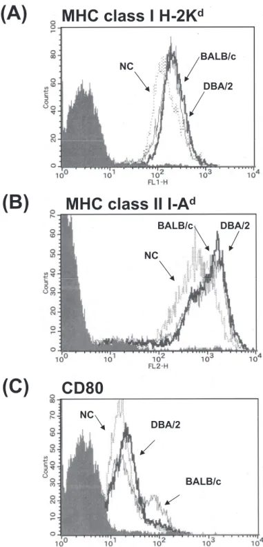
cells (data not shown). Next, cell surface expres-sions of MHC class I, MHC class II, and CD80 were
Fig. 1. OVA specific T cell responses in BALB/c, DBA/2 and NC mice. Mice were immunized with OVA 2 times at a 2 week -interval. Two weeks after the second immunization, spleen cells were stimulated with OVA or anti - CD3 mAb for 72 hr in vitro. Pro-liferation response was determined by counting incorporated [3H]TdR activity. Results are expressed as means!SD. White, black and hatched bars indicate BALB/c, DBA/2 and NC mice, respectively. Data are presented as means!SD. **P!0.01.
Fig. 2. Characterization of NC mouse DCs. DO11.10 CD4+cells were stimulated with OVA in the presence of indicated number of DCs from BALB/c, DBA/2 or NC mice. Ag - presenting ability of DCs was evaluated by incorporated [3H]TdR activity. Levels of in-corporated [3H]TdR were less than 1,500 cpm when DO11.10 CD4+cells or DCs alone were cultured with OVA. Levels of incorporated [3H]TdR were less than 500 cpm when DO11.10 CD4+cells or DCs alone were cultured with medium only. Results are expressed as means!SD. The data are representative of two independent experiments of pooled samples from 3 or 4 mice.
determined by flow cytometric analysis. Expression level of MHC class II in NC mouse CD11c+ cells
was significantly lower that that in BALB/c mouse CD11c+cells. Reduced MHC class II expression was
observed not only in DCs but also in B cells in NC mice (data not shown). In the case of MHC class I and CD80, expression of these molecules in NC
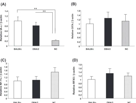
mouse DCs was slightly lower than that in BALB/c and DBA/2 mouse DCs (Fig. 3). Although we also evaluated transcription factors that contribute to MHC class II expression, differences in the expres-sion of CIITA, NF-Yα and RFX5 were not observed between NC, BALB/c and DBA/2 mice (Fig.4 B-D).
Fig. 3. Expression of MHC class I, MHC class II and CD80 in DCs from BALB/c, DBA/2 and NC mice. Expression of MHC class II was determined as described in the “Materials and methods” section. Shaded histograms represent cells stained with an isotype -matched control Ab.
Decreased expression of cathepsin E in NC mouse CD11c+DCs
Cathepsin S is a member of the papain family of cystein proteases (7), and cathepsin E is an intra-cellular aspartic protease of the pepsin superfamily (8). Both molecules have been shown to be crucial for Ag processing and presentation in Ag-presenting
cells (9). Expression of cathepsin S and cathepsin E in DCs was determined by Western blot analysis. A difference in cathepsin S expression was not ob-served between BALB/c, DBA/2 and NC mouse CD11c+cells. In contrast, it was found that
expres-sion of cathepsin E in NC mouse DCs was signifi-cantly decreased compared to that in BALB/c and DBA/2 mouse DCs (Fig. 5). Densitometry analysis
Fig. 4. CIITA, NF - Yα and RFX5 mRNA expression levels in DCs. cDNA was prepared from purified CD11c+cells and analyzed for expression of CIITA (A), NF - Yα (B), RFX5 (C) and β-actin message by quantitative RCR as described in the “Materials and methods” section. Data are presented as means!SD. **P!0.01.
Fig. 5. Expression of cathepsin E and S in DCs. (A) Expression of cathepsin E and cathepsin S was analyzed by Western blotting with anti cathepsin E and anti cathepsin S mAb. To ensure the presence of equal amounts of protein, the membranes were re -probed with an anti -β-actin mAb. (B) Histograms show the cathepsin E/β-actin and cathepsin S/β-actin ratio. Data are presented as means!SD. *P!0.05.
of autoradiograms showed that the relative expres-sion level of cathepsin E in NC mice is one-tenth of that in BALB/c and DBA/2 mice.
DISCUSSION
NC mice provide a new animal model for the study of atopic-like dermatitis and allergy disease. The contribution of possible immunological factors to the development of allergic response in NC mice has been revealed (6, 10-12). In addition to the de-velopment of allergic response, it has been shown that NC mice posses unique immunologic pheno-types [3-5]. In the course of investigating the mecha-nism for the onset of allergic response in NC mice, we found that Ag-specific T cell response following Ag immunization was severely impaired (Fig. 1). To elucidate the mechanism for this phenotype, we focused on DC function in NC mice. Consistent with the impaired induction of Ag-specific T cells, Ag-presenting ability in NC mice was significantly weaker than that in BALB/c and DBA/2 mice (Fig. 2). Furthermore, expression level of MHC class II in NC mouse DCs was about half of that in BALB/c mouse DCs (Fig. 3). The main control ele-ment regulating transcription is a highly conserved 150-bp promoter proximal region situated upstream of the transcription initiation site. Additional pro-moter distal regulatory elements have been identi-fied (13). At first, we examined the nucleotide se-quence in conserved regulatory elements, named distal Y’-S’ motif and proximal S-Y motifs, between NC and BALB/c mice, but a difference was not found. In addition, expression of DNA-binding pro-teins, CIITA, RFX and NF-Y, were examined by real-time PCR assay and no differences in expres-sion levels were found (Fig.4 B-D).
Several lines of evidence suggest that cathepsin E plays a role in Ag processing via the MHC class II pathway (14). Treatment with a cathepsin E-specific inhibitor inhibits Ag processing, resulting in an im-paired Ag-specific T cell response. Similar to the re-sults obtained by using a specific inhibitor, cathepsin E-deficient mouse DCs showed reduced ability of antigen presentation. Cathepsin E is mainly present in the endosome compartment in DCs. In this study, we found that expression of cathepsin E, but not that of cathepsin S, in NC mouse DCs was weak. Re-duced Ag presenting ability of exogenous Ag in NC mouse DCs was similar to that in cathepsin E- defi-cient mouse DCs or DCs treated with a cathepsin
E-specific inhibitor. From these findings, it seems likely that a low level of cathepsin E expression con-tributes to defect in Ag-presenting ability in NC mice. However, it is not known whether a low level of cathepsin E expression directly affects the expres-sion of MHC class II because expresexpres-sion of MHC class II in cathepsin E-deficient mouse DCs was comparable to that in wild - type mice (15). We speculate that decreased expression of MHC class II and cathepsin E synergistically affect Ag-present-ing ability in DCs, resultAg-present-ing in impaired induction of Ag-specific T cell response in NC mice.
NC mice have been shown to develop human atopic-like skin (1, 2). Interestingly, cathepsin E-de-ficient mice develop atopic-like dermatitis. Tsukuba et al. showed that development of atopic dermati-tis in cathepsin E-deficient mice contributes to the systemic accumulation of IL-18 and IL-1β. They also showed cathepsin E activity in erythrocyte ghosts in NC mice and found that cathepsin E ac-tivity in NC mice was lower than that in C57BL/6 mice (16).
In this study, we found a new immunologic phe-notype in NC mice. Low MHC class II and cathep-sin E expression levels in an experimental animal have not previously been reported. Therefore, NC mice will provide a new useful animal model for investigation of induction of Ag-specific immune response and acquired immunity.
CONFLICT OF INTEREST
None of the authors have any conflicts of interest to declare.
ACKNOWLEDGMENTS
This work was partially supported by research Grant-in Aid for Scientific Research.
REFERENCES
1. Matsuda H, Watanabe N, Geba GP, Sperl J, Tsudzuki M, Hiroi J, Matsumoto M, Ushio H, Saito S, Askenase PW, Ra C : Development of atopic dermatitis-like skin lesion with IgE hy-perproduction in NC/Nga mice. Int Immunol 9 : 461-466, 1997.
The NC/Nga mouse : a model for atopic der-matitis. Mol Med Today 6 : 209-210, 2000. 3. Sakai T, Kogiso M, Mitsuya K, Komatsu T,
Yamamoto S : Defect of toll-like receptor 9-mediated activation in NC/Nga mouse macro-phages. Immunol Lett 106 : 91-95, 2006. 4. Sakai T, Kogiso M, Mitsuya K, Komatsu T,
Yamamoto S : Deletion polymorphisms in the promotor region of Fcγ receptor IIB is not asso-ciated with antigen-specific IgG2a and IgG2b antibody responses in NC/Nga Mice. Micro-biol Immunol 50 : 475-480, 2006.
5. Sakai T, Kogiso M, Mitsuya K, Komatsu T, Yamamoto S : Defect of oral tolerance in NC/ Nga mice. J Med Invest 53 : 29-33, 2006. 6. Matsumoto M, Itakura A, Tanaka A, Fujisawa
C, Matsuda H : Inability of IL-12 to down-regu-late IgE synthesis due to defective production of IFN-γ in atopic NC/Nga mice. J Immunol 167 : 5955-5962, 2001.
7. Nakagawa TY, Brissette WH, Lira PD.
Griffiths RJ, Petrushova N, Stock J, McNeish JD, Eastman SE, Howar ED, Clarke SR : Im-paired invariant chain degradation and antigen presentation and diminished collagen-induced arthritis in cathepsin S null mice. Immunity 10 : 207-217, 1999.
8. Zaidi C, Kalbacher H : Cathepsin E : A mini re-view. Biochem Biophys Res Commun 367 : 517-522, 2008.
9. Chapman HA, Riese RJ : Cathepsins and com-partmentalization in antigen presentation. Cur-rent Opin Immunol 12 : 107-113, 2000.
10. Matsumoto M, Itakura A, Tanaka A, Fujisawa C, Matsuda H : Inability of IL-12 to down-regu-late IgE synthesis due to defective production
of IFN-γ in atopic NC/Nga mice. J Immunol 167 : 5955-5962, 2001.
11. Tenda Y, Yamashita M, Kimura M, Hasegawa A, Shimizu C, Kitajima M, Onodera A, Suzuki A, Seki N, Nakayama T : Hyperresponsive Th2 cells with enhanced nuclear factor-κB activa-tion induce atopic dermatitis-like skin lesions in Nishiki-nezumi Cinamon/Nagoya mice. J Allergy Clin Immunol 118 : 725-733, 2006. 12. Vestergaard C, Yoneyama H, Murai M,
Nakamura K, Tamaki K, Terashima Y, Imai T, Yoshie O, Irimura T, Mizutani H, Matsushima K : Overproduction of Th2-specific chemokines in NC/Nga mice exhibiting atopic dermatitis-like lesions. J Clin Invest 104 : 1097-1105, 1999. 13. Mach B, Steimle V, Martinez-Soria E, Reith W : Regulation of MHC class II genes : lessons from a disease. Annu Rev Immunol 14 : 301-331, 1996.
14. Bennett K, Levine T, Ellis J.S, Peanasky RJ, Samloff IM, Kay J, Chain BM : Antigen proc-essing for presentation by class II major histo-compatibility complex requires cleavage by cathepsin E. Eur J Immunol 22 : 1519-1524, 1992.
15. Chain BM, Free P, Medd P, Swetman C, Tabor AB, Terrazzini N : The expression and function of cathepsin E in dendritic cells. J Immunol 174 : 1791-1800, 2005.
16 Tsukuba T, Okamoto K, Okamoto Y, Yanagawa M, Kohmura K, Yasuda Y, Uchi H, Nakahara T, Furue M, Nakayama K, Kadowaki T, Yamamoto K, Nakayama KI : Association of cathepsin E deficiency with development of atopic dermatitis. J Biochem 134 : 893-902, 2003.