INTRODUCTION
As the safety of hepatic resection has improved and its benefits for patients with primary and sec-ondary tumors of the liver have become clearer, a more aggressive approach of the management of liver lesions has been adopted. This has led to the definition of new pathologies and new lesions (1). A solitary necrotic nodule of the liver is an
ex-tremely rare non-malignant lesion described by Shepherd and Lee in 1983 (2). The etiology of the lesion remains uncertain. Despite the develop-ment of imaging modality, a preoperative diagno-sis is very difficult because ultrasound and radio-graphic patterns are similar to those of the metas-tatic lesions (3, 4). Specific patterns of this lesion on preoperative examinations have not yet been identified.
In this study we report about a solitary necrotic nodule with rapid growth located in left lobe of the liver that had the similar presence as that of me-tastatic tumors.
CASE REPORT
Rapid-growing solitary necrotic nodule of the liver
Satoru Imura, Kotaro Miyake, Tetsuya Ikemoto, Yuji Morine, Masahiko Fujii,
Nobuya Sano
1, and Mitsuo Shimada
Department of Digestive and Pediatric Surgery, Institute of Health Biosciences, The University of Tokushima Graduate School, and1
Department of Pathology, Tokushima University Hospital, Tokushima, Japan
Abstract : The solitary necrotic nodule of the liver is an uncommon nonmalignant lesion with an uncertain etiology. The lesion was defined as a nodule with a completely necrotic core and fibrous capsule etc. and without a consistency of viable cells. The characteristic fea-tures of this benign lesion on the imaging modalities are similar to the metastatic tumor. In this paper we discuss the case of a rapid-growing solitary necrotic nodule of the liver occurring in a patient with chronic renal failure on hemodialysis. The lesion located on the left median lobe of the liver had rapidly enlarged in diameter in the last seven months. Despite some examinations by imaging modalities to confirm the preoperative diagno-sis, we were unable to visually confirm. Several histological examinations using a needle biopsy specimen were performed, but the diagnosis was all necrotic tissue. However, we recommended an extended left hepatic lobectomy for this rapid-growing lesion since cholangiocarcinoma with necrosis could be hardly differentiated. Permanent histology revealed that the lesion was solitary necrotic nodule. We consider that permanent his-tology of the entire lesion is possibly the only accurate method of diagnosis. Since the solitary necrotic nodule does not indicate malignancy, hepatic resection should be per-formed. J. Med. Invest. 53 : 325-329, August, 2006
Keywords : solitary necrotic nodule, liver tumor
Received for publication May 31, 2006 ; accepted July 12, 2006. Address correspondence and reprint requests to Satoru Imura, M.D., Ph.D., Department of Digestive and Pediatric Surgery, Institute of Health Biosciences, The University of Tokushima Graduate School, Kuramoto-cho, Tokushima 770-8503, Japan and Fax : +81-88-631-9698.
The Journal of Medical Investigation Vol. 53 2006
CASE
A 62-year-old woman undergoing hemodialysis for chronic renal failure for 15 years was admitted for evaluation of a huge liver tumor, which was dis-covered using abdominal examinations for epigas-tralgia, with rapid enlargement in diameter from 3 cm to over 8 cm for the past 7 months. There are no whats should be mentioned specially in the pre-vious history and the family medical history ex-cluding having received the hemodialysis due to renal failure. The serum CEA and CA 19-9 levels were 4.3 ng/ml (normal range : < 2.5 ng/ml) and 52 IU/ml (normal range: < 47 IU/ml), respectively. The serum alpha-fetoprotein level was normal. Laboratory data on admission was shown in Table 1. The ultrasonography (US) showed the presence of a hypoechoic area measuring 8.5 cm in Cou-inaud’s segment 3 of the liver with reticular calcifi-cation measuring about 3 cm at the center of the hypoechoic area. A dynamic computed tomogra-phy (CT) showed the presence of a lobular shaped low-density area without intratumoral enhance-ment (Fig. 1). A CT during hepatic angiography was performed, but further findings other than that from the dynamic CT scan were not acquired (Fig. 2). Magnet resonance imaging (MRI) showed iso-high intensity lesion with low intensity ring and high intensity central area on T 1 weight image and high intensity lesion with slight high intensity area in center (Fig. 3). However, we could not decide the preoperative diagnosis of this lesion from the find-ings of these imaging modalities. Then we
per-formed a liver biopsy for the purpose of definition of diagnosis, but the pathological findings were “ne-crotic tissue”. Systemic examinations were per-formed to investigate the possibility of metastasis from the digestive tract or gynecological
malig-Fig. 1. Contrast-enhanced CT ; (a) : July 2003, (b) : March
2004
Low-density lesion with central calcification was located in the left median lobe and it had been growing rapidly in diameter in the last 7 months. Intranodular enhancement was not found in any phase of the dynamic studies.
Fig. 2. Angiography and CT during angiography
(a) : Portography (b) : CT during the portography (CT-AP)(c) : Hepatic arteriography (d): CT during hepatic arteriography (CT-A) Findings of the intratumoral enhancement and the vascular en-casement were not detected in all these studies. Only the re-gion around the lobular-shaped nodule was enhanced on CT-A.
Fig. 3. MRI
(a) : T1 weight image (b) : T2 weight image
MRI shows that iso-high intensity lesion with low intensity ring and high intensity central area on T1 weight image. T2 weight MRI shows high intensity lesion with slight high intensity area in center.
Table 1. Laboratory data on admission
WBC 4300/µl AST 22 IU/l RBC 333×104/µl ALT 9 IU/l Hb 11 g/dl LDH 202 IU/l Ht 35% T-Bil 0.5 mg/dl Plt 13.3×10⁴ ALP 407 IU/l γ-GTP 39 IU/l PT 12.1 s AMY 219 IU/l HPT 121.8% BUN 66 mg/dl HBs-Ag (−) Cre 9.13 mg/dl HCV-Ab (−) Na 140 mEq/l K 4.6 mEq/l CEA 4.3 ng/ml Cl 103 mEq/l CA19-9 52 mAU/ml CRP 1.1 mg/dl AFP 4 ng/ml PIVKA-II 18 mAU/ml
S. Imura, et al. Solitary necrotic nodule of the liver
nancy but there were no abnormal findings indi-cating malignancy as a possible origin of metasta-sis. Then, we considered that this lesion with rapid enlargement and malignant possibility was suitable for operative treatment.
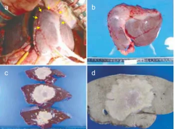
The extended left hepatic lobectomy including a left caudate lobectomy was performed. The resected specimen revealed the presence of a lobular shaped, nonencapsulated nodule, measuring 8.5 cm with a hard consistency. There was a reticular calcification measuring about 3 cm at the center of the nodule (Fig. 4).
A histological examination revealed that the lesion consisted of a homogeneous necrosis without vi-able cells, rimmed by thin granulation tissue (Fig. 5 a, b). No specific inflammatory change was shown throughout the lesion and no infectious agents were detected using Ziel-Neelsen, Giemsa, PAS and Grocott stainings. Immunohistochemistry for cy-tokeratins failed to demonstrate epithelial elements in the necrotic area and circumscribing granula-tion tissue. Although amyloid deposigranula-tion was not confirmed in the lesion, scattered amorphous mi-crocalcification was observed mainly in the center of the necrotic area (Fig. 5c). The surrounding liver did not show significant pathologic changes (Fig. 5 d).
The postoperative course was uneventful and the patient was leaving the hospital on day 15 post-operation. This patient has been followed closely because the serum CEA and CA 19-9 levels were not normalized after surgery.
DISCUSSION
A solitary necrotic nodule of the liver is a rare lesion with fewer than 50 cases reported in the world-wide research literature (2-14). The lesions are not symptomatic and almost all reported cases have been incidental findings at postmortem, operation, or radiological investigation.
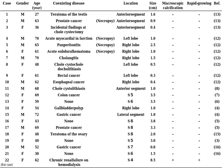
Table 2 shows the comparison of characteristics of our case and that of 21 cases of solitary necrotic nodule of the liver where there was a detailed men-tion about clinical and radiological findings after 1990 (3-8, 12, 13, 15, 16). Although 17 cases had the lesion in the right hepatic lobe, only 5 cases in-cluding our case had the left hepatic lobe. Fifteen out of 22 cases were cases where the tumor size was equal to or less than a diameter 2.0 cm and all lesions were less than 5 cm in diameter except our reported case. In our case the tumor enlarged rap-idly during the course of follow up which is a rare occurrence. In addition, our case was only one that presented macroscopic calcification on US or CT. These findings are particularly different from past reports. The cause that lesion had been rap-idly grown though the central calcification had not been enlarged was unclear.
Recent developments in hepatic surgery have lead to an increase in the number of candidates for partial hepatic resection for metastatic tumors. The major roles of imaging are to provide clear and de-tailed information of hepatic metastasis and to make
Fig. 4. Operative findings
(a) : The lesion was located in the left median lobe (arrows) and palpation revealed the nodule was very hard.
(b)-(d) : Resected specimens
Fig. 5. Histological findings
(a) : The lesion was composed simply of homogeneous necrosis. (b) : The necrotic area was separated from liver parenchyma by thin granulation zone.
(c) : Microcalcification was preferably observed in the central portion of the necrotic area.
(d) : No significant pathologic change was detected in the sur-rounding liver tissue.
a careful preoperative selection of patients, thus avoiding unnecessary surgical exploration. Ad-vances in imaging modality, i.e. US, CT and MRI improved the detection of hepatic metastasis. However, the radiologic features of these modali-ties are not sufficiently specific for the accurate characterization of liver tumors (4). Despite the various examinations there were no findings, which were strongly suspected of having a solitary ne-crotic nodule of the liver, although the view on an imaging specific to a solitary necrotic nodule of the liver had not been established, and metastatic tumor, cholangiocarcinoma and cystadenocarci-noma were considered as a possible diagnosis.
Currently the only reliable way to make the di-agnosis of a solitary necrotic nodule is permanent histology of the entire lesion. Characteristically, this lesion shows a completely necrotic core with a dense hyalinized fibrous capsule containing elastin fibers. Micro calcification is also a characteristic feature, although our case had an obvious central
calcifica-tion on imaging modalities. The Ziel-Neelsen, Gram, and PAS stains did not reveal bacteria or fungi (2). Also the pathological findings of our reported case indicated that the uniform necrotic tissue with focal calcification and invasion of inflammatory cells in the portal area of adjacent liver were compatible. This result was not contradicted with characteris-tics of a solitary necrotic nodule of the liver.
Our reported case had been undergoing hemo-dialysis for chronic renal failure for 15 years. To our knowledge, there was no report that the hemodialysis over a long period of time was participating in the generating of a solitary necrotic nodule of the liver. The view of amyloidosis that the hemodialysis over a long period of time also in pathological study which was considered to be the cause was not seen.
Although a preoperative liver biopsy was performed in this case, we did not suspect solitary necrotic nodule and assumed that the lesion was a cholan-giocarcinoma or metastatic tumor. When a necrotic lesion was suspected from the histological
find-Table 2. Comparison of the characteristics of our and 21 case of solitary necrotic nodule of the liver that there was a detailed mention about clinical and radiological and findings after1990
Case Gender Age (year)
Coexisting disease Location Size
(cm)
Macroscopic calcification
Rapid-growing Ref.
1 M 27 Teratoma of the testis Anteriorsegment 1.6 ‐ ‐ (13)
2 M 63 Prostate cancer (Necropsy) Anteriorsegment 0.9 ‐ ‐ (13)
3 F 36 Incidental findings at chole cystectomy
Anteriorsegment 0.4 ‐ ‐ (13)
4 M 70 Acute myocardial in farction (Necropsy) Left lobe 1.0 ‐ ‐ (12)
5 M 65 Panperitonitis (Necropsy) Right lobe 2.5 ‐ ‐ (12)
6 F 61 Acute subduralhematoma (Necropsy) Right lobe 1.0 ‐ ‐ (12)
7 M 79 Cholangitis Right lobe 1.5 ‐ ‐ (12)
8 F 48 Chole cystochole
docholithiasis
Left lobe 0.5 ‐ ‐ (12)
9 F 61 Rectal cancer Left lobe 0.3 ‐ ‐ (12)
10 M 62 Esophageal cancer Right lobe 0.4 ‐ ‐ (12)
11 M 68 Chole cystolithiasis Anterior segment 1.0 ‐ ‐ (8)
12 F 69 Colon cancer S5 3.5 ‐ ‐ (7)
13 F 59 None S6 3.5 ‐ ‐ (6)
14 F 54 Gallbladderpolyp Right lobe 1.0 ‐ ‐ (4)
15 M 72 Gastric cancer Lateral segment 1.0 ‐ ‐ (4)
16 F 43 None S8 3.8 ‐ ‐ (5)
17 M 69 Prostate cancer S8 3.3 ‐ ‐ (5)
18 F 48 Teratoma of the ovary S8 2.0 ‐ ‐ (15)
19 F 40 None S5 3.0 ‐ ‐ (3) 20 M 52 Gastric cancer S7 0.8 ‐ ‐ (16) 21 F 30 None S6 1.5 ‐ ‐ (16) 22 (Our case) F 62 Chronic renalfailure on hemodialysis S4 8.5 + +
S. Imura, et al. Solitary necrotic nodule of the liver
ings and there was no evidence of a malignant liver tumor or metastatic tumor, a solitary necrotic nodule of the liver should be taken into considera-tion. However, a needle biopsy could not make a clear diagnosis in this case. A permanent histologi-cal examination of the entire lesion may be the only accurate method of diagnosis for a solitary necrotic nodule of the liver.
The solitary necrotic nodule of the liver, which we reported, is mentioned as being very rare to have generated in the left hepatic lobe and to have grown rapidly, in addition to that the patient was a woman undergoing hemodialysis for chronic renal failure.
In conclusion, although a solitary necrotic nodule of the liver is a benign lesion, since the rapid-growing lesion cannot indicate malignancy, he-patic resection should be confirmed as the best treatment.
REFERENCES
1. Nakamura Y : Non-neoplastic lesions in the liver. Pathology 45 : 703-714, 1995
2. Shepherd NA, Lee G : Solitary necrotic nodules of the liver simulating hepatic metastasis. J Clin Pathol 36 : 1181-1183, 1983
3. Koea J, Taylor G, Miller M, Rodgers M, McCall J : Solitary necrotic nodule of the liver : a riddle that is difficult to answer. J Gastrointest Surg 7: 627-630, 2003
4. Yoon KH, Yun KJ, Lee JM, Kim CG : Solitary necrotic nodules of the liver mimicking hepatic metastasis:Report of two cases. Korean J Radiol 1 : 165-168, 2000
5. De Luca M, Luigi B, Formisano C, Formato A, De Werra C, Cappuccio M, Loffredo A, Forestieri P : Solitary necrotic nodule of the liver
misin-terpreted as malignant lesion : Considerations on two cases. J Surg Oncol 74(Suppl): 219-222, 2000
6. Alfieri S, Carriero C, Doglietto GB, Pacelli F, Crucitti F : Solitary necrotic nodule of the liver : diagnosis and treatment. Hepato-Gastroenterology 44 : 1210-1211, 1997
7. Carella R, Fortunato C, Gubinelli M, D’Errico A, Mancini AM : Solitary necrotic nodule of the liver simulating a metastasis. Pathologica 85 : 573-577, 1993
8. Clouston AD, Walker NI, Prociv P : Parasitic origin of a solitary necrotic nodule of the liver. J Clin Pathol 46 : 578, 1993
9. Berry CL : Solitary necrotic nodule of the liver ; a non-existent lesion. J Pathol 146 : 263 A, 1985 10. Berry CL : Solitary“necrotic nodule” of the liver : a probable pathogenesis. J Clin Pathol 198 : 1278-1280, 1985
11. Shepherd NA : Solitary necrotic nodule : J Clin Pathol 43 : 348-349, 1990
12. Tsui WM, Yuen RW, Chow LT, Tse CC : Soli-tary necrotic nodule of the liver : parasitic origin? J Clin Pathol 45 : 975-978, 1992
13. Sundaresan M, Lyons B, Akosa AB : ’Solitary’ necrotic nodules of the liver : an aetiology re-affirmed. Gut 32 : 1378-1380, 1991
14. Desai S, Prabhu SR, Shrividya S:Fibrosing ne-crotic nodule of the liver. Indian J Gastroen-terol 14 : 23-24, 1995
15. Iwase K, Higaki J, Yoon HE, Mikata S, Miyazaki M, Torikai K, Shirai Y, Awai K, Imakita M, Kamiike W: Solitary necrotic nodule of the liver. J Hepatobiliary Pancreat Surg 9 : 120-124, 2002 16. Colagrande S, Politi LS, Messerini L, Mascalchi M, Villari N: Solitary necrotic nodule of the liver: imaging and correlation with pathologic fea-tures. Abdom Imaging 28 : 41-44, 2003