INTRODUCTION
The skin is a physiological barrier against envi-ronmental stressors such as sunlight, pathogens, and chemical/physical insults. One of the predomi-nant environmental stressors for the skin is ultra-violet (UV) radiation, especially ultraultra-violet B (UVB) with a wavelength range between 290 and 320 nm (1). To protect the skin against the DNA-damaging effects of UVB, the skin is endowed with highly complicated cellular programs, including cell-cycle arrest, DNA repair, and apoptosis (2, 3). To provide systemic protection, skin cells also have numerous
defense mechanisms, presumably evolving from a network of intracellular communications coordi-nated by messages from the local neuro - endocrine-immune system (4).
Exposure to UV radiation suppresses systemic immune responses largely by modulating cytokine gene expression in skin keratinocytes (5-8). How-ever, information on the molecular mechanism of the UV - induced response has been provided mostly through the use of cultured cell lines (9 - 11), and those studies in vitro may not evenly reflect local and systemic responses in vivo (12).
The local skin defense machinery communicates with circulating blood leukocytes by producing cy-tokines and other inflammatory mediators (13). Peripheral blood leukocytes express receptors for these mediators and also produce various biologi-cally active molecules. Thus, peripheral blood leu-kocytes may be one of appropriate targets for the
ORIGINAL
Identification of ultraviolet B-sensitive genes in human
peripheral blood cells
Eun Ju Jung*, Tomoko Kawai , Hwan Ki Park , Yoshiaki Kubo*, Kazuhito Rokutan ,
and Seiji Arase*
*
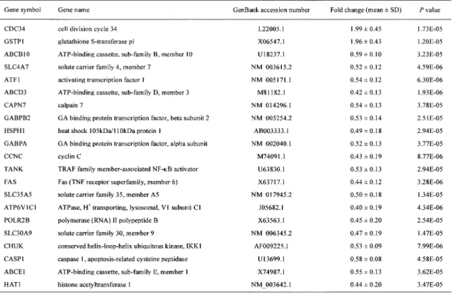
Departments of Dermatology and Department of Stress Science, Institute of Health Biosciences, the University of Tokushima Graduate School ; and Division of Enzyme Pathophysiology, The In-stitute for Enzyme Research, the University of Tokushima, Tokushima, JapanAbstract : Ultraviolet B (UVB) is a serious irritant for the skin and increases a risk for skin cancer. To identify UVB-sensitive genes in peripheral blood, 11 healthy male volun-teers were exposed to 0.3 J/cm2of narrow-band (NB)-UVB, about half of minimal erythema dose (MED) in Japanese, and gene expression in blood was analyzed at 4 h, 24 h, 4 d and 7 d after the irradiation using microarray carrying oligonucleotide probes for 2,000 stress-responsive genes. RNA prepared before the irradiation was used as a reference control. Microarray analysis identified 21 genes as UVB-responsive genes with a peak at 24 h in 6 subjects, and real-time PCR validated the significant down-regulation of 9 (ABCB10,
ATF1, ABCD3, TANK , FAS , SLC30A9, CHUK , CASP1, and ABCE1) out of the 21 genes in
11 subjects. Considering sensitive and characteristic features of 9 marker genes, they may be useful indicators for monitoring systemic response to UVB irradiation.
J. Med. Invest. 55 : 204-210, August, 2008
Keywords : ultraviolet B irradiation, skin damage, peripheral blood cells, gene expression, marker genes
Received for publication March 14, 2008 ; accepted May 1, 2008.
Address correspondence and reprint requests to Seiji Arase, MD, PhD., Department of Dermatology, Institute of Health Biosciences, the University of Tokushima Graduate School, Kuramoto - cho, Tokushima 770 - 8503, Japan and Fax : +81 - 88 - 632 - 0434.
detection and assessment of local or systemic re-sponses to UVB irradiation.
Acute UVB irradiation causes not only sunburn but also fatigue, and decreases local and systemic immunities. Chronic and persistent exposure to UVB is carcinogenic. To prevent these hazardous effects, it is potentially useful to identify markers sensitive to UVB irradiation in peripheral blood. In this study, we recruited healthy male volunteers, exposed them to a sub-clinical dose of UVB, and examined gene expression profiles in peripheral blood cells (PBC) using a microarray carrying oli-gonucleotide (80 mer) probes for 2,000 stress-re-sponsive genes. Here we show 9 genes identified as sensitive markers for UVB stress.
MATERIALS AND METHODS
SubjectsEleven healthy male Japanese volunteers with either skin type III or IV were recruited for the study. They were all in good physical health, non-smokers, and receiving no medications (median age 27.2 years, range 23 - 39 years). The study was approved by the Human Study Committee of Tokushima University Hospital. A written informed consent was obtained from each participant prior to the study.
Narrow-band (NB) UVB irradiation and blood col-lection
Eleven subjects were exposed to NB - UVB of 0.3J/cm2, about half of minimal erythema dose
(MED : the smallest amount of energy to produce a perceptible erythema) in Japanese, on the entire body except the face and genital region in a spe-cial cabinet equipped with 20 NB-UVB bulbs emit-ting a very short range of wavelengths from 311 to 315 nm exclusively (UV7001K-TL01, Waldmann, Villingen - Schwenningen, Germany). The irradia-tion was performed between 13 : 00 to 14 : 00. Pe-ripheral venous blood (12 ml) was taken from each subject immediately before and 4 h, 24 h, 4 d, and 7 d after UVB irradiation. Two ml of blood was used for total and differential leukocyte counts, and remaining 10 ml of the blood was poured into PAXgeneTM Blood RNA tubes (Becton Dickinson,
Franklin Lakes, NJ). Construction of microarray
We had originally developed a complementary
DNA (cDNA) microarray specifically designed to assess stress response in peripheral blood cells (14, 15). This carried cDNA probes for 1,467 genes that were detected in total RNA from peripheral blood by reverse transcriptase (RT) - PCR. In this study, we constructed a new microarray carrying 80 - mer oli-gonucleotide probes for the above 1,467 genes and newly added 400 genes that are categorized into drug metabolism (see http://www.hitachi.co.jp/LS/ for the full list of genes). These oligonucleotide probes were designed using original software from Hitachi, Ltd. (Saitama, Japan). The microarray showed high reproducibility with a mean coefficient of variation of less than 10%, and the dynamic ranges were three orders of magnitude. This microarray was purchased from Hitachi, Ltd.
RNA preparation, amplification, and hybridization RNA was isolated from PAXgeneTM Blood RNA
tubes using a PAXgene blood RNA kit (Qiagen, Hilden, Germany) according to the manufacturer’s protocol. Contaminated DNA was removed using a DNase - kit (Qiagen). The quality of resultant RNA was checked with an Agilent 2100 Bioanalyzer us-ing an RNA 6000 Nano Labchip kit (Agilent Tech-nologies, Palo Alto, CA). Five micrograms of total RNA from randomly selected 6 subjects were re-verse transcribed with an oligo dT primer conju-gating T7 sequence. Yielded first strand cDNA com-plementary to poly (A) RNA was amplified by us-ing MEGAscript T7 in vitro RNA transcription kit (Applied Biosystems, Foster City, CA). Amplified RNA (6 μg) was reverse transcribed by using ran-dom hexamers and aminoallyl - dUTP. The synthe-sized cDNA was labeled with dye (NHS - ester Cy5 or Cy3 ; Amersham Biosciences, Piscataway, NJ). Cy5-cDNAs from each blood sample collected at the indicated times after UV irradiation were mixed with the equivalent amount of Cy3-cDNAs, which was from each subject prior to UV irradiation. Hybridi-zation of the oligonucleotide microarray was per-formed at 62!!for 12 h. After washing, fluorescence intensity at each spot was assayed using a scanner (ScanArray 5000 ; GSI - Lumonics, Billerica, MA). Data analysis
Signal intensities of Cy5 and Cy3 were quanti-fied and analyzed by subtracting backgrounds us-ing QuantArray software (GSI - Lumonics, Billerica, MA). The intensity values for duplicate oligoDNA probes were averaged. Following global normali-zation, we selected 1,095 genes having fluorescence
intensities higher than the cut - off value of 300 for either Cy5 or Cy3 conditions among all 24 - paired samples. In order to consider the quality criteria of our microarray chip, we performed comparative ex-periments by hybridizing the same 2 samples on one microarray chip after labeling Cy5 and Cy3 dye separately (self - self test). Based on results of these experiments, we decided that cut - off values higher than 300 are sufficient for analysis. The rela-tive expression values (Cy5/Cy3) for 1,095 genes were subjected to hierarchical clustering using GeneSpring 7.3 software (Agilent), and similarity was measured by standard correlation. After Cy5/ Cy3 ratios of 1,095 genes were transformed to loga-rithms, statistical significance between UV irradia-tion and non - irradiated controls was examined by the paired t - test using Cyber - T stats program writ-ten in the R stats language (available at http : // visitor.ics.uci.edu/genex/cybert/help/index.html). Statistical significance was defined as a Bonferroni-corrected P value of < 0.05.
Quantitative real-time RT-PCR
Total RNA (0.5 μg) was prepared as a template for cDNA synthesis using the SuperScriptTMIII first
-strand synthesis system for RT - PCR (Invitrogen, Carlsbad, CA). The mRNA levels of target genes based on GenBank association numbers (CDC34, TaqMan assay ID Hs00362082_m1 ; GSTP1, Hs 00168310_m1 ; ABCB10, Hs00429240_m1 ; SLC4A7, Hs00186192_m1 ; ATF1, Hs00270896_m1 ; ABCD3, Hs00161065 _m 1 ; CAPN7, Hs00204734 _ m1 ; GABPB2, Hs00242573_m1 ; HSPH1, Hs00971475_ m1 ; GABPA , Hs01022023 _ m1 ; CCNC , Hs 00193177_m1 ; TANK, Hs00370305_m1 ; FAS, Hs 00531110_m1 ; SLC35A5, Hs00215733_m1 ; ATP6 V1C1, Hs00184625_m1 ; POLR2B, Hs00265358_ m1 ; SLC30A9, Hs00197118_m1 ; CHUK, Hs 00175141_m1 ; CASP1, Hs00169146_m1 ; ABCE1, Hs01003010_g1 ; HAT1, Hs00186320_m1 ; GAPDH, Hs99999905_m1) were analyzed by quantitative real -time PCR using TaqMan gene expression assays and ABI - PRISM 7500 sequence detection system (Applied Biosystems). Glyceraldehyde - 3 - phosphate dehydrogenase (GAPDH) was used as the endo-genous quantity control. Data were analyzed using SDS 2.3 software (Applied Biosystems). The thresh-old cycle (Ct) for each gene was calculated by the standard curve method. Quantity values were fi-nally normalized to GAPDH mRNA expression. Af-ter the relative ratio of each mRNA expression level between pre - and post - irradiation was calculated,
the paired t - test was performed to compare the mean of relative ratios for each gene.
RESULTS
Changes in gene expression in peripheral blood cells after UVB irradiation
A single exposure to 0.3J/cm2NB - UVB did not
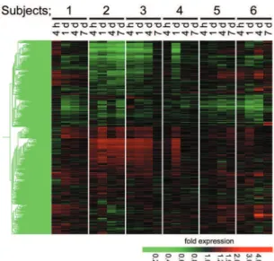
cause any change on the skin in any of the subjects. To detect UVB - sensitive genes, among the 11 vol-unteers exposed to the irradiation, we randomly se-lected 6 subjects and measured time - dependent changes in their gene expression profiles using RNAs prepared prior to the irradiation as individ-ual controls. As shown in the hierarchical cluster-ing map of the 1,095 genes (Fig. 1), 2 subjects (Nos. 2 and 3) had a similar expression change. This change was also detected at one time point (24 h) in subject 4. However, response of gene expression differed among the other subjects (Nos. 1, 5, and 6), suggesting a significant individual variability of sen-sitivity and gene expression response to NB - UVB ir-radiation. In all of the cases, most profound changes in gene expression were observed 24 h after the ir-radiation. In spite of the variations, we could iden-tify 21 genes whose mRNAs were significantly up-or down - regulated in all 6 subjects at 24 h after the irradiation (Table 1). None of the genes were
sig-Fig. 1. Hierarchical clustering of gene expression pattern in peripheral blood cells after irradiation by 0.3 J/cm2NB - UVB.
Whole blood RNA collected at the indicated times after irradia-tion by 0.3 J/cm2NB - UVB (Cy5) were applied to
oligonucleo-tide microarray together with reference RNA collected imme-diately before irradiation (Cy3) in each 6 volunteer. Relative ex-pression values (Cy5/Cy3) were subjected to a hierarchical clus-tering and shown in intensity of red and green.
nificantly regulated in all 6 subjects at 4 h or 7 d after the irradiation. Only one gene (HSPH1) in-cluded in the 21 genes significantly changed its ex-pression level at 4 d after the irradiation. The iden-tified 21 genes were likely to be sensitive universal marker genes, and most were detected as down-regulated genes to the lowest levels at 24 h (Fig. 2, upper panel).
Even at about half MED, NB-UVB irradiation may leads to a shift in the subpopulation of leuko-cytes ; therefore, the changes found in mRNA lev-els of whole blood might reflect a shift between leu-kocyte subpopulations. However, we could not de-tect any common, significant changes in total and differential leukocyte counts (data not shown). We had already purified neutrophil-, monocyte-, T cell-, and B cell - enriched fractions and identified genes preferentially expressed in each fraction using a whole human genome microarray carrying 44,000 oligonucleotide probes (Agilent) (see supplemen-tary Table S1 in ref. (13)). The microarray used in this study carries 3 neutrophil markers, 4 monocyte markers, 8 B cell markers, and 9 T cell markers. We tested how mRNA levels of these 24 cell fraction -specific genes were modified after the UVB irradia-tion. T cell fraction - specific genes were seemed to be generally down - regulated (Fig. 2 lower panel), while none of them was included in the commonly and significantly changed genes.
Fig. 2. Identification of 21 marker genes after irradiation by 0.3 J/cm2NB-UVB. The paired t-test (Bonferroni corrected p value
! 0.05) revealed that 21 genes were commonly and significantly changed between before and 24 h after irradiation by 0.3 J/cm2
NB-UVB in 1,095 genes. Time-dependent changes in relative ex-pression levels of 21 genes are shown in the upper panel. Each leukocyte marker genes are referred in the lower panel (Mo ; monocyte, PMN ; neutrophil). Gene symbols are listed on the right.
Validation of microarray data by quantitative real-time PCR
Based on microarray findings, we performed quantitative real - time PCR to validate the putative UVB - sensitive 21 genes. In this case, mRNA levels were compared between those immediately before (control) and 24 h after NB - UBV irradiation (0.3 J/ cm2) in all 11 subjects. Real - time PCR reproduced
the findings by microarray of a significant reduction in the mRNA level of 9 genes (ABCB10, ATF1,
ABCD3, TANK , FAS , SLC30A9, CHUK , CASP1, and ABCE1) in response to a half erythemogenic dose
of UVB, while we could not confirm any significant changes in the other 12 mRNA levels by real - time PCR (Fig. 3). Among the down - regulated 9 genes, 4 genes encode essential regulators of nuclear fac-tor (NF)-κB and apoptosis ; these are a conserved helix - loop - helix ubiquitous kinase (CHUK ), TNF receptor - associated factor (TRAF) family member-associated nuclear factor (NF) - κB activator (TANK ), Fas (FAS ), and caspase 1 (CASP1).
DISCUSSION
In addition to wavelength, UV - induced injury de-pends on many variables including dose, skin types, and characteristics of the skin tissue (16). Individ-ual sensitivity, probably due to genetic factors, is also an important determinant of the susceptibility to UV irradiation.
In this study, after a single exposure to a half MED of NB - UVB (0.3 J/cm2), time - dependent
changes in gene expression of peripheral blood cells were precisely examined up to 7 d in 6 subjects us-ing RNA prepared from each subject immediately before the irradiation as an individual reference control. The routine of daily lives of our subjects was similar, and they had no regular outdoor ac-tivities. Even these relatively homogenous subjects displayed inter - individual variations following ex-posure to the same UVB dose. Among the hetero-geneous responses, we identified 21 genes as com-monly UVB - sensitive genes. Although there was inconsistency between microarray and real - time PCR analyses, 9 genes were confirmed to be sig-nificantly down - regulated in response to the irra-diation of 0.3 J/cm2NB - UVB by both analyses.
At present, two different technologies are com-monly used for preparation of RNA from whole blood and its leukocyte compartments (17). RNA from whole blood cells contains a large amount of hemoglobin mRNAs that disturb specific and sen-sitive hybridization reactions (18). However, the in
vitro handling required for isolation of distinct
leu-kocyte compartments stimulates ex vivo expression and degradation of mRNAs for a number of genes, particularly stress - responsive genes (14, 19 - 20). Therefore, to detect UVB-sensitive genes, we used the whole blood RNA preparation method.
UV light activates distinct signaling pathways and transcription factors, leading to changes in gene expression in the skin (12, 17, 21 - 24). Upon UVB stress, keratinocytes produce numerous cytokines and inflammatory mediators, including production of interleukin (IL)-1β, tumor necrosis factor (TNF)-α and reactive oxygen species (25). These media-tors subsequently activate NF - κB, JunD/c-Fos and p53 (26). In turn, activation of NF - κB has been re-ported to result in transactivation of a number of target genes, including those for IL - 1, IL - 6, IL - 8, GRO - 1, TNF - α and COX - 2 (27). UVB also sup-presses trafficking of lymphocytes and functions of T and natural killer cells (28). Since UV does not effectively penetrate into the dermis where blood vessels are located, inflammatory mediators pro-duced by skin cells are believed to act on PBC and evoke systemic response (5 - 8). Among the 9 genes confirmed by microarray and real - time PCR to be significantly down - regulated, ATF1 encodes acti-vating transcription factor 1, and this transcription factor together with the cAMP response element binding protein regulates the stress - induced tran-scription of several immediate early genes (29). It should be noted that among the confirmed 9 genes,
Fig. 3. Validation of UVB - responsive 21 genes. The mRNA levels of putative UVB - responsive 21 genes listed in Table 1 were measured in sample of 24 h after irradiation of NB - UVB by quantitative real - time PCR. GAPDH mRNA was used as an en-dogenous quantity control. The fold changes in relative ratios (each mRNA level/GAPDH mRNA) are shown. Values are means!S. E. M. (n = 11).
*Significantly different compared with that before the irradia-tion (P!0.05 by the paired t -test)
4 genes (CHUK , TUNK , CASP1, and FAS ) encode molecules essential for activation of NF - κB or apop-tosis. CHUK encodes an inhibitor of NF - κB kinase α(IKK - α). IKK - α, a component of a cytokine - acti-vated protein complex that is an inhibitor of the NF - κB complex, phosphorylates IκB leading to the degradation of the inhibitor via the ubiquination pathway, thereby activating NF - κB. TUNK encodes a RAF family member - associated NF - κB activator (TRAF2 ; TNF receptor - associated factor 2). TRAF2 associates with and transduces signals from mem-bers of the TNF receptor superfamily. TRAF2 binds to the TNF receptor death domain (TRADD) and recruits NF - κB -inducing kinase (NIK) leading to phosphorylation of IκB. CASP1 encodes caspase 1, apoptosis - related cysteine peptidase (IL - 1β conver-tase). Fas is a member of the TNF - receptor super-family containing the death domain and plays a cen-tral role in the regulation of apoptosis. This receptor also activates NF - κB, MAPK3/ERK1, and MAPK8/ JNK. The 9 genes also included SLC30A9, encod-ing a recently identified nuclear receptor coactiva-tor interacting with β - catenin (30) and 3 genes (ABCB10, ABCD3, and ABCE1) encoding members of the superfamily of ATP - binding cassette (ABC) transporters.
Because the 4 identified genes (CHUK , TUNK ,
CASP1, and FAS ) were closely related to activation
of NF - κB and apoptosis, we additionally performed real - time PCR measurement of mRNA levels of other NF - κB activation associated genes such as Toll - like receptor (TLR) family members (TLR1,
TLR2, TLR4, and TLR5) and 2 apoptosis - regulating
factors (BCL2A1 and BCL2L11) all of which are ex-pressed in peripheral leukocytes. We also measured changes in mRNA levels of HSP60 and stress- in-ducible HSP70 to assess stress response. However, none of these 8 genes changed their mRNA levels after exposure to 0.3 J/cm2 of NB - UVB (data not
shown), suggesting the high sensitivity of 9 marker genes to UVB irradiation.
Dermatologists recommend a variety ways to limit sun exposure to avoid UV- induced injury. Despite the extensive use of sunscreens, the incidence of skin cancer has been increasing. The efficacy of sunscreens is generally assessed using the sun pro-tection factor (SPF) as an indicator. Evaluation of SPF is based solely on the ability to prevent UV -induced perceptible erythema. However, our results suggest that even about half MED of UVB may evoke significant changes in the expression of dis-tinct genes in PBC. Reliable indicators should
evalu-ate not only the protective effect against erythema, but also potency of protection against UV - induced systemic immunological and mutational effects. In this context, sensitive and characteristic features of 9 marker genes may be useful indicators for moni-toring systemic response to UVB irradiation.
REFERENCES
1. Hahn WC, Weinberg RA : Modelling the mo-lecular circuitry of cancer. Nat Rev Cancer 2 : 331 - 341, 2000
2. Chow J, Tron VA : Molecular aspects of ultra-violet radiation - induced apoptosis in the skin. J Cutan Med Surg 9 : 289 - 295, 2005
3. Yaar M, Gilchrest BA : Photoageing : mecha-nism, prevention and therapy. Br J Dermatol 157 : 874 - 887, 2007
4. Slominski A, Wortsman J : Neuroendocrinology of the skin. Endocr Rev 21 : 457 - 487, 2000 5. Rivas JM, Ullrich SE : Systemic suppression
of delayed - type hypersensitivity by superna-tants from UV - irradiated keratinocytes : An es-sential role for keratinocyte - derived IL - 10. J Immunol 149 : 3865 - 3871, 1992
6. Rivas JM, Ullrich SE : The role of IL - 4, IL - 10, and TNF - α in the immune suppression in-duced by ultraviolet radiation. J Leukoc Biol 56 : 769 - 775, 1994
7. Schwarz T, Urbanski A, Luger TA : Ultraviolet light and epidermal cell derived cytokines. In : T.A. Luger, T. Schwarz (Eds), Epidermal growth factors and cytokines, Marcel Dekker Inc, New York, 1994, pp.303 - 363
8. Kondo S, Kooshesh F, Sauder DN : Penetra-tion of keratinocyte - derived cytokines into basement membrane. J Cell Physiol 171 : 190 -195, 1997
9. Ullrich SE, Schmitt DA : Exposure to ultraviolet radiation causes dendritic cells/macrophages to secrete immune - suppressive IL - 12p40 ho-modimers. J Immunol 165 : 3162 - 3167, 2000 10. Komura K, Hasegawa M, Hamaguchi Y, Saito
E, Kaburagi Y, Yanaba K, Kawara S, Takehara K, Seki M, Steeber DA, Tedder TF, Sato S : Ultraviolet light exposure suppresses contact hypersensitivity by abrogating endothelial in-tercellular adhesion molecule - 1 up - regulation at the elicitation site. J Immunol 171 : 2885 -2862, 2003
Stephan N, Parg S, Krammer PH : Ultraviolet irradiation suppresses T cell activation via blocking TCR-mediated ERK and NF - κB sig-naling pathways. J Immunol 175 : 2132 - 2143, 2005
12. Enk CD, Jacob - Hirsch J, Gal H, Verbovetski I, Amariglio N, Mevorach D, Ingber A, Givol D, Rechavi G, Hochberg M : The UVB - induced gene expression profile of human epidermis
in vivo is different from that of cultured
kerati-nocytes. Oncogene 25 : 2601 - 2614, 2006 13. Duthie MS, Kimber I, Norval M : The effect
of ultraviolet radiation on the human immune system. Br J Dermatol 140 : 995 - 1009, 1999 14. Morita K, Saito T, Ohta M, Ohmori T, Kawai
K, Teshima - Kondo S, Rokutan K : Expression analysis of psychological stress - associated genes in peripheral blood leukocytes. Neuro-sci Lett 381 : 57 - 62, 2005
15. Kawai T, Morita K, Masuda K, Nishida K, Shikishima M, Ohta M, Saito T, Rokutan K : Gene expression signature in peripheral blood cells from medical students exposed to chronic psychological stress. Biol Psychol 76 : 147 -155, 2007
16. Heck DE, Gerecke DR, Vetrano AM, Laskin JD, Solar ultraviolet radiation as a trigger of cell signal transduction. Toxicol Appl Phar-macol 195 : 288 - 297, 2004
17. Fan H, Hegde PS : The transcriptome in blood : challenges and solutions for robust expression profiling. Curr Mol Med 5 : 3 - 10, 2005
18. Feezor RJ, Baker HV, Mindrinos M, Hayden D, Tannahill CL, Brownstein BH, Fay A, MacMillan S, Laramie J, Xiao W, Moldawer LL, Cobb JP, Laudanski K, Miller - Graziano CL, Maier RV, Schoenfeld D, Davis RW, Tompkins RG : Whole blood and leukocyte RNA isolation for gene expression analyses. Physiol Genom-ics 19 : 247 - 254, 2004
19. Debey S, Schoenbeck U, Hellmich M, Gathof BS, Pillai R, Zander T, Schultze JL : Compari-son of different isolation techniques prior gene expression profiling of blood derived cells : im-pact on physiological responses, on overall ex-pression and the role of different cell types. Pharmacogenomics J 4 : 193 - 207, 2004
20. Thach DC, Lin B, Walter E, Kruzelock R, Rowley RK, Tibbetts C, Stenger DA : Assess-ment of two methods for handling blood in col-lection tubes with RNA stabilizing agent for surveillance of gene expression profiles with high density microarrays. J Immunol Meth-ods 283 : 269 - 279, 2003
21. Sesto A, Navarro M, Burslem F, Jorcano JL : Analysis of the ultraviolet B response in pri-mary human keratinocytes using oligonucleo-tide microarrays. Proc Natl Acad Sci USA 99 : 2965 - 2970, 2002
22. Enk CD, Shahar I, Amariglio N, Rechavi G, Kaminski N : Gene expression profiling of in
vivo UVB - irradiated human epidermis.
Photodermatol Photoimmunol Photomed 20 : 129 -137, 2004
23. Pisarchik A, Wortsman J, Slominski A : A novel microarray to evaluate stress - related genes in skin : effect of ultraviolet light radiation. Gene 341 : 199 - 207, 2004
24. Murakami T, Fujimoto M, Ohtsuki M, Nakagawa H : Expression profiling of cancer -related genes in human keratinocytes follow-ing non - lethal ultraviolet B irradiation. J Der-matol Sci 27 : 121 - 129, 2001
25. Garssen J, van Loveren H : Effects of ultravio-let exposure on the immune system. Crit Rev Immunol 21 : 359 - 397, 2001
26. Dröge W : Free radicals in the physiological control of cell Function. Physiol Rev 82 : 47 -95, 2002
27. Karin M, Cao Y, Greten FR, Li ZW : NF - κB in cancer : from innocent bystander to major cul-prit. Nat Rev Cancer 2 : 301 - 310, 2002
28. Morison WL, Parrish JA, Bloch KJ, Krugler JI : In vivo effect of UVB on lymphocyte func-tion. Br J Dermatol 101 : 513 - 519, 1979 29. Wiggin GR, Soloaga A, Foster JM, Murray
-Tait V, Cohen P, Arthur JSC : MSK1 and MSK2 are required for the mitogen - and stress - in-duced phosphorylation of CREB and ATF1 in fibroblasts. Mol Cell Biol 22 : 2871 - 2881, 2002 30. Chen YH, Yang CK, Xia M, Ou CY, Stallcup
MR : Role of GAC63 in transcriptional activa-tion mediated by β - catenin. Nucl Acids Res 35 : 2084 - 2092, 2007