Dengue and Japanese Encephalitis Antibody Levels in Sera from Dengue Haemorrhagic Fever
Patients in Rangoon, Burma
MAY LA LINH1), K. BUNDO, A. IGARASHI
D epartment of Virology, Institute for Tropical Medicine Nagasaki University
SOE THEIN and MI MI KHIN
Virology Research Division, Department of Medical Research Ministry of Health, Rangoon, Burma
Abstract: Enzyme-linked immunosorbent assay (ELISA) was compared with haemaggluti- nation inhibition (HI) test for determination of anti-dengue and anti-Japanese encephalitis (JE) antibodies in paired sera from 168 patients with clinical diagnosis of dengue haemor- rhagic fever (DHF) in Rangoon, Burma. Using paired sera, HI serology gave 60.7%
positive results. IgG ELISA antibodies showed cross reaction between dengue and JE, and also not so efficient to detect significant antibody rise. By IgM capture ELISA 60.5%
positive results was obtained using acute sera and dengue 4 antigen alone for diagnosis.
When paired sera and all 4 types of dengue were used, 82.1% positive results were obtained by the IgM-capture ELISA, and none of the test sera had diagnostic levels of IgM-ELISA antibodies against JE.
The results showed that IgM capture ELISA is a useful method for rapid diagnosis of dengue infections and has advantages over HI in terms of its diagnostic efficiency even by a single serum specimen, rapidity, simplicity and differential diagnosis with other related flavivirus infections like JE.
Key words*. Dengue haemorrhagic fever, Antibody levels, ELISA, Serodiagnosis
INTRODUCTION
Dengue infections have been implicated as the most important arbovirus disease in South East Asia resulting in a large number of hospitalization with occasional shock and death (Halstead, 1966). The disease is caused by 4 serotypes of dengue viruses, belonging
Received for Publication, February 20, 1985
Contribution No. 1641 from the Institute for Tropical Medicine, Nagasaki University.
0Present address: Virology Research Division, Department of Medical Research, Ministry of
Health, Rangoon, Burma
to the family Flaviviridae sharing commonantigens with other flaviviruses such as Japanese encephalitis (JE) and yellow fever viruses (Shope, 1979). Serodiagnosis and seroepidemiolo- gical survey on dengue infections have been carried out mostly by haemagglutination- inhibition (HI) test (Clarke and Casals, 1958). Recently enzyme-linked immunosorbent assay (ELISA) has been applied for detection of humoral antibodies to disease agents and proved to be simple, rapid and sensitive (Engvall and Permian, 1971'; Voller et al. , 1976).
In this laboratory, Igarashi et al. (1981) and Bundo et al. (1982a7 b) had developed ELISA for serodiagnosis and seroepidemiological studies on JE and extended that method to den- gue infections (Bundo and Igarashi, 1983, 1984). We assayed anti-JE and anti-dengue
immunoglobulin G (IgG) and immunoglobulin M (IgM) antibodies in sera from DHF pati- ents in Burma. Comparison were made between standard HI test and ELISA in terms of their diagnostic efficiency on dengue and differential diagnosis between dengue and JE.
MATERIALS AND METHODS
Test sera. Paired sera were obtained from 168 patients clinically diagnosed as DHF at Children's Hospital in Rangoon, Burma, during epidemic season of 1982. The age of the patients ranged from under 1 year up to 12 years old. Patients were grouped into grades according to the severity of their clinical manifestations using the criteria of
Nimmannitya et al. (1962).
HIserology. The HI test was carried out by the method of Clarke and Casals (1958) in a microtiter system using sucrose-acetone extracted antigen from infected suckling mousebrains and goose red blood cells. The results were classified into primary, secondary, presumptive secondary, and not dengue cases according to the guide line by the Working Group of World Health Organization (WHO, 1983).
ELISA antigens. Dengue viruses, type 1 Hawaiian strain (Dl), type 2 New Guinea B strain (D2), type 3 H-87 strain (D3), and type 4 H-241 strain, were grown in Aedes albopictus clone C6/36 cell cultures (Igarashi, 1978) at 28°C and maintained in Eagle's medium (Eagle, 1959) containing 2% heat inactivated fetal calf serum supplemented with 0.2 mM each of nonessential amino acids. Infected culture fluids were harvested on day 7 and stored at -70°C until used for IgM-capture ELISA. For IgG-ELISA, Dl antigen was further purified by polyethylene glycol precipitation and ultracentrifugation (Bundo and Igarashi, 1983). Formalin inactivated and purified JE vaccine concentrate (Takaku et al.
1968) was used both for IgM-capture and IgG-ELISA.
IgG ELISA. Indirect micromethod of Voller et al. (1976) was followed with modi-
fication as described before (Igarashi etal. 9 1981 ; Bundo et al., 1982a, b), using purified
Dl and JE antigens as mentioned above. ELISA-OD at 490 nm was measured using Micro
ELISA autoreader (Dynatech, USA). The titer of test serum was calculated by comparing
the OD of each specimen with those developed by serial dilution of a.standard positive
serum with known endpoint titer (Igarashi et al., 1981 ; Morita et al., 1982).
Preparation of antiflavivirus IgG and labeling with horseradish peroxidase. Hy- perimmune anti-flavivirus IgG was prepared from high-titered DHF patients* sera. Pooled sera were dialysed overnight against 0.005 M sodium phosphate buffer, pH 8.0, at 4°C and passed through DEAE Sephacel column (Pharmacia, Uppsala, Sweden). IgG eluted
by the same buffer was collected in 5 ml fractions and OD28o was measured, and high OD fractions were pooled and concentrated by negative-pressure dialysis. Concentrated antiflavivirus human IgG was labeled with horseradish peroxidase (Sigma, Type VI) using
Nakane and Kawaoi's method (1974).
IgM capture ELISA, IgM ELISA antibody levels against all 4 dengue serotypes and JE antigen were measured by IgM capture ELISA method as deacribed before (Bundo et al., 1984). ELISA OD were recorded and titer for each specimen was calculated as described above.
Reagents. Anti-human IgM (^-chain specific) goat IgG and peroxidase-conjugated anti-human IgG (heavy and light chain) goat IgG were the products of Cappel Laboratories, USA. Latex globulin was obtained from Kyowa Pharmaceutical Co. and used for the agglutination test to detect rheumatoid factors in the sera.
RESULTS Serodiagnosis by the HI.
The results of the HI test was interpreted according to the WHO guide line (1983) and the specimens were categorized into 18 primary, 24 secondary, 60 presumptive seconda- ry, and 66 not dengue cases. The ratio of possible secondary infections (secondary and presumptive secondary cases) to primary infection was similar to the cases in Thailand
Table 1. The number of DHF patients according to their age groups, clinical grades, and serodiagnosis by the HI
Serodiagnosis by HI
££) Pá"y S-^ f£S±T Notdengue á"
I* II III IV I II III IV I II III IV I II III IV I II III IV Total
<1 2 1 1 4 1 5
1-4 4 1 1 3 4 6 1 5 ll 13 2 7 7-10 19 23 30 3 75
5-9. 2 3 1 4 2 2 7 9 8 13 13 9 26 27 20 73
10- à" 1 2 1 1 1 2 1 5 1 2 4 5 4 1 14
total" 9 6 2 8 7 8 1 13 21 23 3 23 21 22 53 55 55 4 167
Total 17** 24 60 66 167
* I, II} III, IV represent clinical grades.
** One of the primary cases did not have the record of clinical grade.
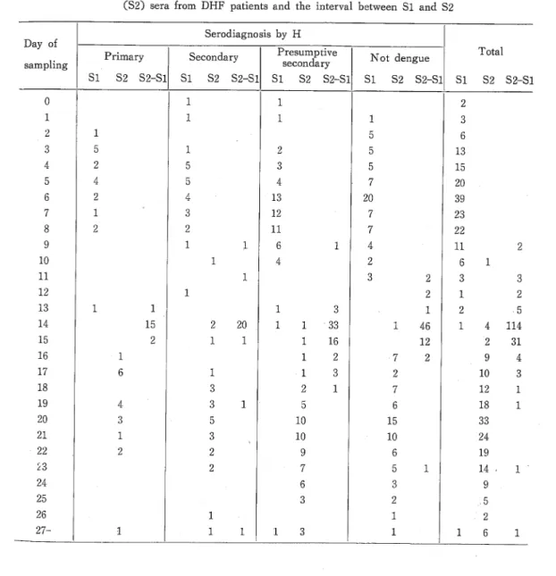
(Nimmannitya et al., 1969). Table 1 shows the number of patients according to their age group, clinical grades, and serodiagnosis by the HI. Most of the patients were in 1-4, followed by those in 5-9 years old group, similar to the report in Thailand (Nimmanni tya et al., 1969). Severe clinical manifestions in the grades III and IV were considered as dengue shock syndrome (DSS) according to the WHO Technical Guides (1975), and their numbers were compared in each of the HI serology and age group as shown in Table 2. The percentage of DSS in DHF cases was lower in the primary (12%) than those in the secondary (38%) or presumptive secondary (43%), or even not dengue cases (33%), Table 3 shows sampling day of acute (Si), and convalescent (S2) phase sera after onset of the disease, and also the interval between Si and S2 sera. On average, acute sera (Si) was collected on day 5.87, with range between 0-14 days. Convalescent phase sera (S2) were collected between 14 to 36 days with average of day 20.2. Most of the paired sera were obtained with interval of 14 days. Sampling times of the presumptive secondary or not dengue specimens were almost similar to those of the primary or secondary cases, indicating that later sampling did not result in the inefficient serodiagnosis by the
HI.
IgG-ELISA.
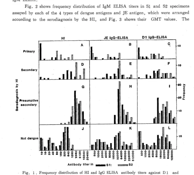
The IgG-ELISA titers against JE and Dl antigens were measured for the test specimens and their frequencies were shown together with those by the HI in each category of HI serology (Fig 1). The titers of presumptive secondary group, both in Si and S2, distributed in high titered ranges compared with other groups, although the S2 titer of secondary cases was also high. In all types of HI antibody response, the geometrical meantiters (GMT) of IgG ELISA assayed by JE antigen were higher than those by Dl antigen (Table 4). Significant change (4 fold or more rise) in GMT of IgG ELISA was found only in the primary type of HI antibody response. The GMTs of IgG ELISA in
Table 2. Proportion of DSS among DHF cases arranged by their age groups and serodiagnosis by the HI
A Serodiagnosis by the HI
Age group ° J_
Total
(years) Primary Secondary P£^j^* Not dengue
< 1 0/2 (0) 0/1 (0) . 1/2 (50) 1/5 (20)
1 - 4 1/6 (17) 7/14 (50) 15/31 (48) 10/24 (42) 33/75 (44)
5 - 9 1/6 (17) 2/8 (25) 8/24 (33) 9/35 (26) 20/73 (27)
10 - 0/3 (0) 0/1 (0) 3/5 (60) 2/5 (40) 5/14 (36)
Total . 2/17 (12) 9/24 (38) 26/60 (43) 22/66 (33) 59/167(35)
Figures in the parentheses: percentages of DSS in DHF cases
the primary cases were significantly lower than those in other groups. Table 5 shows titer changes detected between paired sera by the IgG ELISA. When 4 fold or more rise in Dl IgG ELISA antibody titers in paired sera was taken as significant, 31 pairs (18.5%) could be diagnosed as dengue infections. The value rose up to 58 pairs when the significant change was taken as 2 fold or more rise. Nineteen out of 31 pairs with 4 fold,rise in Dl IgG ELISA also showed significant rise in JE IgG ELISA. Significant changes in JE IgG ELISA titers between Si and S2 was found in 25 cases of DHF.
Table 3. Sampling day after onset of the disease of acute (Si) and convalescent (S2) sera from DHF patients and the interval between Si and S2
Serodiagnosis by H
Day of -- _ .
Primary Secondary Presumptive Not dengue Total
sampling secondary l ucug uc
Si S2 S2-S1 Si S2 S2-S1 Si S2 S2-S1 Si S2 S2-S1 SI S2 S2-S1
0 1 1 2
1 1 1 1 3
2 1 5 6
3 5 1 2 5 13
4 2 5 3 5 15
5 4 5 4 - 7 20
6 2 4 13 20 39
7 1 3 12 7 23
8 2 2 ll 7 22
9 1 1 6 1 4 ll 2
10 1 4 2 6 1
H 1 3 2 3 3
12 1 2 1 2
13 1 1 1 3 . 1 2 .5
14 15 2 20 1 1 33 1 46 1 4 114
15 2 1 1 1 16 12 à" 2 31
16 1 1 2 7 2 9 4
17 6 1 1 3 2 10 3
18 3 2 1 7 12 1
19 4 3 1 5 6 18 1
20 3 5 10 15 33
21 1 3 ( 10 10 24
22 2 2 9 6 19
£3 2 7 5 1 14- 1 '
24 6 3 9
25 3 2 ;, 5
26 1 1 2
27- 1 1 1 1 3 1 1 6 1
IgM ELISA.
Fig. 2 shows frequency distribution of IgM ELISA titers in Si and S2 specimens assayed by each of the 4 types of dengue antigens and JE antigen, which were arranged according to the serodiagnosis by the HI? and Fig. 3 shows their GMT values. The
Primary
Secondary
«
.2 Presumptive i
£ secondary
Not dengue
Antibody titerin ^HMHSI: uszzzzzmSZ
Fig. 1. Frequency distribution of HI and IgG ELISA antibody titers against D1 and JE antigens in sera from DHF patients with various serodiagnosis by the HI. Closed col- umns represent acute (S i ), and thatched columns convalescent (S 2 ) sera, respectively.
Table 4. Geometrical mean titer of IgG ELISA in sera from DHF patients,
Assay antigen JE Dl
Serum specimen Si S2 Si S2
Primary 1022.9 4537.8 854.5 3487. 5
Secondary 21372. 7 67711. 6 7895. 4 22980.0
Presumptive secondary 61296. 1 77060. 2 24682. 9 35406. 9
Not dengue 10875.0 13798. 1 4919.6 6179.9
GMT of primary infection was relatively lower than those of the secondary or presumptive secondary infections. In the cases of primary and secondary infections, the titer against dengue antigens increased from acute (Si) to convalescent (S2) sera. On the contrary, the titer decreased from Si to S2 in the cases of presumptive secondary and not dengue
Table 5. Percent positives of serodiagnosis by IgM ELISA on DHF patients using different types of dengue antigens
Test serum Assay antigen
SI S2 S1+S2
Dl 47.5 28.1 52.9
D2 61.0 56.9. 70.6
D3 50.1 38.9 61.7
D4 60.5 61.7 76.0
All 4 types 66.9 73.9 82.1
Assay antigen Primary
Secondary
à"- Presumptive
° secondary
Not dengue
IgM-ELiSAliter in *~ BiS1: ^S2
Fig. 2. Frequency distribution of IgM-ELISA antibody titers against each of,the 4 types of dengue and JE antigens in sera from DHF patients with various serodiagnosis by the HI. Closed columns represent acute (S i ), and thatched columns convalescent (S-
2 ) sera, respectively.
cases. However, the titer against JE remained at low level both in Si and S2 in every case. Since sampling time did not differ significantly between those in the primary or secondary and those in the presumptive secondary or not dengue cases (Table 3), the latter groups appear to consist of the patients whose antibody response were more rapid than those in the former groups.
Geometrical Mean liter (GMT) of IgM-ELISA in Acute (SO,
& Convalescent (S2) Sera from DHF Patients in Rangoon, Burma, as Measured by JE & Dengue Antigens
Primary infection
Secondary infection
Presumptive secondary
infection
Not dengue
(/>
0
>
&
CO o c O) GJ à"5 o i- o
CO
300 200 100
600 400 200 100
CM CO 00 IUU
1400 CO
800 c
600 £
400 **
200 2'
UJ
100^
£?
M- o
200 H
^
100 O
JE D1 D2 D3 04
Assay antigen in IgM-ELISA
Fig. 3. Geometrical mean titer of IgM-ELISA in sera from DHF patients as measured
by JE and dengue antigens. Closed columns represent acute (S i ) and thatched columns
convalescent (S 2 ) sera, respectively.
Positive Results by IgM-ELISA on Acute (S1) &
Convalescent (S2) Sera from DHF Patients in Rangoon, Burma, with Different Serodiagnosis by HI
Primary infection
n:18
Secondary infection
n:24
Presumptive secondary
infection
n:59
Not dengue
n:66
>
JD
CO O c D) 03 à"5 o u 0
CO
80 60
40 ra 20S +
80 CO
06
60
40 CM
CO 20
I
80 co c
60 -
<
40 2
LU 20 "
o>
80 60
>
.Q
CO CD
>
40 CO o
Q.
20 *
D1 D2 D3 D4 All4types
Assay antigen in IgM-ELISA
Fig. 4. Dengue infection-positive results by IgM ELISA on DHF patients sera as me-
asured by each ^of the 4 types of dengue antigens. Closed columns represent acute (Si),
thatched columns convalescent (S 2 ), and dotted columns S 1 +S 2 sera, respectively.
Fig. 4 shows the positive results obtained by the IgM ELISA on Si and S2 sera using each of the 4 types and all the 4 types of dengue antigens, according to the diagnostic criteria set up by Bundo and Igarashi (1984). Fifteen out of the 18 primary, all the 24 secondary7 and all the 60 presumptive secondary infections were diagnosed as dengue infections by the IgM capture ELISA method. Moreover, 39 out of the 66 "not dengue" cases could be diagnosed as dengue infections by the IgM capture ELISA method. Total of 82.1% test specimens were considerd as positive with dengue infections
Table 6. Dengue infection positive cases by the IgM ELISA on DHF patients with different serodiagnosis by the HI on various days after the onset of the disease
Days after Serodiagnosis by the HI
°nSet Primary Secondary SS^ Notdengue
0 0/1 0/1 0/1 CO)
1 0/1 1/1 0/1 1/3 (33)
2 1/1 1/5 2/6 (33)
3 1/5 0/1 2/2 3/5 6/13 (46)
4 1/2 4/5 2/3 1/5 8/15 (53)
5 2/4 2/5 4/4 1/7 9/20 (45)
6 0/2 4/4 12/13 ll/20 27/39 (69)
7 0/1 3/3 12/12 5/7 20/23 (87)
8 2/2 2/2 ll/ll 5/7 20/22 (91)
9 1/1 6/6 3/4 10/ll (91)
10 3/4 2/2 5/6 (83)
ll 2/3 2/3 (67)
12 1/1 1/1 (100)
13 0/1 0/1 0/2 (0)
14 2/2 2/2 1/1 5/5 (100)
15 1/1 0/1 1/2 (50)
16 1/1 1/1 2/7 4/9 (44)
17 4/6 1/1 0/1 2/2 7/10 (70)
18 3/3 2/2 1/7 6/12 (50)
19 4/4 3/3 3/5 3/6 13/18 (72)
20 3/3 5/5 10/1 1 9/15 27/34 (79)
21 1/1 2/3 8/10 6/10 17/24 (70)
22 2/2 2/2 9/9 4/6 17/19 (90)
23 2/2 6/7 4/5 12/14 (86)
24 5/6 2/3 7/9 (78)
25 2/3 2/2 4/5 (80)
26 1/1 0/1 1/2 (50)
27- . 1/1 4/4 0/1 5/7 (71)
Total 22/36 (61) 40/48 (83) 105/120 (88) 70/132 (53) 237/336 (71)
Figures in the parentheses: percent positives
by the IgM ELISA. Table 6 shows percent positives of DHF patients' sera by the IgM ELISA using different types of dengue antigens. The result shows that D4 antigen gave the highest positives; 60.5% with Si, 61.7% with S2, and 76% when both Si and S2 sera were used. None of the DHF patients examined were diagnosed as JE because of their low IgM antibody levels. AH the sera had no detectable levels of rheumatoid factors.
DISCUSSIONS
In Burma, DHF has been a major health problem since 1970 when the first outbreak took place followed by yearly monsoon outbreaks. For serodiagnosis of DHF, HI test had been used, which required paired acute and convalescent sera to obtain meaningful results and the serological cross reactions between antibodies to dengue and other flaviviruses sometimes caused uninterpretable results. The results of the IgG ELISA appeared to give cross-reactions as shown in our results and also in the previous studies on DHF pateints' sera in Thailand (Bundo and Igarashi, 1983) and the test was less efficient than the HI to detect significant antibody rise between Si and S2. Thus, the IgG-ELISA is not an efficient method for the serodiagnosis on DHF.
Reports had appeared on the use of more type-specific IgM ELISA for diagnosis of acute flavivirus infections (Dittmar et al. , 1979 ; Schmitz and Emmerich, 1984 ; Summers et al. 1984). Our results showed that IgM ELISA antibodies of DHF patients sera were more specific to dengue antigens and cross reactions with JE antigen was not virtually observed. It had been reported that rheumatoid factors may give false positive results in
£100
til 2
2à"
>
.Q
O
>
150 O a a o on
0) c 0 Q
Days after onset of the disease
Fig. 5. Dengue infection positive results by IgM-ELISA obtained on various days after
onset of the disease.
the IgM EL.ISA (Burke and Nisalak, 1982). Since none of our DHF patients' sera showed detectable levels of rheumatoid factors, the results of IgM capture ELISA could be considered to represent virus-specific IgM antibodies. Table 5 shows the number of specimens diagnosed as dengue infections according to their samlping day after the onset and their serodiagnosis by the HI. In the case of secondary and presumptive secondary cases, almost all the cases were positively diagnosed as dengue infections after 6 and 5 days of the disease. Fig. 5 shows the time course of positive diagnosis for total DHF specimens.
When IgM ELISA antibody level against single dengue antigen was used for diag- nosis, D4 antigen gave the highest positive results, 60.5% using only Si sera taken on the average day 5.8, while HI serology gave 60.7% definite results using paired SI and S2 sera. Since a single dengue antigen is more simple and inexpensive in practical test than using all the 4 types of dengue antigens and the diagnostic positive rate obtained by D4 antigen was 83 to 92% of the results using all the 4 types of dengue antigens, we may be able to propose IgM capture ELISA on Si using only D4 antigen without losing so many positive cases. When the test sera did not show significant levels of IgM antibodies against D4, then the sera should be retested against all the other 3 types of dengue and JE antigens using both Si and S2 sera when they are available.
Our results appear to indicate that the IgM capture ELISA has advantages over the HI in terms of its diagnostic efficiency, rapidity, simplicity, capacity of getting results by single serum sample and differential diagnosis with other related flavivirus infections like JE. Therefore, the application of IgM capture ELISA is a valuable extension of the diagnostic tools for rapid diagnosis on dengue infections including DHF.
ACKNOWLEDGEMENTS
The authors are grateful to Dr. Aung Than Batu, Director General of the Department of Medical Research, Ministry of Health, Rangoon, Burma, for his support. Excellent Technical assistance of Ms. N. Segawa and S. Neriishi are also gratefully acknowledged.
The first author was supported by JICA (Japan International Corporation Agency) under the Medical Cooperation Project for the Research on Infectious Diseases and the Application of its Achievement for their Control in Burma, for her visit to Japan,
REFERENCES
1 ) Bundo, K. , Matsuo, S. , and Igarashi, A. (1981): Enzyme-linked immunosorbent assay (ELISA) on Japanese encephalitis virus. II. Antibody levels in the patient sera. Trop. Med. 23, 135-
148.
2 ) Bundo, K. , Morita, K. , and Igarashi, A, (I982a): Enzyme-linked immunosorbent assay (ELI SA>
on Japanese encephalitis virus. Ill, Assay on antibody titer on swine sera. Trop. Med. 24, 87-
102.
3) Bundo, K., Igarashi, A., Morita, K., Hayashi, K., Yamada, A., Goto, I., Douke, S., Sakai, S., Katsuki, K. Watanabe, K., and Ishii, K. (I982b): Enzyme-linked immunosorbent assay (ELISA) on Japanese encephaHtis virus. V. Antibody levels among inhabitants in endemic and non-endemic areas. Trop. Med., 24, 139-150.
4) Bundo, K. , and Igarashi, A. (1983): Enzyme-linked immunosorbent assay (ELISA) on sera from dengue haemorrhagic fever patients in Thailand. pp. 478-484. In T. Pang and R. Pathmathan
(ed. ), Proceedings of the International Conference on Dengue/Dengue Haemorrhagic Fever. Kuala Lumpur, Malaysia.
5 ) Bundo, K. , and Igarashi, A. (1984): Antibody capture ELISA for detection of immunoglobulin M antibodies in sera from Japanese encephalitis and dengue haemorrhagic fever patients. J. Virol.
Methods., in press.
6 ) Burke, D. S. , and Nisalak, A. (1982): Detection o£ Japanese encephalitis virus immunoglobulin M antibodies in serum by antibody capture radioimmunoassay. J. Clin. Microbiol., 15, 353-
361.
7 ) Clarke, D. H, , and Casals, J. (1958): Techniques for haemagglutination and haemagglutination- inhibition with arthropod-borne viruses. Amer. J. Trop. Med. Hyg., 7, 561-573.
8) Dittmar, D.? Cleary, D. J., and Castro, A. (1979): Immunoglobulin G- and M- specific enzyme- linked immunosorbent assay for detection of dengue antibodies. J, Clin, Microbiol. , 9, 498-502.
9) Eagle, H. (1959): Amino acid metabolism in mammalian cell cultures. Science, 130, 432-437.
10) Engvall, E. , and Perlmann, D. (1971): Enzyme-linked immunosorbent assay (ELISA). Quantita- tive assay of immunoglobulin G. Immunochemistry, 8, 871-874.
ll) Halstead, S. B. (1966): Mosquito-borne haemorrhagic fevers of South and Southeast Asia. Bull.
WHO., 35, 3-15.
12) Igarashi, A. (1978): Isolation of a Singh's Aedes albopictus cell clone sensitive to dengue and chikungunya viruses. J. Gen. Virol., 40, 531-544.
13) Igarashi, A., Bundo, K., Matsuo, S., Makino, Y., and Lin, W-J. (1981): Enzyme-linked immunosorbent assay (ELISA) on Japanese encephalitis virus. I. Basic conditions of the assay on
human immunoglobulin. Trop. Med. 23, 49-59.
14) Morita, K. , Bundo, K. , and Igarashi, A. (1982): Enzyme-linked immunosorbent assay (ELISA) on Japanese encephalitis virus. IV. A computer system to calculate ELISA end-point titer from ELISA-OD at a single dilution of test sera. Trop. Med., 23, 131-137.
15) Nakane, P. K., and Kawaoi, A. (1974): Peroxidase-labeled antibody. A new method of con- jugation. J. Histochem. Cytochem. , 22, 1084-1091.
16) Nimmanmtya, S., Halstead, S. B., Cohen, S. N., and Margiotta, M. R. (1969): Dengue and
chikungunya virus infection in man in Thailand, 1962-1964. I. Observations on hospitalized pa- tients with hemorrhagic fever. Am. J. Trop. Med. Hyg., 18, 954-971.
17) Schmitz, H., and Emmerich, P. (1984): Detection of specific immunoglobulin M antibody to different flavivirus by use of enzyme-labeled antigens. J. Clin, Microbiol. , 19, 664-667.
18) Shope, R. E., and Sather, G. E. (1979): Arbovirus. pp. 767-814. In E. H. Lennette, and N.
J. Schmidt (ed.). Diagnostic Procedures for Viral and Rickettsial Infections. 5th. ed. , American Public Health Association, Inc.
19) Summers, P. L., Eckels, K. H., Darlymple, J. M., Scott R. M., and Boyd, V. A. (1984):
Antibody response to dengue 2 vaccine measured by two different radioimmunoassay methods. J.
Clin. Microbiol., 19, 651-659.
20) Takaku, K., Yamashita, T., Osanai, T., Yoshida, Y., Kato, M.., Gdda, H., Takagi, M.,
Hirota, T., Amano, T., Fukai, K., Kunita, N., Inoue, K., Shoji, K., Igarashi, A., and
Ito, T. (1968): Japanese encephalitis purified vaccine. Biken J., ll, 25-39.
21) Voller, A. , Bidwell, D., and Bartlett, A. (1976): Microplate enzyme immunoassays for immune- diagnosis of virus infections, pp. 506-512. In N. K. Rose and N. Friedman (ed.). Manual of Clinical Immunology, American Society for Microbiology, Washington, D... C.
22) World Health Organization. Technical Advisory Committee on Dengue Haemorrhagic Fever for the South East Asia and Western Pacific Regions. (1983): Guide for diagnosis, treatment and control of dengue haemprrhagic fever, pp. 23-24.
ビルマ,ラングーンにおけるデング出血熱患者血清中のデングおよび日本脳炎抗体価 May La Linn,分藤桂子,五十嵐章(長崎大学熱帯医学研究所ウイルス学部門) Soe Thein, Mi Mi Khin (ビルマ,ラングーン,保健省,医学研究局ウイルス研究部)
ビルマ,ラングーンにおけるデング出血熱患者から得られた168対の血清中のデングと日本脳炎 ウイルスに対する抗体価を免疫酵素測定法(ELISA)と血球凝集抑制反応(HI)により測定し た.IgG ELISAはデングと日本脳炎に交叉反応性が高く,対血清において有意の抗体価上昇を 示す例も少ないために血清診断上あまり有用ではなかった.IgM捕捉ELISAではデング4型 抗原を用い急性期の血清のみでも60.5%が陽性と判定された.対血清と4つの型のデング抗原す べてを用いた場合のIgM捕捉ELISAでは82.1%に陽性の結果が得られたが,すべての検体は 日本脳炎に対するIgM ELISAは陰性であった.この結果,IgM捕捉ELISAは,単一血清で も判定が可能であること,迅速性,簡便性,日本脳炎のようにデングと関連性のある他のフラビ ウイルス感染症との鑑別の点で,従来のHIに優るデング感染の優れた診断法であると云える.
熱帯医学 第27巻 第1号, 23‑36見1985年3月