Acta Med. Nagasaki 59: 67−71−
Introduction
Gefitinib is a molecularly targeted drug for oral administration, a selective epidermal growth factor receptor (EGFR) tyrosine kinase inhibitor. It is effective for patients with lung cancer who have EGFR mutation and enables long-term survival of patients with advanced disease (1-3). When patients survive for a long period of time, their complications becomes an important problem, not only control of their cancer. Here,
we report a rare complication of infectious cerebral thromboembolism caused by endocarditis in a patient with lung cancer during long-term administration of gefitinib.
Case Presentation
An 84-year-old Asian female with no smoking history was admitted with a 2-month history of cough, appetite loss,
MS#AMN 07158
Case Report
Infectious cerebral thromboembolism in a patient with lung cancer during long-term administration of gefitinib: a case report
Midori S
himada1, Minoru F
ukuda2, Yumi O
nn3, Noriko A
be4, Junji I
rie4, Takeshi K
itazaki1, Kohji H
ashiguchi1, Masaaki F
ukuda1, Yoichi N
akamura5, Naofumi S
uyama4, Kazuto A
shizawa2, Shigeru K
ohno51 Department of Respiratory Medicine, Japanese Red Cross Nagasaki Genbaku Hospital, Nagasaki, Japan
2 Nagasaki University Hospital Clinical Oncology Center, Nagasaki, Japan
3 Department of Radiology, Chikugo City Hospital, Fukuoka, Japan
4 Nagasaki Harbor Medical Center City Hospital, Nagasaki, Japan
5 Second Department of Internal Medicine, Nagasaki University Hospital, Nagasaki, Japan
Introduction
Gefitinib is a molecularly targeted drug for oral administration, a selective epidermal growth factor receptor tyrosine kinase inhibitor (EGFR-TKI). It is effective for patients with lung cancer who have EGFR mutation and enables long-term survival of patients with advanced disease.
Case presentation
An 84-year-old Asian female with lung cancer (adenocarcinoma, cT4N3M1b stage IV PUL BRA OSS ADR) was treated by gefitinib for two years. The primary tumor was markedly reduced, and bone metastasis had almost disappeared. However, the patient became unconsciousness with general edema and died two days after falling unconsciousness. In the autopsy, it was diagnosed as infectious cerebral thromboembolism due to infectious endocarditis. The cause of death was cardiac tamponade.
Conclusions
While long-term survival can be expected in patients with advanced lung cancer by molecular-targeted agents, cautious management is warranted for complications, the same as for the primary disease.
ACTA MEDICA NAGASAKIENSIA 59: 67−71, 2014 Key words: lung cancer, infectious endocarditis, gefitinib
Address correspondence: Minoru Fukuda, MD, PhD Nagasaki University Hospital Clinical Oncology Center 1-7-1 Sakamoto, Nagasaki 852-8501, Japan
Phone: +81-(95)-819-7779; Fax: +81-(95)-819-7776, E-mail: [email protected] Received May 9, 2014; Accepted June 12, 2014
68 Midori Shimada et al.: Infectious cerebral thromboembolism
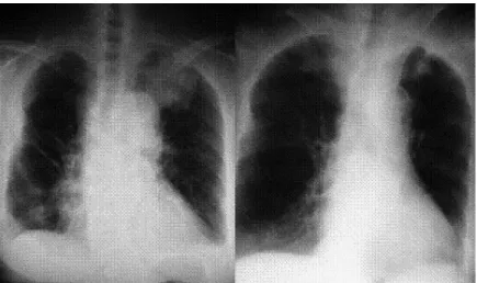
general malaise, lumbago, keeping her bedbound. Chest X-ray and computed tomographic imaging of the chest demonstrated a bulky tumor in the upper lobe of the left lung (Figures 1A and 2A) and small nodules as pulmonary metastases. Bone scintigram revealed multiple bone metastases (Figure 3A). A diagnosis of lung cancer (adenocarcinoma, cT4N3M1b stage IV PUL BRA OSS ADR) was made based on the findings of a trans-bronchial lung biopsy. Chemotherapy with gefitinib was started as therapy. After three weeks, she could walk and left the hospital. The therapeutic response was partial response (PR) with no severe adverse effects.
After leaving hospital, gefitinib therapy was continued, her activities of daily living improved, she could resume her previous life, and returned for a follow-up visit still able to walk. One year and 3 months after onset, she was admitted emergently to hospital because of acute pyelonephritis. As a result of repeated urinary tract infection, she became bedbound because of pyogenic spondylitis as a result of methicillin-resistant Staphylococcus aureus (MRSA) and was moved to the Department of Orthopedics in our hospital.
One year and 11 months after onset, she was moved to the
Department of Internal Medicine again for the purpose of lung cancer follow-up. The primary tumor was markedly reduced on chest X-ray (Figure 1B) and computed tomography (Figure 2B), and bone metastasis had almost disappeared (Figure 3B). Tumor markers were in the normal range.
Course of CEA was presented in Figure 4. After three weeks of hospitalization, diarrhea, vomiting, cough, sputum, hematuria and fever were observed and CRP was increased on a blood test. After four weeks of hospitalization, she became unconsciousness with general edema. It was highly suspicious of cerebral accident including brain metastasis and investigated about brain but not done about cardio-vascular and lung.
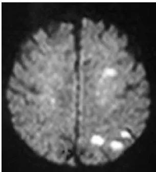
Brain MRI revealed multiple cerebral thromboses (Figure 5), and the patient died two days after falling unconsciousness, two years after onset.
In the autopsy, 600 ml fresh blood was observed in the pericardium, and was considered cardiac tamponade.
Verruciform adhered to the heart inside the left ventricle outflow at the aortic valve (Figure 6A), and was diagnosed as infectious endocarditis (Figure 6B). On the surface above the bleeding in the epicardium and right atrium from the
Figure 1. Chest X-ray at onset (A, left) and one year and 11 months after onset (B, right).
Figure 2. Computed tomography at onset (A, left) and one year and 11 months after onset (B, right).
Figure 3. Bone scintigram at onset (A, left) and one year and 11 months after onset (B, right).
69 Midori Shimada et al.: Infectious cerebral thromboembolism
Figure 4. Course of CEA before and during treatment.
0 100 200 300 400 500 600 700
0 2 12 24
Figure 4
Time (months)
CEA (ng/ml)
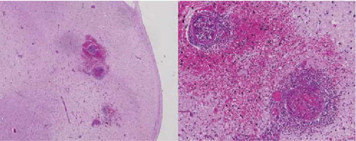
aortic valve side, a verruca was observed on an aortic valve, and an image considered to show a rupture was seen in the right atrium wall, which is equivalent to the back of the verruca. On Gram staining, many Gram-positive cocci were observed in infectious vegetation and the cardiac muscle wall (Figure 6C). An inflammatory cell-invasive lesion that contained neutrophils in blood vessels was observed in the cerebrum and fibrin was deposited in the blood vessels, which was diagnosed as infectious cerebral thromboembolism (Figure 7). Thus, repeated urinary tract infection had resulted in pyogenic spondylitis, bacteremia, and infectious endocarditis, and finally brought on infectious cerebral thromboembolism and cardiac tamponade, resulting in death. The cause of death was cardiac tamponade. The sudden loss of consciousness was caused by multiple infectious cerebral thromboembolisms.
Although lung cancer progression was not found by a pathological approach, marked adenocarcinoma remained as part of the primary lesion in the left upper lobe and did not show metastasis to other areas. General edema was caused by heart failure. Although a small infarcted lesion of cerebral denaturation by infectious thrombotic embolism, which occurs frequently, was observed, a large infarcted lesion was not recognized.
Discussion
Gefitinib is clearly effective for patients with lung cancer who have EGFR mutation compare with cytotoxic chemotherapy in progression-free survival and response rate (1-3). To evaluate the impact of gefitinib against overall survival, Japanese patients with EGFR mutations who began
Figure 5. Diffusion-weighted image of brain MRI at falling unconsciousness. Multiple peripheral high in- tensity areas were observed in left cerebral hemispheric dominant but also right occipital lobe and both cerebel- lum.
first-line systemic therapy was investigated before and after gefitinib approval in Japan (4). It was significantly longer among the EGFR-mutant patients treated after gefitinib approval and median survival time prolonged to 27.2 months compared with the before gefitinib approval of 13.6 months.
While some cancer patients show marked prolongation of life, complete recovery is rare. The present case showed an effect of gefitinib therapy on the tumor, and improved from PS 3 to 1. As urinary tract infection developed, PS worsened again, and death from cardiac tamponade and infectious cerebral thromboembolism occurred as a result of infectious endocarditis. The responsible bacteria were considered to be MRSA, because many Gram-positive cocci were observed in part of the verrucous vegetation, which was the same as in pyogenic spondylitis. Infectious endocarditis is a disease characterized by bacterial inflammation in the endocardium.
Disordered cardiac blood flow makes fissures in the
endocardium, which usually has a smooth surface, and
bacteria in the blood stream attach to it. Bacterial proliferation
forms verrucous vegetation. Once vegetation develops at a
cardiac valve, it destroys the valve and the surrounding
tissue, and causes regurgitation and heart failure. If some of
the vegetation separates and enters the blood stream, it will
move to the brain etc. and cause an embolism. Although it is
not a frequent infection, 10 to 50 persons/million/year,
infectious endocarditis has a high fatality rate of 16-25%
70 Midori Shimada et al.: Infectious cerebral thromboembolism
Figure 7. HE stain of the cerebrum in autopsy. Low power field (A, left) and high power field (B, right).
3
Figure 5
Figure 6A
(A)
Figure 6. Macroscopic image of interior surface of heart (A), HE stain of heart insection (B), and Gram stain of verruca (C) in autopsy. Infectious verruca (arrow) adhered at aortic valve of left ventricular outflow tract.
(B) (C)
71 Midori Shimada et al.: Infectious cerebral thromboembolism
(5-6). In particular, it becomes critical when infectious endocarditis is complicated with cerebral embolism. In an observational cohort study of 1779 patients with infectious endocarditis, Staphylococcus aureus was the leading cause and included 25% of MRSA (7). We did not diagnose infectious endocarditis while the present patient was alive.
Because the prognosis of patients with lung cancer has been extending, we should carefully auscultate and examine by ultrasound when an abnormal cardiac murmur is found.
Moreover, early treatment of bacteremia is needed.
Conclusion
We have reported a patient with lung cancer who developed cerebral thromboembolism from infectious endocarditis during gefitinib therapy. While long-term survival can be expected in patients with stage IV lung cancer by molecular- targeted agents, cautious management is warranted for complications, the same as for the primary disease.
Abbreviations
EGFR, epidermal growth factor receptor; MRSA, methicillin- resistant Staphylococcus aureus;
References
1) Mok TS, Wu YL, Thongprasert S, et al. Gefitinib or carboplatin-pacli- taxel in pulmonary adenocarcinoma. N Engl J Med 361(10): 947-57, 2) Maemondo M, Inoue A, Kobayashi K, et al. North-East Japan Study 2009 Group: Gefitinib or chemotherapy for non-small-cell lung cancer with mutated EGFR. N Engl J Med 362(25): 2380-8, 2010
3) Mitsudomi T, Morita S, Yatabe Y, et al. Gefitinib versus cisplatin plus docetaxel in patients with non-small-cell lung cancer harbouring muta- tions of the epidermal growth factor receptor (WJTOG3405): An open label, randomised phase 3 trial. Lancet Oncol 11(2): 121-8, 2010 4) Takano T, Fukui T, Ohe Y, et al. EGFR mutations predict survival benefit
from gefitinib in patients with advanced lung adenocarcinoma: An his- torical comparison of patients treated before and after gefitinib approval in Japan. J Clin Oncol 26(34): 5589-95, 2008
5) Moreillon P, Que YA. Infective endocarditis. Lancet 363(9403): 139-49, 6) Laupland KB, Gregson DB, Zygun DA, et al. Severe bloodstream infec-2004
tions: A population-based assessment. Crit Care Med 32(4): 992-7, 2004 7) Fowler VG Jr, Miro JM, Hoen B, et al. Staphylococcus aureus endocardi- tis: A consequence of medical progress. JAMA 293(24): 3012-21, 2005