REVIEW
67(5(27$&7,&%2'<5$',27+(5$3<)25/81*780256%$6,&
35,1&,3/(6$1'&/,1,&$/5(68/76
Masahiko Aoki
1),Yoshiomi Hatayama
1),Hideo Kawaguchi
1),Mariko Sato
1),Mitsuki Tanaka
1), Ichitaro Fujioka
1),Koji Ichise
1),Katsumi Hirose
1,2),and Yoshihiro Takai
2)Abstract In stereotactic body radiotherapy (SBRT), extracranial primary tumors or metastases are treated with high doses of radiation in a few fractions. The precise and accurate delivery of multiple radiation beams to the target maximizes tumor cell death while keeping the dose to the surrounding normal tissue to a minimum. Much of the technology to overcome the barriers to applying this treatment to moving tumors was developed in Japan. This review defines SBRT and presents the history of its technical development for safe and effective administration, as well as the clinical results of using SBRT to treat early-stage non-small cell lung cancer and lung metastases.
Hirosaki Med.J. 69:1―9,2019
Key words: stereotactic body radiotherapy; non-small cell lung cancer; lung metastases;
oligo-recurrence.
1) Department of Radiology and Radiation Oncology, Hirosaki University Graduate School of Medicine, 5 Zaifu-cho, Hirosaki, Aomori, 036-8562, JAPAN
2) Department of Radiation Oncology, Southern Tohoku BNCT Research Center, 7-10, Yatsuyamada, Koriyama, Fukushima 963-8052, JAPAN
Correspondence: M. Aoki
Received for publication, January 24, 2018 Accepted for publication, February 9, 2018
Introduction
Stereotactic body radiotherapy (SBRT) is a treatment technique with high efficacy for relatively small tumors, such as early-stage lung cancer and lung metastases. Stereotactic irradiation is differentiated from conventional radiotherapy mainly by the administration of high doses in hypofractionation, with the expectation of a high biological effect. To min- imize adverse effects on normal tissues, it is important that the high-dose region is matched to the shape of the tumor and that the dose around the tumor diminishes sharply. SBRT now provides an alternative treatment to surgery for medically inoperable patients with early-stage non-small cell lung cancer (NSCLC)
or lung metastases. Japan is one of the leading countries in the development and use of this high-precision external beam radiotherapy.
This review defines SBRT and describes the history of its technical development for safe and effective administration. It also summarizes the clinical results for SBRT in the treatment of early-stage NSCLC and lung metastases.
'H¿QLWLRQRI6%57
SBRT is a technique in which the target is precisely irradiated from multiple directions; this is to improve local control and reduce adverse effects on the surrounding normal tissue. The technique is used for small tumors localized in the trunk. Specifically, SBRT is defined by the following three criteria
1):
1. Three-dimensional irradiation from a linear accelerator is used.
2. The deviation of the position of the irradiation center is noted and confirmed to be within 5 mm for every irradiation.
3. Either the patient is immobilized to prevent
scopic real-time tumor-tracking system in 1999;
this allowed the location of a metallic marker in a tumor to be detected in three dimensions to an accuracy of within 2 mm. This dramatic improvement in the localization of a moving tumor made it possible to irradiate the tumor at a favorable phase of respiration. Kamino et al.
11)developed a four-dimensional image-guided radiotherapy system in 2006, which, uniquely, had an innovative gimbaled X-ray head; this enabled small-angle rotations along the two orthogonal gimbals, allowing accurate irradiation without stopping respiration. The majority of devices for SBRT developed in Japan have been commercialized and are currently widely used clinically.
&OLQLFDOVWXGLHVRI6%57DSSOLHGWR16&/&
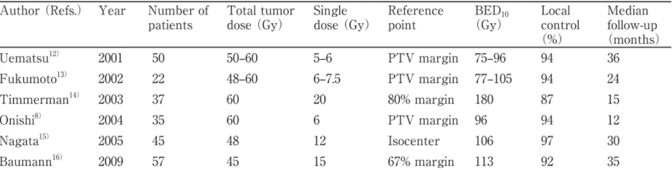
NSCLC is a leading cause of mortality world- wide. With the recent increase in the use of CT examinations, NSCLC is now often detected at an early stage. The first-choice treatment for early-stage NSCLC is surgical resection; however, many patients are considered to be inoperable because of their advanced age, poor respiratory function or other chronic illness, the risk of com- plications, or refuse surgery. SBRT provides an alternative treatment for such patients. Table 1 lists the irradiation methods and local control rates for several institutions that performed SBRT for primary stage-I NSCLC
8,12-16). Although these institutions used different prescribed doses and reference points, the initial data appear prom- ising, with local control rates of 87 % to 97 %.
Nagata et al.
17)surveyed the current status of SBRT in Japan and reported fractionation schedules. According to their survey, the most common schedules for primary lung cancer were 48 Gy administered in four fractions (used at 22 institutions), 50 Gy administered in five fractions (11 institutions), and 60 Gy admin- istered in eight fractions (four institutions).
The reason the first of these schedules is the motion, such as by using a fixed frame or shell,
or the irradiation is synchronized with the pa- tientʼs motion by tracking physiological respi- ratory movement or the movement of internal organs, providing precision control.
7KHWHFKQLFDOGHYHORSPHQWRI6%57
SBRT was developed against the background of the great success of stereotactic irradiation for intracranial tumors. Several investigators have reported the efficacy of stereotactic radiosurgery or radiotherapy for patients with intracranial malignancies
2-5). The success of these techniques resulted, in the mid-1990s, in considerable interest in their application for extracranial tumors. SBRT using CT-guided lin- ear accelerator treatment, also called a FOCAL unit, was first pioneered in 1996 by Uematsu et al.
6)for the adjustment of tumor position.
The FOCAL unit was composed of a linear accelerator, an X-ray simulator, a CT scanner, and a treatment table. It was confirmed that using the FOCAL unit reduced the set-up error to almost zero (within 0.5 mm)
7). However, even when the location of a tumor can be ac- curately identified, the problem of its respira- tory motion remains. Onishi et al.
8)developed a novel technique for lung cancer irradiation that combined a linear accelerator and CT with patient-controlled end-inspiratory breath-hold and radiation beam switching. The advantages of this technique included reduced set-up and internal margins, reduced tumor motion during irradiation without the need for a respiratory monitoring device, improved dose-volume histograms (DVHs) because of the breath- hold, and shorter treatment times. In addition, Onishi et al.
9)developed a simple respiratory monitoring device, the so-called Abches that did not include any electronic components.
Many further devices related to SBRT were
developed in Japan. Shirato et al.
10)developed
a linear accelerator synchronized with a fluoro-
most common in Japan may be related to the impact of a Japanese phase II clinical trial
18)(JCOG0403). This was the first Phase II clinical trial in the world for a medically operable case group. In JCOG0403, 48 Gy administered in four fractions was prescribed for the isocenter.
Sixty-five patients were included between July 2004 and January 2007.The median observation period was 45 months, the 3-year overall survival rate was 76 % and the 3-year locally progression-free rate was 69 %. No cases of treatment-related toxicity of grade 4 or above were identified. However, many different SBRT fractionation schedules are currently used in other institutions in Japan, and there is a lack of consensus regarding the optimal fractionation schedule. Biologically effective dose (BED)
values for tumoral and normal tissues have been used to compare the efficacy of various fractionation schedules, with many investigators reporting its utility
15, 19).
There have been several reports of a cor- relation between BED
10and local control. BED
10is defined as nd [1 + d / (
Į/
ȕ)], where n and d represent the number of fractions and fraction size, respectively, and Į/ȕ is assumed to be 10 Gy for tumors. Onishi et al.
19)evaluated clinical outcomes following stereotactic hypofractionat- ed high-dose irradiation of stage-I NSCLC and found that local control rates were better with BED
10t100 Gy than with BED10
<100 Gy (91.9
% vs. 73.6 %, respectively). Similar findings regarding the importance of BED
10for local control have been reported by Nagata et al.
15). BED
10also appears to be useful for comparing the efficacy of treatment protocols with differ- ent fraction sizes and total doses. Conversely, Shibamoto et al.
20)highlighted issues with the use of a linear-quadratic (LQ) model and BED for estimating the efficacy of radiation schedules in SBRT. The LQ model has utility for the conversion of the relatively low radiation doses used in conventional radiotherapy, but it may not be applicable to higher daily doses or small- er fraction numbers
21). Further research is need- ed, focusing on the development of alternative mathematical models for SBRT.
&OLQLFDOVWXGLHVRI6%57DSSOLHGWROXQJ metastases
Distant cancer metastases, which may, for example, form as a result of hematogenous me- tastases of the cancer, help define the advanced- stage disease. They are often associated with a poor prognosis and limited life expectancy.
However, some patients have distant metastasis in only a few sites. In 1995, Hellman and Weichselbaum proposed an intermediate state of metastasis, which they called oligometastasis, in which there were only a limited number of metastatic tumors and sites
22). The lungs are among the most common sites of metastasis fol-
Table 1. Summary of studies reporting the use of stereotactic body radiotherapy for primary lung cancer Author (Refs.) Year Number of
patients
Total tumor dose (Gy)
Single dose (Gy)
Reference point
BED10
(Gy)
Local control
(%)
Median follow-up
(months)
Uematsu12) 2001 50 50‒60 5‒6 PTV margin 75‒96 94 36
Fukumoto13) 2002 22 48‒60 6‒7.5 PTV margin 77‒105 94 24
Timmerman14) 2003 37 60 20 80% margin 180 87 15
Onishi8) 2004 35 60 6 PTV margin 96 94 12
Nagata15) 2005 45 48 12 Isocenter 106 97 30
Baumann16) 2009 57 45 15 67% margin 113 92 35
BED10, biologically effective dose; PTV, planning target volume.
lowing the radical treatment of a primary can- cer. SBRT has been widely used as a treatment option for lung oligometastases worldwide
23). Recently, Niibe et al.
24)addressed the states of oligo-recurrence, in which a patient shows one to several distant metastases/recurrences in one to several organs, and disease control at the primary cancer site. Several studies have reported favorable outcomes following SBRT for oligo-recurrence in the lungs. Inoue et al.
25)reported the results of SBRT for 22 patients with lung oligo-recurrence; with a median follow- up period of 25 months, the 3-year local control and overall survival rates were 100 % and 72
%, respectively. In a study of SBRT treatment for 42 patients with lung oligo-recurrence, Takahashi et al.
26)reported that, with a median follow-up of 20 months, the 2-year local control and overall survival rates were 87 % and 65 %, respectively.
Oligo-recurrence in the lungs following prima- ry colorectal cancer has widely been considered a worse prognostic factor for local control than that following other primary cancers. Takeda et al.
27)compared outcomes for patients with oligometastatic lung tumors following colorectal cancer (21 tumors) and following other primary cancers (23 tumors); all were treated with SBRT of 50 Gy in five fractions. The 2-year local control rates for colorectal oligometastases and the oligometastases from other origins were 72
% and 94 %, respectively (P < 0.05). It is not known why the local control rate after SBRT is worse for lung metastases from colorectal cancer than from other cancer types, but there have been several further similar reports, including one from our institution
28-30). Recently, Jingu et al.
31)reported an analysis of 93 patients that showed that dose escalation improved the local control rate of pulmonary oligometastases from colorectal cancer after SBRT. The median observation period was 28 months, the 3-year local control rates for higher BED
10(t130 Gy
for isocenter) and lower BED
10(<130 Gy for isocenter) were 95 % and 60 %, respectively
(P = 0.011). For
t 130 Gy BED10prescribed with the isocenter, standard prescribed doses for primary lung cancer in Japan (e.g. 48Gy/4, 50 Gy/5 or 60 Gy/8 fractions) are insufficient for pulmonary oligometastases from colorectal cancer. However, future studies are needed to establish the required extent of the dose increase.
$GYHUVHHYHQWVDIWHU6%57IRUOXQJWXPRU
SBRT is associated with excellent local control and minimal toxicity; however, excessive pulmonary toxicity following SBRT has been reported with the use of hypofractionated regimens, especially for centrally located tumors.
Timmerman et al.
32)reported the results of RTOG 0236, a phase II trial of SBRT in medically inoperable patients with T1 or T2 tumors who were treated with 60‒66 Gy in three fractions of 20‒22 Gy. The study enrolled 70 patients; Grade 3 to 5 toxicity occurred in 14 of these patients. The analysis of those 14 patients suggested that tumor location (hilar/
pericentral vs. peripheral) was a strong predic- tor of toxicity. The authors suggested that this regimen should not be used for patients with tumors near the central airways because of the excessive toxicity.
Conversely, there have been several reports of adverse events after SBRT for peripherally located lung tumors. The largest study of ra- diation pneumonitis after SBRT to date found frequencies of around 10 % and 2 %‒4 % for Grade 2 and Grade 3 radiation pneumonitis, respectively
33). Radiation pneumonitis following SBRT usually appears after 2 to 7 months, mostly only as an image finding that is as- ymptomatic or involves only a mild cough.
Other reported side effects of SBRT include
radiation dermatitis
34), chest wall pain
35), and rib
fractures
36); however, high-grade toxicity (Grade
3 to 5) is rare.
For SBRT candidates with lung tumors, at- tention should be paid to the presence of comor- bid interstitial pneumonia, even when findings are minimal. Takeda et al.
37)reported the case of a primary lung cancer patient with slight fo- cal honeycomb changes of the lung on CT, who experienced acute exacerbation of subclinical idiopathic pulmonary fibrosis following SBRT. In their survey of SBRT in Japan, Nagata et al.
17)reported the frequency of Grade 5 radiation pneumonitis to be 0.5 %.
&OLQLFDOH[SHULHQFHRI6%57IRUOXQJWXPRUVDW our institution
At Hirosaki University Hospital, we started using SBRT for lung tumors in May 2003. Our eligibility criteria for treatment with SBRT are as follows: (1) primary lung cancer (T1‒
2N0M0), or no more than three lung metastases without active primary cancer; (2) tumor size <50 mm across the maximum diameter;
(3) tumor visible by fluoroscopy; and (4)
performance status d 2 according to the Eastern Cooperative Oncology Group performance scale.
We have previously reported our initial clinical experience of SBRT in patients with early- stage NSCLC and lung metastasis, using a total dose of 54 Gy administered in nine fractions
38), and we have subsequently performed a dose escalation study of SBRT for localized lung tumor with increases in fraction size of 1 Gy.
We started our dose escalation study with a fraction size of 6 Gy. Although a fraction size of 12 Gy is now commonly used in Japan
18), the optimal fractionation schedule for SBRT was unknown at the time the study commenced.
Table 2 summarizes the 3-year local control rates and adverse events according to almost uniform doses with five treatment schedules at our institution. Regardless of the fractionation schedule, SBRT with total doses between 50 and 56 Gy administered over five to nine fractions achieved acceptable tumor control without any severe complications
39).
The current treatment schedules for SBRT at our institution are as follows: a total dose of 50 Gy administered in five fractions for tumors <3 cm in diameter; 60 Gy administered in six fractions for tumors >3 cm in diameter or for lung metastasis from colorectal cancer, regardless of tumor size.
Between May 2003 and December 2017, 395 patients with 445 lung tumors (primary lung cancer, n = 327; metastases, n = 118)
were treated by SBRT in our institution. The median follow-up period for all the patients was 27.5 months. The 3-year local control rates for stage-I lung cancer and lung metastases were 90.1 % and 87.1 %, respectively (Figure 1). The frequency of radiation pneumonitis after SBRT was 1.5 % (six patients) at Grade 2 and 0.3 %
(one patient) at Grade 3. We observed no other adverse events after SBRT at Grade 2 or more
Table 2. Three-year local control rates and adverse events following stereotactic body radiotherapy, by fractionation schedule39)
Fraction size Total dose BED10 Tumor size (n) 3-year LC Radiation pneumonitis (n)
(Gy) (Gy) (Gy) d 3 cm > 3 cm (%) Grade 1 Grade 2
6 54 86.4 12 8 90 13 0
7 56 95.2 19 1 95 13 0
8 56 100.8 19 1 95 15 1
9 54 102.6 18 2 95 13 1
10 50 100.0 18 2 100 16 0
BED10, biologically effective dose; LC, local control.
Figure 1 Kaplan-Meier curves of local control rates for stage-I lung cancer (n = 327) and lung metastases (n = 118)
after stereotactic body radiotherapy (SBRT). The curves include all such tumors at Hirosaki University Hospital between May 2003 and December 2017.
Figure 2 Clinical course of a patient treated with stereotactic body radiotherapy (SBRT). The patient was an 80-year- old woman with primary lung cancer (adenocarcinoma; cT1N0M0). (A) Pre-treatment CT scan with the dose distribution. (B‒F) CT scans acquired at 3 months (B), 6 months (C), 1 year (D), 2 years (E), and 5 years
(F) after the SBRT. Radiation pneumonitis was observed 6 months after the SBRT, which changed to radiation fibrosis. The tumor was controlled more than 5 years after the SBRT.
A B C
D E F
in our series. A typical case of a patient with early-stage NSCLC who responded well to this treatment is shown in Figure 2.
Conclusion
SBRT offers a high degree of local control
with minimal toxicity for patients with early-
stage NSCLC and lung oligo-recurrence. It is our
intention to refine this technology and adapt it for other malignant tumors, and we will continue to use it for the treatment of many patients.
5HIHUHQFHV
1) Onishi H. Guideline for stereotactic body radio- therapy. J Jpn Soc Ther Radiol Oncol. 2006;18:1- 17.
2) Flickinger JC, Kondziolka D, Lunsford LD, Coffey RJ, Goodman ML, Shaw EG, Hudgins WR, et al.
A multi-institutional experience with stereotactic radiosurgery for solitary brain metastasis. Int J Radiat Oncol Biol Phys. 1994;28:797-802.
3) Shirato H, Takamura A, Tomita M, Suzuki K, Nishioka T, Isu T, Kato T, et al. Stereotactic irra- diation without whole-brain irradiation for single brain metastasis. Int J Radiat Oncol Biol Phys.
1997;37:385-91.
4) Sneed PK, Lamborn KR, Forstner JM, McDermott MW, Chang S, Park E, Gutin PH, et al. Radiosur- gery for brain metastases: Is whole brain radio- therapy necessary? Int J Radiat Oncol Biol Phys.
1999;43:549-58.
5) Aoki M, Abe Y, Hatayama Y, Kondo H, Basaki K.
Clinical outcome of hypofractionated conventional conformation radiotherapy for patients with single and no more than three metastatic brain tumors, with noninvasive fixation of the skull without whole brain irradiation. Int J Radiat Oncol Biol Phys. 2006;64:414‒8.
6) Uematsu M, Shioda A, Tahara K, Fukui T, Yamamoto F, Tsumatori G, Ozeki Y, et al. Focal, high dose, and fractionated modified stereotactic radiation therapy for lung carcinoma patients: a preliminary experience. Cancer. 1998;82:1062-70.
7) Uematsu M, Fukui T, Shioda A, Tokumitsu H, Takai K, Kojima T, Asai Y, et al. A dual computed tomography linear accelerator unit for stereotactic radiation therapy: a new approach without cranially fixated stereotactic frames. Int J Radiat Oncol Biol Phys. 1996;35:587-92.
8) Onishi H, Kuriyama K, Komiyama T, Tanaka S, Sano N, Marino K, Ikenaga S, et al. Clinical outcomes of stereotactic radiotherapy for stage
I non-small cell lung cancer using a novel irra- diation technique: patient self-controlled breath- hold and beam switching using a combination of linear accelerator and CT scanner. Lung Cancer.
2004;45:45-55.
9) Onishi H, Kawakami H, Marino K, Komiyama T, Kuriyama K, Araya M, Saito R, et al. A simple respiratory indicator for irradiation during volun- tary breath holding: A one-touch device without electronic materials. Radiology. 2010;255:917-23.
10) Shirato H, Shimizu S, Kitamura K, Nishioka T, Kagei K, Hashimoto S, Aoyama H, et al. Four- dimensional treatment planning and fluoroscopic real-time tumor tracking radiotherapy for moving tumor. Int J Radiat Oncol Biol Phys. 2000;48:435- 42.
11) Kamino Y, Takayama K, Kokubo M, Narita Y, Hirai E, Kawawda N, Mizowaki T, et al.
Development of a four-dimensional image-guided radiotherapy system with a gimbaled X-ray head.
Int J Radiat Oncol Biol Phys. 2006;66:271-8.
12) Uematsu M, Shioda A, Suda A, Fukui T, Ozeki Y, Hama Y, Wong JR, et al. Computed tomography- guided frameless stereotactic radiotherapy for stage I non-small-cell lung cancer : A 5-year expe- rience. Int J Radiat Oncol Biol Phys. 2001;51:666- 70.
13) Fukumoto S, Shirato H, Shimizu S, Ogura S, Onimaru R, Kitamura K, Yamazaki K, et al.
Small-volume image-guided radiotherapy using hypofractionated, coplanar, and noncoplanar multi- ple fields for patients with inoperable Stage I non- small cell lung carcinomas. Cancer. 2002;95:1546- 53.
14) Timmerman R, Papiez L, McGarry R, Likes L, DesRosiers C, Frost S, Williams M. Extracranial stereotactic radioablation: results of a phase I study in medically inoperable stage I non-small cell lung cancer. Chest. 2003;124:1946-55.
15) Nagata Y, Takayama K, Matsuo Y, Norihisa Y, Mizowaki T, Sakamoto T, Sakamoto M, et al.
Clinical outcomes of a phase I/II study of 48 Gy of stereotactic body radiotherapy in 4 fractions for primary lung cancer using a stereotactic body frame. Int J Radiat Oncol Biol Phys. 2005;63:1427-31.
16) Baumann P, Nyman J, Hoyer M, Wennberg B,
Gagliardi G, Lax I, Drugge N, et al. Outcome in a prospective phase II trial of medically inoperable stage I non-small-cell lung cancer patients treated with stereotactic body radiotherapy. J Clin Oncol.
2009;27:3290-6.
17) Nagata Y, Hiraoka M, Mizowaki T, Narita Y, Matsuo Y, Norihisa Y, Onishi H, et al. Survey of stereotactic body radiation therapy in Japan by the Japan 3-D Conformal External Beam Ra- diotherapy Group. Int J Radiat Oncol Biol Phys.
2009;75:343-7.
18) Nagata Y, Hiraoka M, Shibata T, Onishi H, Kokubo, M. Karasawa K, Shioyama Y, et al.
Prospective trial of stereotactic body radiation therapy for both operable and inoperable T1N0M0 non-small cell lung cancer: Japan clinical oncology group study (JCOG0403). Int J Radiat Oncol Biol Phys. 2015;93:989-96.
19) Onishi H, Araki T, Shiato H, Nagata Y, Hiraoka M, Gomi K, Yamashita T, et al. Stereotactic hypofractionated high-dose irradiation for stage I nonsmall cell lung carcinoma. Clinical outcomes in 245 subjects in a Japanese multiinstitutional study. Cancer. 2004;101:1623-31.
20) Shibamoto Y, Otsuka S, Iwata H, Sugie C, Ogino H, Tomita N. Radiobiological evaluation of the radia- tion dose as used in high-precision radiotherapy:
Effect of prolonged delivery time and applicability of the linear-quadratic model. J Radiat Res. 2012;
53:1-9.
21) Kirkpatrick JP, Meyer JJ, Marks LB. The linear- quadratic model is inappropriate to model high dose per fraction effects in radiosurgery. Semin Radiat Oncol. 2008;18:240-3.
22) Hellman S, Weichselbaum RR. Oligometastases. J Clin Oncol. 1995;13:8-10.
23) Lewis SL, Porceddu S, Nakamura N, Palma DA, Lo SS, Hoskin P, Moghanaki D, et al. Definitive stereotactic body radiotherapy (SBRT) for extracranial oligometastases: an international survey of >1000 radiation oncologists. Am J Clin Oncol. 2017;40:418-22.
24) Niibe Y, Hayakawa K. Oligometastases and oligo- recurrence: the new era of cancer therapy. Jpn J Clin Oncol. 2010;40:107-11.
25) Inoue T, Katoh N, Onimaru R, Shirato H. Clinical outcomes of stereotactic body radiotherapy for patients with lung tumors in the state of oligo- recurrence. Pulm Med. 2012;2012:369820.
26) Takahashi W, Yamashita H, Niibe Y, Shiraishi K, Hayakawa K, Nakagawa K. Stereotactic body radiotherapy for metastatic lung cancer as oligo- recurrence: an analysis of 42 cases. Pulm Med.
2012;2012:454107.
27) Takeda A, Kunieda E, Ohashi T, Aoki Y, Koike N, Takeda T. Stereotactic body radiotherapy
(SBRT) for oligometastatic lung tumors from colorectal cancer and other primary cancers in comparison with primary lung cancer. Radiother Oncol. 2011;101:255-9.
28) Hamamoto Y, Kataoka M, Yamashita M, Shinkai T, Kubo Y, Sugawara Y, Inoue T, et al. Local control of metastatic lung tumors treated with SBRT of 48 Gy in four fractions: in comparison with primary lung cancer. Jpn J Clin Oncol. 2010;40:125-9.
29) Yamamoto T, Jingu K, Shirata Y, Koto M, Matsushita H, Sugawara T, Kubozono M, et al. Outcomes after stereotactic body radiotherapy for lung tu- mors, with emphasis on comparison of primary lung cancer and metastatic lung tumors. BMC Cancer. 2014;14:464.
30) Aoki M, Hatayama Y, Kawaguchi H, Hirose K, Sato M, Akimoto H, Miura H, et al. Stereotactic body radiotherapy for lung metastases as oligo- recurrence: a single institutional study. J Radiat Res. 2016;57:55-61.
31) Jingu K, Matsuo Y, Onishi H, Yamamoto T, Aoki M, Murakami Y, Yamashita H, et al. Dose escalation improves outcome in stereotactic body radiotherapy for pulmonary oligometastases from colorectal cancer. Anticancer Res. 2017;37:2709-13.
32) Timmerman R, McGarry R, Yiannoutsos C, Papiez L, Tudor K, DeLuca J, Ewing M, et al. Excessive toxicity when treating central tumors in a phase II study of stereotactic body radiation therapy for medically inoperable early-stage lung cancer. J Clin Oncol. 2006;24:4833-9.
33) Guckenberger M, Baier K, Polat B, Richter A, Krieger T, Wilbert J, Mueller G, et al. Dose- response relationship for radiation-induced pneu- monitis after pulmonary stereotactic body radio-
therapy. Radiother Oncol. 2010;97:65-70.
34) Baumann P, Nyman J, Lax I, Friesland S, Hoyer M, Rehn Ericsson S, Johansson KA, et al. Factors important for efficacy of stereotactic body ra- diotherapy of medically inoperable stage I lung cancer. A retrospective analysis of patients treated in the Nordic countries. Acta Oncol. 2006;45:787-95.
35) Woody NM, Videtic GM, Stephans KL, Djemil T, Kim Y, Xia P. Predicting chest wall pain from lung stereotactic body radiotherapy for different fractionation schemes. Int J Radiat Oncol Biol Phys. 2012;83:427-34.
36) Aoki M, Sato M, Hirose K, Akimoto H, Kawaguchi H, Hatayama Y, Ono S, et al. Radiation-induced rib fracture after stereotactic body radiotherapy with a total dose of 54-56 Gy given in 9-7 fractions for patients with peripheral lung tumor: impact of maximum dose and fraction size. Radiat Oncol.
2015;10:99.
37) Takeda A, Enomoto T, Sanuki N, Nakajima T, Takeda T, Sayama, K, Kunieda E. Acute exacer- bation of subclinical idiopathic pulmonary fibrosis triggered by hypofractionated stereotactic body radiotherapy in a patient with primary lung can- cer and slightly focal honeycombing. Radiat Med.
2008;26:504-7.
38) Aoki M, Abe Y, Kondo H, Hatayama Y, Kawaguchi H, Fujimori A, Suzaki K, et al. Clinical outcome of stereotactic body radiotherapy of 54 Gy in nine fractions for patients with localized lung tumor using a custom-made immobilization system.
Radiat Med. 2007;25:289-94.
39) Aoki M, Hatayama Y, Kawaguchi H, Hirose K, Sato M, Akimoto H, Fujioka I, et al. Clinical outcome of stereotactic body radiotherapy for primary and oligometastatic lung tumors: a single institutional study with almost uniform dose with different five treatment schedules. Radiat Oncol.
2016;11:5.