Temporal Attenuation of Iodine Content and its Effect on the Antibacterial Activity of
Iodine‑Supported Titanium Implants
著者 加藤 貴士
著者別表示 Kato Takashi journal or
publication title
博士論文本文Full 学位授与番号 13301甲第4587号
学位名 博士(医学)
学位授与年月日 2017‑06‑30
URL http://hdl.handle.net/2297/00052033
doi: 10.4172/1948-5948.1000298
Temporal Attenuation of Iodine Content and its Effect on the Antibacterial Activity of Iodine-Supported Titanium Implants
Takashi Kato
1, Toshiharu Shirai
1, Norio Yamamoto
1*, Hideji Nishida
1, Katsuhiro Hayashi
1, Akihiko Takeuchi
1, Shinji Miwa
1, Kaori Ohtani
2and Hiroyuki Tsuchiya
11
Department of Orthopaedic Surgery, Graduate School of Medical Science, Kanazawa University, 13-1 Takaramachi Kanazawa, Japan
2
Department of Bacteriology, Kanazawa University, 13-1 Takaramachi Kanazawa, Japan
*Corresponding author: Yamamoto N, Department of Orthopaedic Surgery, Graduate School of Medical Science, Kanazawa University, 13-1 Takaramachi Kanazawa, Japan, Tel: +81-76-265-2374; E-mail: [email protected] Received May 25, 2016; Accepted June 28, 2016; Published July 08, 2016 Citation: Kato T, Shirai T, Yamamoto N, Nishida H, Hayashi K, et al. (2016) Temporal Attenuation of Iodine Content and its Effect on the Antibacterial Activity of Iodine-Supported Titanium Implants. J Microb Biochem Technol 8: 285-289. doi:
10.4172/1948-5948.1000298
Copyright: © 2016 Kato T, et al. This is an open-access article distributed under the terms of the Creative Commons Attribution License, which permits unrestricted use, distribution, and reproduction in any medium, provided the original author and source are credited.
Keywords: Iodine-supported titanium, Antibacterial activity, Tem- poral attenuation, Implant infection
Abbreviations: E. coli: Escherichia coli PBS: phosphate-buffered saline
S. aureus: Staphylococcus aureus
Introduction
A wide variety of implants is currently used in orthopedic procedures. Most conventional implants do not possess antimicrobial activity and implant-related infections continue to be a significant complication. Implant-related infections occur in 0.7% to 4.2% of patients after orthopedic surgery and are a particular concern after prosthetic replacement arthroplasties [1-5]. The risk of infection is particularly high in patients receiving chemotherapy for orthopedic conditions, such as bone soft tissue tumors and rheumatoid arthritis.
Indeed, the infection rate is 1–2% in patients with total knee arthroplasty, but 14.6–17% for cases of bone soft tissue tumors of the knee that underwent reconstruction with implants [6-10].
The treatment of orthopedic implant-related infections is challenging because bone infections require the long-term administration of antibacterial agents, irrigation, multiple surgeries, and implant removal or replacement. The prevention of such infections is therefore very important. Various biomaterial surface modifications of stainless steel and titanium orthopedic implants have been developed for prophylaxis against implant-related infections. The covalent attachment of polycationic groups, implantation of Ca+, N+, and F+
ions with antimicrobial agents, and alloying and surface processing of silver have been investigated. We have developed a technique to form a highly porous anodic oxide film on the surface of titanium implants and impregnate iodine into the pores; this strategy imparts antimicrobial
Abstract
Infections related to orthopedic implants often require prolonged therapy and complex interventions; accordingly, the development of implants with a low risk of infection is a high priority. Iodine-supported titanium implants with antibacterial activity are safe and effective for prophylaxis against implant-related infections. However, temporal changes in the iodine content of implants and the relationship between iodine content and antibacterial activity have not been investigated. Temporal changes in iodine for 10–12 μg/cm
2iodine-supplemented titanium implants were investigated in vitro and in vivo using rabbit models (subcutaneous soft tissue, intra-articular, and endo-osseous sites). In vitro antibacterial activity against Staphylococcus aureus and E. coli were also investigated using implants with various iodine contents (0%, 20%, 50%, 60%, and 100%, where 100% corresponds to 13 μg/cm
2iodine based on current implants in clinical use). The residual iodine after 1 year of implantation and the minimum effective iodine concentration required for antibacterial activity were determined. In vitro and in vivo experiments showed a similar temporal pattern of initially rapid and subsequently slow attenuation of iodine in the implants, with approximately 30% of the initial iodine content remaining at 1 year. Pure titanium implants and implants with a 0% oxide layer did not exhibit antibacterial activity. Titanium implants supplemented with 20%, 50%, 80%, and 100% iodine showed in vitro antibacterial activity that varied in a dose-dependent and duration-dependent manner. Implants with
≥20% iodine achieved complete clearance of S. aureus and E. coli colonies by 24 hours of incubation. Implants with iodine contents of ≥20% demonstrated sufficient antibacterial activity, indicating that current iodine-supported titanium implants possess adequate antibacterial activity to prevent implant-related infections, even after 1 year of implantation. These results support the clinical use of iodine-supported titanium implants to prevent orthopedic implant-related infections.
activity to the implants [11-19]. In general, iodine-supported titanium implants have a 5–10-μm-thick oxide film with >50,000 pores/mm
2loaded with 10–12 μg/cm
2iodine. In our previous studies, we have demonstrated the in vitro antibacterial activity, in vitro cytocompatibility, and in vivo safety characteristics of iodine- supported titanium implants [18]. Early results from ongoing clinical trials have also demonstrated the safety and effectiveness of these implants in patients [20-23]. We have observed that the iodine content on the implant surface decreases over time; however, temporal changes in the iodine content and the relationship between iodine content and antibacterial activity have not been investigated. The purpose of this study was to examine the time course of the iodine content using in vitro and in vivo experimental models and to demonstrate the relationship between iodine content and antibacterial activity.
Materials and Methods Study design
Iodine-supported titanium implants were produced in various
shapes and with different quantities of iodine. in vitro antibacterial
Citation: Kato T, Shirai T, Yamamoto N, Nishida H, Hayashi K, et al. (2016) Temporal Attenuation of Iodine Content and its Effect on the Antibacterial Activity of Iodine-Supported Titanium Implants. J Microb Biochem Technol 8: 285-289. doi: 10.4172/1948-5948.1000298
activity against S. aureus and E.coli was investigated using implants with different iodine contents to determine the minimum effective iodine content required for antibacterial activity. The temporal change in iodine content in the implants over 1 year was also investigated using in vitro and in vivo rabbit models.
Iodine-supported implants
Iodine-supported titanium implants were produced by Prostec Instruments Company (Sabae, Japan) using a technique described previously [18,19]. Briefly, a 5–10-μm-thick anodic oxide film with
>50,000 pores/mm
2was formed on the surface of titanium implants.
Ionized iodine was electrodeposited within these pores to achieve the desired iodine content (0–13 μg/cm
2iodine). Four types of iodine- supported titanium implants were developed as follows: (1) square plates (20 mm × 20 mm × 2 mm); (2) circular discs (6 mm diameter, 2 mm thick); (3) washers (outer diameter 16 mm, inner diameter 6.5 mm, 2 mm thick); and (4) screws (6.5 mm diameter, 16 mm length).
The iodine content on implants was measured by X-ray fluorescence spectroscopy (Prostec Instruments Company).
In vitro temporal assessment of iodine contents in implants Square implants with iodine contents of 10–12 μg/cm
2(n = 3 per condition) were immersed in 30 mL of PBS in a 50-mL centrifuge tube and incubated at 37
°C for various durations up to 1 year. After the specified periods (3 hours, 12 hours, 24 hours, 10 days, 50 days, and 1 year), the implants were removed and the iodine content was measured by X-ray fluorescence spectroscopy (Prostec Instruments Company) and averaged for the three implants. Residual iodine is expressed as a percentage of the iodine content in the fresh implant.
In vivo temporal assessment of iodine contents in implants A previously described rabbit model was used for the in vivo analysis [18]. All animal experiments were conducted with the approval of the Institutional Animal Care and Use Committee and carried out in strict accordance with its regulations. Mature female Japanese white rabbits (n = 15) weighing 2.5–3.0 kg were anesthetized by an intramuscular injection of ketamine hydrochloride (50 mg/kg; Warner–Lambert,
Morris Plains, NJ, USA) and an intravenous injection of pentobarbital sodium (40–50 mg/kg). Each animal received three iodine-supported titanium implants (one circular disc, one washer, and one screw; iodine content, 10–12 μg/cm
2) at three different anatomical sites (subcutaneous soft tissue, intra-articular, and endo-osseous) relevant for orthopedic implants (Figure 1).
For the subcutaneous soft tissue model, a circular disc was implanted in the dorsal aspect of the rabbit. The knee joint was used to model the joint and bone sites. A screw was inserted through a washer into the distal intra-articular end of the femur, with the screw and washer representing the bone and joint site, respectively (Figure 1). At specific postoperative periods (14, 28, 90, 200, and 365 days), the animals were killed (n=3 per time point), implants were retrieved, and residual iodine was measured by X-ray fluorescence spectroscopy (Prostec Instruments Company). Values obtained for three animals at each time point were averaged for the analysis.
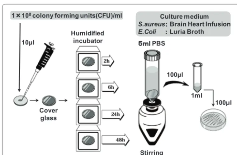
In vitro antibacterial activity
Circular disc implants (diameter: 20 mm; thickness: 2 mm) were used. Five types of implants were fabricated with different iodine content: 0 μg/cm
2(0% oxide layer), 3 μg/cm
2(20%), 7 μg/cm
2(50%), 10 μg/cm
2(80%), and 13 μg/cm
2(100%). Note that 13 μg/cm
2was considered 100% because it is the standard amount in clinical implants [18]. Pure titanium implants were used as controls.
The implants were exposed to gram-positive S. aureus strain 25923 (ATCC, Manassas, VA, USA) or gram-negative E. coli strain MG1655 (ATCC). The antibacterial activity of the implants was measured using a method approved by Japanese Industrial Standards as previously described [18]. Approximately 10
6colony-forming units were inoculated on an autoclaved circular implant placed in a sterile glass Petri dish, which was then covered and incubated at 37°C for 2, 6, 24, or 48 hours (n = 3 per implant type). At each time point, each implant was washed with 5 mL of phosphate-buffered saline (PBS). The washed eluate was diluted 1:50 with PBS and 100 μL of the dilute eluate was incubated in Brain Heart Infusion broth for S. aureus and LB broth (1% w/v tryptone, 0.5% w/v yeast extract, 0.5% w/v NaCl) for E. coli at 37°C. The number of bacterial colonies was counted after 24 hours and the average number of colonies for the triplicate implants at different incubation periods (2–48 hours) was used to determine antibacterial activity against S. aureus and E. coli (Figure 2).
Washer Screw Circular
Implant
Figure 1: Study design for in vivo temporal assessments of the iodine content in implants. Each rabbit received three implants—a subcutaneous circular implant, an intra-articular washer, and an endo-osseous screw. Animals were euthanized at various time points over 1 year and implants were retrieved to measure the residual iodine
10μl
Cover glass
Humidified incubator 1 × 10
8colony forming units(CFU)/ml
5ml PBS
Stirring 100μl
1ml 100μl Culture medium S.aureus : Brain Heart Infusion E.Coli : Luria Broth
Figure 2: Study design for in vitro antibacterial activity assessment using a
modified version of the Japanese Industrial Standards method.
For the statistical analysis, Student's t-tests were performed for parametric tests and differences among means were consider statistically significant when p < 0.05.
Results
In vitro temporal assessment of iodine quantities in implants Residual iodine in the implants decreased gradually over the duration of the in vitro study. The average residual iodine was 68.7% at 3 hours, 62.7% at 12 hours, 53.5% at 24 hours, 41.6% at 10 days, 24.8%
at 50 days, and 29.8% at 365 days (Figure 3).
In vivo temporal assessment of iodine quantities in implants Residual iodine in the implants also decreased gradually over time in the in vivo study at all three anatomical locations (Figure 4).
In the subcutaneous soft tissue (circular implant), the average residual iodine was 59.7% at 14 days, 54.4% at 28 days, 34.1% at 90 days, 23.1% at 200 days, and 27.3% at 365 days. In the intra-articular location (washer implant), the average residual iodine was 54.7% at 14 days, 51.1% at 28 days, 53.7% at 90 days, 42.7% at 200 days, and 31.5%
at 365 days. Similarly, in the intra-osseous location (screw implant), the
average residual iodine was 47.4% at 14 days, 47.1% at 28 days, 34.9% at 90 days, 32.4% at 200 days, and 30.4% at 365 days.
In vitro antibacterial activity
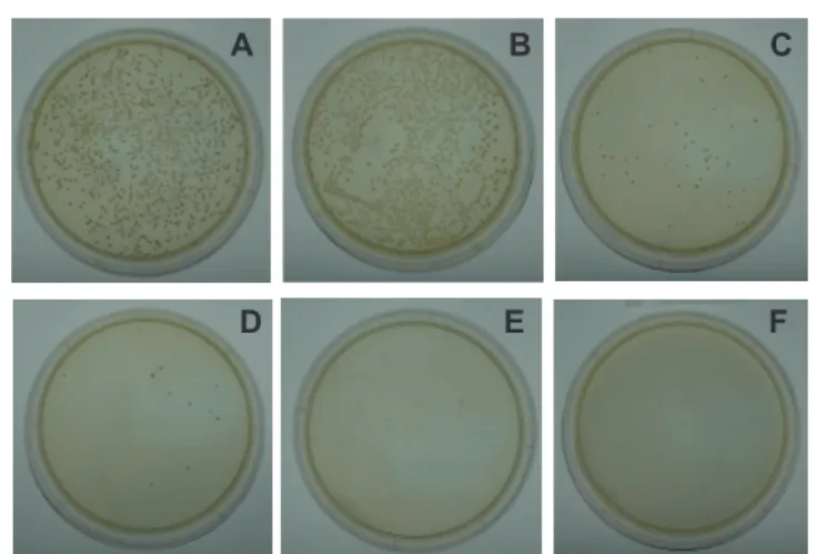
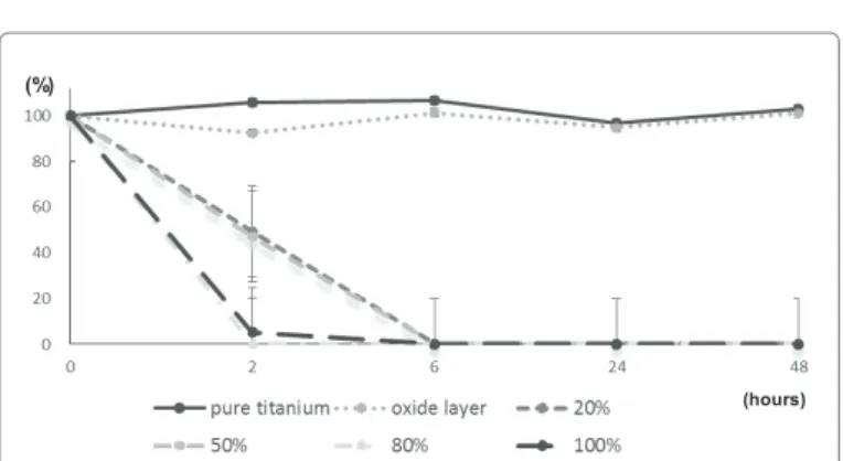
There was no reduction in the number of bacterial colonies in the pure titanium implants and the 0% oxide layer groups over 48 hours of incubation, and there was no significant difference between these two groups. However, all four iodine-supported implant groups (20%, 50%, 80%, and 100% iodine contents) showed significantly fewer S. aureus and E. coli colonies than the control groups, starting 2 hours after incubation (p<0.05). Longer incubation periods resulted in greater decreases in the number of bacterial colonies in all iodine-supported implants, with complete disappearance of S. aureus and E. coli colonies by 6 hours in ≥80% iodine groups (Figure 5). Implants with ≥20%
iodine completely inhibited S. aureus colonies after 24 hours and E. coli colonies after 6 hours (Figure 6 and 7).
10 50 100 365
(Days)(%)
Figure 3: In vitro temporal profile of iodine content in implants, showing an initially rapid attenuation, followed by a more gradual attenuation during incubation in phosphate-buffered saline (PBS). Implants retained approximately 30% of the initial iodine content at the end of 1 year.
The vertical axis is for iodine content.
(days)
(%)
14 28 90 200 365
Figure 4: In vivo temporal profile of iodine content in implants, showing an initially rapid attenuation, followed by a more gradual attenuation at all three anatomical sites. All implants retained approximately 30% of the initial iodine content at the end of 1 year.
The vertical axis is for iodine content.
A B C
F E
D
Figure 5: In vitro antibacterial activity of titanium implants with different iodine contents on S. aureus after 6 hours of incubation. A: pure titanium implant, B: oxide layer with 0% iodine, C: 20%, D: 50%, E: 80%, and F: 100% iodine contents (where 100% corresponds to 13 μg/cm2 iodine). The number of bacterial colonies was high in pure titanium implant and implants with an oxide layer with 0% iodine. In contrast, the number of colonies decreased gradually as the iodine content in implants increased.
(hours)
(%)
Figure 6: In vitro antibacterial activity of titanium implants with different iodine contents on S. aureus. While pure titanium implants and 0% iodine implants had no antibacterial activity, implants with ≥20% iodine showed a significant decrease in the number of colonies compared with the control group beginning at 2 hours (p<0.05). Implants with 100% iodine exhibited significantly greater and faster decreases in the number of colonies compared to 20% iodine implants (p<0.05). Implants with ≥20% iodine showed complete inhibition of bacterial colonies after 24 hours.
The vertical axis is for bacterial colonies.
Citation: Kato T, Shirai T, Yamamoto N, Nishida H, Hayashi K, et al. (2016) Temporal Attenuation of Iodine Content and its Effect on the Antibacterial Activity of Iodine-Supported Titanium Implants. J Microb Biochem Technol 8: 285-289. doi: 10.4172/1948-5948.1000298
Discussion
The in vitro and in vivo experiments showed similar temporal patterns of attenuation of iodine contents in the implants. In the in vitro analysis, an initially rapid attenuation was followed by a more gradual decrease, with approximately 30% of the initial iodine maintained at 1 year. In the in vivo analysis, the iodine content in implants similarly decreased rapidly to 50–60% at 28 days, followed by a more gradual decline, with approximately 30% of initial iodine remaining in the implants at 1 year. We speculated that initial period of rapid decline can be attributed to the superficial pores, while the prolonged release is explained by the deep pores in the oxide layer.
The in vitro antibacterial activity of iodine-supported titanium implants was related to the iodine quantity in a dose-dependent and duration-dependent manner. While implants with high iodine contents (≥80%) completely inhibited S. aureus and E. coli colonies by 6 hours, implants with lower iodine contents (≥20%) also completely inhibited S. aureus colonies by 24 hours and E. coli colonies by 6 hours. These results suggest that implants with iodine contents of ≥20% of the levels typically used in clinical applications possess sufficient antibacterial activity to prevent implant-related infections.
Based on the observed antibacterial activity and the temporal attenuation profile of the implants, currently used iodine-supported titanium implants possess adequate iodine to maintain antibacterial activity, even after 1 year of implantation. Further, the attenuation was similar at subcutaneous, intra-articular, and endo-osseous sites, suggesting that the implants effectively prevent infections in each of these locations.
The onset of implant-related infections can be early (1 to 3 months after implantation), delayed (within several months to 1 year after implantation), or late (beyond 1 year; infection carried by the blood from a non-surgical site infection) [24]. Therefore, it is crucial that the antibacterial implant retains antibacterial activity over long periods.
Iodine-supported titanium implants are therefore particularly useful as they not only prevent early and late postoperative infections, but also likely prevent delayed infections. The long-term antibacterial activity of iodine-supported titanium implants is beneficial for implants used to treat fractures. Iodine-supported titanium implants are particularly
useful in cases of open fractures, which are associated with a higher risk of infection, and in cases with a risk of poor bone union, which may require plates and intramedullary nails for periods exceeding a year.
Several other surface treatment strategies, such as coating with antibiotics or silver ions, have also been used to impart antimicrobial properties to implant surfaces. However, many antibiotics act against a narrow spectrum of bacteria, and the emergence of resistant bacteria is an important risk [25]. Furthermore, the rapid release of antibiotics (80% in 60 minutes) limits their ability to effectively prevent infections over long periods [26]. Silver-coated orthopedic implants also show a rapid release of silver ions in the first three days, with only about 30%
remaining after 14 days and antibacterial activity lasting for about 28 days [27,28]. Furthermore, toxicity resulting from high silver ion concentrations in vivo remains a concern. In particular, toxicity to bone cells and the accumulation of silver ions in the kidney, spleen, liver, and brain have been reported [29-32].
Iodine-supported titanium implants offer distinct advantages over implants with antibiotics and silver ion surface modifications. First, iodine has a wide antibacterial spectrum, is active against bacteria, viruses, fungi, and spores, and does not induce resistance in bacteria.
Second, iodine has proven safety as a disinfectant and contrast agent [33]. Third, iodine-supported titanium implants possess long-term antibacterial activity that lasts longer than 1 year. In our previous in vitro and clinical studies, we demonstrated that iodine-supported titanium implants have sufficient antibacterial activity, but no cytotoxicity or deleterious effects on thyroid function [18,33]. We have also shown that iodine-supported titanium implants possess similar high bone affinity and osteoconductive properties to those of pure titanium implants [18].
This study had a few limitations. First, to examine antibacterial activity, we prepared titanium implants with low iodine contents to approximate the attenuated iodine content in implants after various periods. However, the in vivo release profile and antibacterial activity of these low-iodine-content titanium implants may differ from those of actual implants. Second, a relatively small number of implants were used per group. However, low variability within groups and repeated measurements from the same specimens enabled us to detect statistically significant differences.
In summary, iodine-supported titanium implants retain approximately 30% of the iodine content at 1 year in both in vitro and in vivo conditions. As implants with iodine contents of ≥20% demonstrated sufficient antibacterial activity, current iodine-supported titanium implants can be expected to possess adequate antibacterial activity to prevent implant-related infections, even after one year of implantation, and should be strongly considered for orthopedic applications.
Acknowledgement
This work was supported by JSPS KAKENHI Grant Number 25670640. We would like to express our sincere gratitude to late Dr. Tohru Shimizu, Professor at Department of Bacteriology, Kanazawa University, for sharing his pearls of wisdom with us during the course of this research. Editorial support, in the form of medical writing based on authors’ detailed directions, collating author comments, copyediting, fact checking, and referencing, was provided by Cactus Communications.
References
1. Bauer TW, Schils J (1999) The pathology of total joint arthroplasty. I.
Mechanisms of implant fixation. Skeletal Radiol 28: 423-432.
2. Bauer TW, Schils J (1999) The pathology of total joint arthroplasty II Mechanisms of implant failure. Skeletal Radiol 28: 483-497.
3. Periti P, Mini E, Mosconi G (1998) Antimicrobial prophylaxis in orthopaedic surgery: the role of teicoplanin. J Antimicrob Chemother 41: 329-340.
(hours)