Table 1 Laboratory data on admission
【Serology】
【CBC】
mg/dl 10.1 /µl CRP
436 × 104 RBC
mg/dl 891 IgG
g/dl 13.4 Hb
mg/dl 331 IgA
% 40.0 Hct
mg/dl 127 IgM
/µl 26.3 × 10⁴ Plt
【Pleural effusion】
/µl 8,100 WBC
1.033 Gravity
% 71.1 Neut.
g/dl 4.7 Protein
% 16.9 Lymph.
IU/L 2,684 LDH
% 10.9 Mono
IU/L 66.9 ADA
【Blood chemistry】
mm 67/104 ESR
g/dl 7.3 TP
mm 0 × 0/20 × 15 PPD
g/dl 3.1 Alb
IU/L 15 GOT
IU/L 12 GPT
IU/L 393 LDH
mg/dl 12 BUN
mg/dl 0.76 Cr
序 文
日常診療では胸水貯留症例の診断に苦慮するこ とが多い.今回われわれは,気管支ファイバース コープを用いた胸腔鏡下胸膜生検を施行し,結核 性胸膜炎の診断を得た 1 例を経験したので報告す る.
症 例
症例:27 歳,男性.
主訴:発熱,右胸背部痛.
現病歴:平成 13 年 2 月末頃より 38℃ 台の発熱 が出現.右胸背部痛も伴い次第に呼吸困難を自覚 するようになり 3 月 2 日当院外来を受診.その際,
胸部 X 線写真で右胸水貯留が認められたため,同 日精査加療目的で入院となった.
既往歴:特記すべきことなし.
家族歴:特記すべきことなし.
現症:身長 160cm,体重 51kg,体温 39.3℃,血 圧 136! 84mmHg,脈拍 80! 分・整,呼吸回数 16!
分,貧血・黄疸なし,心音・清,呼吸音・右下肺 野で減弱,腹部平坦かつ軟,神経学的異常所見な し.
入院時検査所見を Table 1 に示す.
CRP が 10.1mg ! dl と上昇し,血沈は 1 時間値 67 mm と亢進していた.ツベルクリン反応は陽性で
あった.
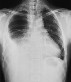
入 院 時 胸 部 X 腺 写 真 お よ び 胸 部 CT 写 真 を Fig. 1 および Fig. 2 に示す.中等量の右胸水貯留 が認められたが,肺野に浸潤影を認めなかった.
入院後経過を Fig. 3 に示す.入院時に施行した 胸腔穿刺の結果, 胸水の性状は滲出性であったが,
胸水のグラム染色,抗酸菌染色および抗酸菌の PCR 検査は陰性であった.しかし,胸水中のアデ ノシン・デ・アミナーゼ(以下 ADA)値が 66.9 IU ! l と高値であることから結核性胸膜炎を疑っ た.そこで,入院 5 日後より INH 0.3g! 日,RFP
気管支鏡下胸膜生検により診断しえた結核性胸膜炎の 1 例
東邦大学第一内科
北條 貴子 木村 一博 外山 勝弘 杉野 圭史 佐野 剛 山田 浩之 磯部 和順 廣井 眞弓 梁 英富
(平成 14 年 8 月 9 日受付)
(平成 14 年 9 月 27 日受理)
別刷請求先:(〒143―8541)東京都大田区大森西 6―
11―1
東邦大学第一内科 北條 貴子
Key words: tuberculous pleuritis, fiberoptic bronchoscopy, pleural biopsy
38感染症学雑誌 第77巻 第 1 号
0.45g ! 日の内服を開始したところ,自覚症状およ び血液データーの改善をみた.経過中,Cope 針を
用いて胸膜生検を施行したが採取されたものは壊 死組織のみであった.そこで,確定診断目的に戸 野塚らの方法
1)2)に準じて胸腔鏡下胸膜生検を施行 した.気管支鏡は Olympus BF type 200 (オリンパ ス光学社製)を用いた.胸水の排液目的で挿入中 であった胸腔ドレーンを大気に解放し,胸腔ド レーン孔近くに挿入したサーフロー針を通して空 気で置換した.ドレーンを抜去後,同孔および気 管支鏡挿入部を充分に消毒してから気管支鏡を挿
Fig. 1 A Chest X-ray film obtained on admission.Fig. 3 Clinical coarse
Fig. 2 A chest CT obtained on admission.
気管支鏡下生検で診断しえた結核性胸膜炎 39
平成15年 1 月20日
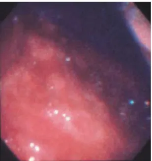
入し胸腔内を観察したところ,胸膜上に白色で滑 らかな小結節性病変が散見された (Fig. 4) .生検は 病変部と思われる壁側胸膜を数カ所採取し,臓側 胸膜の生検は施行しなかった.
胸膜生検の組織像を Fig. 5 に示す.中心部に乾 酪壊死を認め,周囲に類上皮細胞とラングハンス 型巨細胞を伴うことから肉芽腫病変であると考え られた.画像上では浸潤影を認めず,喀痰,胃液 検査でも抗酸菌が検出されなかったことから特発 性結核性胸膜炎と診断し,INH+RFP+PZA+EB による 4 者併用療法を開始したところ,臨床症状 の改善をみたため入院 22 日後に退院となった.
考 察
結核性胸膜炎の診断は,滲出性胸水を認め,喀 痰,胸水,胸膜の生検材料などから結核菌を細菌 学的に証明するか,胸膜生検の検体中に病理学的 に 特 異 的 肉 芽 腫 を 認 め る こ と に よ っ て 確 定 す る
3).しかし, 結核性胸膜炎症例では胸水培養での 結核菌の陽性率は 7.9〜20%
4)〜6)と低く,胸膜生検 による病理学的診断が重要となる.しかし,Cope 針などを用いた胸膜生検は盲目的に行われるた め,その診断率も 47.4〜60%
4)〜6)にとどまるのが現 状である.残りの症例は,胸水検査や臨床症状な
どから臨床診断されるが,その際に使用される補 助診断として,胸水中のアデノシンデアミナーゼ
(以下 ADA)値
7)8),胸水中のインターフェロン‐
γ 値7)〜10),ADA のアイソザイムである T 細胞由 来の ADA2 値
10)などが使用されている.
胸膜生検時の胸腔鏡の有用性についてはすでに いくつかの報告がある.木村ら
11)は,先端がフレキ シブルである胸腔鏡を用いた胸膜生検の結果,結 核性胸膜炎における胸膜生検の陽性率が 75% で あったと報告している.また石井ら
12)は,結核性胸 膜炎の胸腔鏡所見について,灰白色の小結節が壁 側胸膜や横隔膜上に多数存在し,とくに肋間筋上 に密に集簇するのが特徴であると述べている.今 回の症例でもほぼ同様の所見が観察された.
われわれは胸腔鏡として軟性気管支鏡を使用し たが,軟性気管支鏡使用時の利点としては,特別 な器具を必要とせず,局所麻酔下に内科医が手軽 に施行できるといった点にある.その一方で,市 販されている胸腔ビデオスコープなどと比較する と,軟性気管支鏡には硬性部分が存在しないため に,肺容積のコントロールができない局所麻酔下 での検査では,胸腔内でのオリエンテーションお よび視野の確保が困難であることが難点である.
今後,胸腔鏡検査に占める内科医の役割はさら に大きいものになると思われる.われわれも局所 麻酔下での胸腔鏡検査に習熟し,胸膜生検の診断 をさらに確実なものにしていく必要がある.
Fig. 4 White nodular lesions were sporadically ob- served on the pleura.
Fig. 5 A histological image of the pleural biopsy tis- sues. Granuloma formation presenting palisade.
北條 貴子 他 40
感染症学雑誌 第77巻 第 1 号
文 献
1)戸野塚博,鈴木 一,秋沢孝則,成島道昭,田中 一正,金重博司,他:気管支鏡を用いた胸腔内検 査.日胸疾会誌 1988;26:936―41.
2)木村一博,外山勝弘,吉田眞弓,北條貴子,梁 英富,中田正幸,他:胸腔鏡下胸膜生検によりリ ウ マ チ 結 節 を 診 断 し え た 1 例.日 呼 吸 会 誌 1998;36:994―7.
3)青木正和:結核性胸膜炎,結核性膿胸.井村裕夫,
尾形悦郎,高久史麿,垂井清一郎編,最新内科学 大系 61,中山書店,東京,1994;p. 104―7.
4)木村一博,杉野圭史,佐野 剛,山田浩之,磯部 和順,外山勝弘,他:当院における結核性胸膜炎 の臨床的検討.感染症誌 2002;76:18―22.
5)中村栄一,芳賀敏彦:国立療養所における結核性 胸膜炎の現状.―国療化研第 29 次 A 研究報告―.
結核 1990;65:205―21.
6)芥川光夫:最近の結核性胸膜炎.肺と心 1978;
25:10―6.
7)Valde"s L, Jose"ES, Alvarez D, Sarandeses A, Pose A, Chomo"n B,et al.:Diagnosis of tuberculous
pleurisy using the biologic parameters adenosine deaminase , lysozyme , and interferon gamma . Chest 1993;103:458―65.
8)Ferrer J:Pleural tuberculosis. Eur Resp J 1997;
10:942―7.
9)So!derblom T, Nyberg P, Teppo A-M, Klockars M, Riska H, Pettersson T:Pleural fluid interferon-γ and tumour necrosis factor-αin tuberculous and rheumatoid pleurisy. Eur Resp J 1996;9:1652―
5.
10)Valde"s L, Alvarez D, Jose"ES, Penela P, Valle JM, Garc!a-Pazos JM,et al.:Tuberculous pleurisy. A study of 254 patients. Arch Intern Med 1998 ; 158:2017―21.
11)木村 丹,米山浩英,中村淳一,富澤貞夫,安達 倫文,川西正泰,他:胸水貯留例の臨床的検討―
ことに胸腔鏡を用いた胸膜生検の有用性につい て.日胸疾会誌 1990;28:882―8.
12)石井芳樹:胸腔鏡検査.北村 諭編,別冊・医学 の歩み・呼吸器疾患,医歯薬出版株式会社,東京,
1999;p. 173―6.
A Case Report of Tuberculous Pleuritis Diagnosed by Thoracoscopy Using Fiberoptic Bronchoscope
Takako HOJYO, Kazuhiro KIMURA, Katsuhiro TOYAMA, Keishi SUGINO, Gou SANO, Hiroyuki YAMADA, Kazutoshi ISOBE, Mayumi HIROI
& Hidetomi RYO
The First Department of Internal Medicine, Toho University School of Medicine
A 27-year-old male visited the outpatient clinic of our hospital with the chief complaints of fever, right chest pain and shortness of breath. He was admitted to our hospital for detailed examination of the right hydrothorax. The pleural effusion obtained by thoracocentesis was exudative and negative for Mycobacterium tuberculosis. Since the titer of adenosine deaminase in the pleural effusion was ab- normally high, antitubercular therapy was started under suspicion of tuberculous pleuritis. Thereaf- ter, the patient s subjective symptoms and blood parameters improved. Necrotic tissues were ob- tained by pleural biopsy using the Cope needle. In order to make a definitive diagnosis, pleural biopsy was performed thoracoscopically. White tubercular lesions with a smooth surface were sparsely dis- tributed on the pleura. Histopathologically, these lesions were characterized by central areas of case- ous necrosis surrounded by epithelial cells and Langhans giant cells. Therefore, they were consid- ered to be granulomatous lesions. The patient was given a diagnosis of idiopathic tuberculous pleuri- tis, and was treated with four antitubercular drugs in combination. His clinical signs subsided, and he was discharged. This case indicates that the examination of the inside of the pleural cavity with a flexible bronchoscope, instead of thoracoscope, under local anesthesia is useful to diagnose patients having tuberculous pleuritis.
〔J.J.A. Inf. D. 77:38〜41, 2003〕
気管支鏡下生検で診断しえた結核性胸膜炎 41
平成15年 1 月20日