北海道医療大学学術リポジトリ
Genome‑wide analysis of DNA methylation array and microarray in human oral epithelial cells stimulated by arecoline for prolong period ‑ Involvement of aberrant expression of DUSP4 gene in betel quid chewing related oral cancer
著者 Bhoj Raj Adhikari
学位名 博士(歯学)
学位授与機関 北海道医療大学
学位授与年度 平成30年度
学位授与番号 30110甲第306号
URL http://id.nii.ac.jp/1145/00064676/
1
Genome-wide analysis of DNA methylation array and microarray in human oral epithelial cells
stimulated by arecoline for prolong period
-Involvement of DUSP4 gene in arecoline induced oral cancer-
平 成 3 0 年 度
北 海 道 医 療 大 学 大 学 院 歯 学 研 究 科
ADHIKARI Bhoj Raj
2
Genome-wide analysis of DNA methylation array and microarray in human oral epithelial cells
stimulated by arecoline for prolong period
-Involvement of DUSP4 gene in arecoline induced oral cancer-
November 2018 Graduate School of Dentistry, Health Sciences University of Hokkaido
ADHIKARI Bhoj Raj
i Abstract
1. Introduction
Oral cancer is the eighth most common human cancer. Both environmental and genetic factors induce the development of oral cancer. Although several studies have shown the factors involved in betel quid chewing related oral cancer, the mechanism of causing this type of oral cancer remains elusive. Genetic mutations and aberrant DNA methylations have been suggested to be involved in causing of oral cancer. Unlike genetic mutations, DNA hypermethylations are reversible and can be diagnostic and therapeutic targets. The purpose of this study is detection of novel DNA hypermethylation that can be applied to diagnostic, predictive, interceptive and therapeutic approaches for betel quid chewing related oral cancer.
2. Method and materials
a. Genome-wide analysis
HGEPs were cultured and treated alternating 3 days with 50 μg/ml arecoline and 3 days without arecoline for 1 month. Untreated samples were used as controls. For methylation array, genomic DNA were analyzed using a DNA microarray scanner (Agilent technology).
For microarray analysis, a global analysis of mRNA expression was carried out using DNA microarray (human SurePrint G3 Human GE Microarray).
b. Confirmation of the reproducibility in the five candidate genes
In order to confirm the reproducibility of the microarray data, DNA samples were treated with sodium bisulfite and quantitative Methylation Specific PCR (qMSP). Expression of mRNA was analyzed by quantitative reverse transcription polymerase chain reaction (qRT-PCR). Results were compared by Mann-Whitney U test with P-value <0.05 accepted as statistically significant.
c. DUSP4 analysis in Oral cancer cell line
To confirm the DUSP4 expression in oral cancer, four cancer cell lines HSC4, SAS,
ii
BSC-OF and HSY were cultured and treated with 50 μg/ml arecoline alternating 3 days or untreated for 7 days.
d. Pathway analysis
To examine any mechanism in additional to hyper-methylation, phosphorylated JNK (pJNK) in the HGEPs treated with or without arecoline were determined using cell based ELISA.
e. Histological and immunohistochemical and CpG methylation in tissue samples Twenty tissue samples were obtained from the patients involved in betel quid chewing habit in Sri Lanka. Twenty-two tissue samples (13 OSCC, 9 fibrous polyp) for betel quid non-chewer were retrieved from the archives of HSUH, Japan. Immunohistochemical examination of the tissue samples was performed using anti-DUSP4 antibody. DUSP4 immunoreactivity was assessed in nucleus of epithelial cells. Genomic DNA was extracted from the tissue samples. DNA samples were treated with sodium bisulfite and qMSP was performed.
f. Statistical analysis
The results were compared using the Mann-Whitney U test with p-value <0.05 accepted as statistically significant.
3. Results
CpG island methylation array and global analysis of mRNA reveled 8638 and 455 genes
that were more than 2 times hyper-methylated, and their mRNA downregulated respectively
in samples stimulated with arecoline for a period of 1 month. Twenty-one of them were tumor
suppressor genes, within -1000bp from promoter region. Four candidate genes HNRNPH3,
BCL2L11, TFAP2A and DUSP4 with the highest amount of promoter region
hypermethylation with downregulated mRNA expression were selected. qMSP showed
DUSP4 genes were significantly hypermethylated in HGEPs treated with arecoline for a
period of 7 days and 1 month. mRNA expression of DUSP4 was down-regulated in HGEPs
iii
treated with arecoline for 7 days and 1 month. Methylation level of DUSP4 was significantly higher in all cancer cell lines (HSC4, SAS, BSC-OF and HSY) without arecoline treatment as compared to HGEPs. mRNA expression of DUSP4 in untreated BSC-OF cell line was not significantly different from that of HGEPs. BSC-OF showed downregulated DUSP4 mRNA expression after arecoline treatment.
The amount of pJNK was elevated in samples treated with arecoline over a period of 1 month.
IHC staining reveled that DUSP4 expression level was significantly higher in betel quid chewers (66.85 3.0) than in non-chewer OSCC (20.34 4.7). qMSP analysis showed that DUSP4 CpG island was significantly hypermethylated in oral cancer samples obtained from betel quid chewers (89.8 2.6) as compared to those from non-chewer oral cancer cases (44.7
10.6) and the healthy control cases (49.4 13.1).
4. Conclusion
In the present study, genome-wide DNA hypermethylation and global mRNA microarray
analysis were carried out using CpG island DNA methylation array and DNA microarray in
HGEPs stimulated with arecoline. This is the first report that shows betel quid related
hypermethylation of DUSP4 followed by its down regulated expression. DUSP4
hypermethylation can be applied as a target for diagnostic, predictive, interceptive and
therapeutic approaches for betel quid chewing related oral cancer.
iv
Table of contents
Abstract i
Table of Contents iv
1. Introduction 1
1.1 Oral Cancer 1
1.2 Betel Quid 2
1.3 Arecoline 2
1.4 DNA methylation in Oral Cancer 3
1.5 Purpose of the Study 4
2. Materials and Methods 5
2.1 Genome-wide analysis 5
2.1.1 Cell Culture (HGEPs) 5
2.1.2 Methylation array 5
2.1.3 Microarray analysis 6
2.2 Confirmation of the reproducibility in the five candidate genes 6
2.2.1 Quantitative methylation specific polymerase chain reaction 6
2.2.2 Quantitative reverse transcription polymerase chain reaction 7 2.3 DUSP4 analysis in Oral cancer cell line by qRT-PCR and qMSP 7 2.4. Pathway analysis: phosphorylated c-Jun N-terminal kinase (pJNK) 8
2.4.1 JNK inhibitor treatment 8
2.4.2 Cell based Enzyme-Linked Immunosorbent Assay (cell ELISA) 8 2.5 Histological and immunohistochemical evaluation of tissue samples 9
2.5.1 Clinical characteristic of the patients 9
2.5.2 Histopathological and Immunohistochemical (IHC) analysis 9
2.5.3 Scoring of positive cells 10
2.6 qMSP in Clinical samples 10
2.7 Statistical analysis 11
3.
Results 12
3.1 Genome-wide DNA methylation analysis, identification of candidate genes 12 3.2 Reproducibility of microarray data by qMSP and qRT-PCR 12
3.3 Evaluation of DUSP4 in oral cancer cells 13
v
3.4 Cell ELISA to determine level of pJNK after arecoline treatment 13 3.5 Immunohistochemical staining for DUSP4 in tissue samples obtained from
betel quid chewers 14
3.6 Analysis of DUSP4 CpG island in betel quid chewers 14
4. Discussion 15
4.1 Genome-wide DNA methylation analysis 15
4.2 Identification of candidate genes 16
4.3 DUSP4 gene 17
4.3.1 Evaluation of DUSP4 methylation in cancer cell lines 17
4.3.2 CpG island methylation and Immunohistochemical study of DUSP4 in
tissue samples obtained from betel quid chewers 18
4.3.3 Loss of DUSP4 by arecoline and interaction with JNK pathway 18
4.5 Clinical implication 19
5. Conclusion 20
References 21
Tables 29
Figures legends 34
Figures legends 38
1 1. Introduction
1 2
1.1. Oral cancer 3
Oral cancer is the eighth most common human cancer, with more than 500,000 new cases 4
being diagnosed globally every year. While it represents just over 2% of the global cancer 5
incidence, its 50% fatality rate is a major cause of concern (Siegel et al., 2017). Both 6
environmental and genetic factors induce the development of oral cancer. Association of 7
tobacco smoking, alcohol consumption, diet, living habits, microbial infections and the 8
exposure to a variety of exogenous or endogenous carcinogens are well documented with oral 9
carcinoma (Asthana et al., 2018; Petti, 2009). Not all individuals exposed to these risk factors 10
develop oral carcinoma, additional genetic factors may also contribute to oral carcinoma 11
susceptibility. The environmental factors often induce posterior genetic modifications such as 12
mutations and epigenetics leading to oral cancer. Several kinds of genetic changes are seen in 13
oral cancer such as single nucleotide polymorphism (as in ADH1B, ALDH2, MMP1, FN1, 14
CCNA2, CA9, VEGFC and FAT1) and mutation (as in p53) (Lee et al., 2018; Li et al., 2018;
15
Lin et al., 2018). A number of epigenetic mechanisms including DNA methylation, functional 16
non-coding RNA and histone modifications regulate the expression of different genes. The 17
promoter hypermethylation-mediated silencing of certain tumor suppressor genes occurs 18
without genetic change. Methylation-associated RARB, C3orf14, GPR27, ZNF717 gene 19
silencing and histone H3K9 methylation, are few examples of epigenetic modification (Lai et 20
al., 2014; Lando et al., 2015).
21
The prevalence of oral cancer is extremely high at approximately 8.7% of cancer incidence 22
in the South and Southeast Asian countries, with 2% worldwide incidence, being the highest 23
globally (Gupta et al., 2016). The habit of betel quid chewing is a major cause of oral cancer in 24
those countries (Lee et al., 2011). Although several studies have shown the factors involved in 25
betel quid chewing related oral cancer, the mechanism of causing this type of oral cancer is 26
still unclear. In a large cohort study in India, all oral cancer developed from potentially 27
malignant oral disorders or precancerous lesions, including oral submucous fibrosis, oral 28
2
leukoplakia, erythroplakia, and oral lichenoid lesions; and were seen among users of betel 29
quid, areca nut, or tobacco-based products, or all of these (Gupta et al., 1980).
30 31
1.2. Betel quid 32
Betel quid, a combination of fresh areca nut, slaked lime, fresh betel leaf, and partially 33
dried tobacco, is widely preferred in South and Southeast Asian countries (Fig. 1). The Asian 34
Betel Quid Consortium study found that the prevalence of betel quid and areca nut chewing 35
in the adult population varied from 10·3% in Malaysia to 43·6% in Nepal for men, and from 36
2·3% in mainland China to 47·8% in Indonesia among women (Lee, et al., 2011). Betel quid 37
have been classified as carcinogenic to human beings (Group 1) by the International Agency 38
for Cancer Research (WHO IARC, 2004). Epidemiological studies have related the habit of 39
betel quid chewing with oral cancer and oral premalignant disorders (Jeng et al., 2001; Lee, et 40
al., 2011). Since arecoline is a major component of betel quid that can be a carcinogen, 41
previous studies have mainly focused on arecoline to elucidate the mechanism of 42
carcinogenesis with betel quid.
43 44
1.3. Arecoline 45
Arecoline (1,2,4,5-tetrahydro-1-methyl-pyridinecarboxylic acid) is a nicotinic acid-based 46
alkaloid, an active component of areca nut, found in betel quid (Fig. 2) (Sharan et al., 2012).
47
Arecoline is genotoxic and might contribute to oral carcinogenesis by facilitating error-prone 48
DNA repair (Ji et al., 2012). DNA repair machinery is an important part of maintaining 49
genomic integrity. Dysregulation of DNA repair resulting in genomic instability is a hallmark 50
of cancer cells that can be associated with arecoline stimulation (Chiba et al., 1998; Kannan et 51
al., 1999). In addition to genetic changes in the tumor-related genes, epigenetic modification 52
plays a vital role in oral carcinogenesis (Takeshima et al., 2008; Tsai et al., 2008). Areca nuts 53
extracts and arecoline induce epigenetic modifications. It has been found that promoter region 54
hypermethylation was followed by loss of mRNA expression of RARB and MGMT in arecoline 55
associated oral cancer (Huang et al., 2010; Lai, et al., 2014). Histone modification is another 56
3
epigenetic change by arecoline. Histone protein H3K9 has been shown to be modified by 57
arecoline in human K-562 cell lines and is associated with oral carcinoma (Lin et al., 2011).
58
Collectively, these pieces of evidence suggest the association of betel quid and arecoline to oral 59
carcinogenesis via various epigenetic mechanisms.
60 61
1.4. DNA methylation in oral cancer 62
DNA methylation is one of the several epigenetic mechanisms that cells use to control gene 63
expression. Epigenetic is described as changes in the pattern of gene expression not involving 64
the DNA sequence. Epigenetic events act through chemical modification of DNA and by 65
selectively activating or inactivating genes to determine their expression. DNA methylation 66
and histone modification are two major mechanisms of epigenetic alteration in human cells 67
(Irimie et al., 2018; Shen & Laird, 2013). DNA methylation is an enzymatically catalyzed 68
covalent modification of DNA, occurring typically in the context of 69
cytosine-phosphate-guanine (CpG) dinucleotides. In general, CpG islands, the regions with 70
high CpG content, are demethylated in normal cells. In contrast, regions with an 71
intermediate or low density of CpGs are differentially methylated in some tissues, but not in 72
others (Bird et al., 1985). Changes in methylation of DNA in cancer were first recognized by 73
Feinberg in 1983. In the early days of its recognition, it was thought that epigenetics was 74
linked to a general disruption of the cell cycle, an effect rather than the cause of malignancy 75
(Feinberg & Vogelstein, 1983). The discovery that downregulation of tumor suppressor genes 76
in the absence of a detectable genetic change have led to greater research emphasis on cancer 77
epigenetics (Feinberg, 2001).
78
Epigenetics refers to the chemical modifications of the DNA leading to selective activation 79
and inactivation of genes thereby influencing their expression. The attachment of the 80
5-methylcytosine-binding protein to methylated cytosine bases interferes with the binding of 81
transcriptional proteins to gene promoters, halting the expression of that gene. Genes 82
commonly found to be hypermethylated in cancer include tumor suppressors and 83
metastasis-related genes. DNA methylations, unlike genetic mutation, are reversible and can 84
4
be a diagnostic and therapeutic target (Ushijima & Asada, 2010). Although several DNA 85
hypermethylation has been detected in oral cancer, little information about it is in the betel 86
quid related oral cancers.
87 88
1.5. Purpose of the study 89
Epigenetic changes are the drivers of oral cancer progression. Earlier studies have analyzed 90
methylation changes in oral precancerous diseases being highly potent for malignant 91
transformation (Cao et al., 2009; Gasco et al., 2002; Kresty et al., 2002; Long et al., 2008).
92
Unlike genetic mutation epigenetic modification is reversible and may be a useful therapeutic 93
target. Hypermethylation on oral epithelium can be a valuable biomarker for prediction of 94
malignant potential of the lesion. Excision of the lesion has been a widely accepted surgical 95
measure to treat these early changes. A key step to improving oral cancer outcomes is 96
identifying the molecular factors driving disease initiation and progression, as these factors 97
represent attractive candidates for targeted therapies (Lubbert et al., 2001; Mack, 2006; Niwa 98
et al., 2013; Schneider & Peek, 2013; Shen et al., 2010; Silverman et al., 2002). A number of 99
approaches exist that enable the large-scale DNA methylation analysis. All of these 100
approaches are based upon any of the three techniques: bisulfite conversion, digestion with 101
methylation sensitive restriction enzymes, and affinity purification of methylated DNA 102
(Zilberman & Henikoff, 2007). DNA microarrays and high-throughput DNA sequencing are 103
commonly used methods of genome-wide study for rapid identification of candidate genes that 104
are associated with carcinogenesis (Kreil et al., 2006). The purpose of this study is the 105
detection of novel DNA hypermethylation that can be applied to diagnostic, predictive, 106
interceptive and therapeutic approaches for betel quid chewing related oral cancer.
107
5 2. Materials and Methods
108 109
2.1. Genome-wide analysis 110
Genome-wide analysis in Human Gingival Epithelial Progenitors, pooled (HGEPs) induced 111
by prolonged stimulation with arecoline.
112 113
2.1.1. Cell culture (HGEPs) 114
HGEPs (CELLnTEC advanced cell systems AG, Switzerland) were cultured in CnT-Prime 115
epithelial cell culture medium (CELLnTEC, advanced cell systems AG) containing antibiotics 116
(5% penicillin-streptomycin; Sigma-Aldrich, USA) at 37˚C in an incubator supplied with 5%
117
CO2. All cells used in this study were at 3 to 5 passages. These samples were treated 118
alternating 3 days with 50 μg/ml arecoline hydrobromide (hereinafter called as arecoline;
119
Sigma-Aldrich, Japan) and 3 days without arecoline for 1 month according to the established 120
method (Fig. 3) (Takai et al., 2016; Uehara et al., 2017). Untreated samples were used as 121
controls.
122 123
2.1.2. Methylation array 124
For methylation array, genomic DNA was extracted from the HGEPs treated with arecoline 125
for 1 month using DNeasy® Blood and Tissue kit (Qiagen, Tokyo, Japan), and sonicated to 126
produce random fragments. One gram of sonicated DNA was incubated with 2 µg of MBD2bt 127
protein. This complex was precipitated using pre blocked nickel magnetic beads. The 128
methylated DNA-enriched DNA fraction was purified using a Qiaquick PCR purification kit 129
(Qiagen) followed by labeling with either cytidine 5-dUTP (Cy 5) or cytidine 3-dUTP (Cy 3).
130
Labeled DNA probes were then mixed and simultaneously hybridized to the human CpG 131
island 224 K array. The DNA samples were analyzed using a DNA microarray scanner 132
(Agilent technology, Santa Clara, USA).
133 134 135
6
2.1.3. Microarray analysis 136For Microarray analysis, total RNA was extracted by the acid guanidine thiocyanate/phenol 137
chloroform method using TRIzol Reagent (Invitrogen Corporation, USA). A global analysis of 138
mRNA expression was carried out using DNA microarray (human SurePrint G3 Human GE 139
Microarray).
140 141 142
2.2. Confirmation of the reproducibility in the candidate genes 143
From Genome-wide analysis, four candidate genes, named HNRNPH3, BCL2L11, TFAP2A, 144
and DUSP4 were taken. In order to confirm the reproducibility of the methylation array and 145
microarray data, genomic DNA and total RNA were extracted from the cells on 7th and 30th 146
days.
147 148
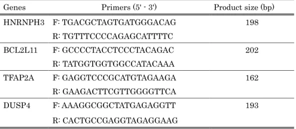
2.2.1. Quantitative methylation-specific polymerase chain reaction 149
In order to analyze CpG island hypermethylation, the methylation profiles were assessed 150
using a quantitative methylation-specific polymerase chain reaction (qMSP) method. Five 151
hundred ng of genomic DNA was subjected to sodium bisulfite conversion using EpiTect® Fast 152
Bisulfite Conversion kit (Qiagen, USA). For qMSP, two sets of qMSP primers were designed 153
using the MethPrimer (http://urogene.org/methprimer/) (Li & Dahiya, 2002) (Table 1).
154
Bisulfite-converted DNA and a pair of either methylated or unmethylated primers were 155
mixed with KAPA SYBR Fast qPCR Kit (NIPPON Genetics). qMSP was performed on Light 156
Cycler® Nano System (Software version 1.1, Roche Diagnostics, Germany). The qMSP 157
conditions included denaturation at 95ºC for 10 min and 45 cycles of denaturation at 95ºC for 158
10 sec and annealing at 60ºC for 30 sec. The percentage of DNA methylation in each sample 159
was estimated using the following formula:
160
Methylated DNA (%) =(M+U)M × 100 = (1+U/M)1 × 100 = (1+2−ΔCt)1 × 100 161
where M and U are the copy number of methylated and unmethylated DNA respectively, and 162
ΔCt = CtU - CtM (Lu et al., 2007; Takai, et al., 2016). Each experiment was conducted in 163
7
triplicate on five biologically different culture systems. The result is presented as the 164
percentage of mean standard errors of the mean (SE) of DNA methylation.
165 166
2.2.2. Quantitative reverse transcription polymerase chain reaction 167
The expression levels of mRNA in the cultured cells were analyzed by quantitative reverse 168
transcription polymerase chain reaction (qRT-PCR). Two µg of RNA was reverse transcribed 169
(SuperScript Ⅱ Reverse Transcriptase; Invitrogen), according to the manufacturer's 170
instructions using Oligo(dT)12-18 primers (Invitrogen). For qRT-PCR, a set of qRT-PCR primer 171
was designed using Primer3 (http://bioinfo.ut.ee/primer3-0.4.0/) (Table 2). cDNA and a pair of 172
primers were mixed with KAPA SYBR Fast qPCR Kit (NIPPON Genetics, Tokyo, Japan).
173
qRT-PCR was performed on Light Cycler® Nano System (Software version 1.1, Roche 174
Diagnostics, Germany). The PCR conditions included denaturation at 95ºC for 10 min and 45 175
cycles of denaturation at 95ºC for 10 sec and annealing at 60ºC for 30 sec. The expression level 176
of target gene mRNA was standardized against GAPDH mRNA. The relative mRNA 177
expression levels of each sample were calculated as the Ct (the value obtained by subtracting 178
the Ct value of the GAPDH mRNA from the Ct value of the target mRNA) using the ΔΔCt 179
method (Livak & Schmittgen, 2001; Takai, et al., 2016). Each experiment was conducted in 180
three technical and four biological cultures per target gene. Data are expressed as mean SE 181
of the ratio of target mRNA to GAPDH mRNA.
182 183
2.3. DUSP4 analysis in Oral cancer cell line by qRT-PCR and qMSP 184
To confirm the DUSP4 expression in oral cancer, cancer cell lines derived from human 185
tongue squamous cell carcinoma (HSC4 and SAS), human basaloid squamous cell carcinoma 186
of the floor of the mouth (BSC-OF) and human papillomavirus-related endocervical 187
adenocarcinoma (HSY) were cultured in Dulbecco’s modified eagle’s medium (DMEM, 188
Sigma-Aldrich, USA) containing antibiotics (2% penicillin-streptomycin) 10% fetal bovine 189
serum (FBS: Gibco, Invitrogen Corporation, CA) at 37˚C in an incubator supplied with 5%
190
CO2. Cancer cells were treated with 50 μg/ml arecoline alternating 3 days or untreated for 7 191
8
days. Genomic DNA and total RNA were extracted from each sample on the 7th day as 192
described above. qRT-PCR and qMSP were performed in a similar manner to that of HGEPs.
193
To determine the amount of methylation and mRNA expression in cancer cell lines without 194
arecoline treatment, untreated HGEPs were used as controls. In order to determine any 195
further changes in methylation and mRNA expression in cancer cell lines after arecoline 196
treatment, untreated cancer cell lines were used as the control.
197 198
2.4. Pathway analysis: phosphorylated c-Jun N-terminal kinase (pJNK) 199
To examine any mechanism in additional to hypermethylation, cell based Human c-Jun 200
N-terminal kinase (human JNK) phosphorylation ELISA kit (RayBiotech, Inc, USA) was used 201
to detect the phosphorylated JNK (pJNK) in the HGEPs treated with or without arecoline.
202 203
2.4.1. JNK inhibitor treatment 204
HGEPs were cultured in CnT-Prime epithelial cell culture medium for a period of 7 days or 205
1 month. These cells were divided into four groups based on either treated or untreated with 206
arecoline and/or SP600125 (hereinafter called as JNK inhibitor, Sigma-Aldrich Japan, Tokyo, 207
Japan). JNK inhibitor, at a concentration of 20 µM, was added 1 hour prior to 50 µg/ml 208
arecoline treatment (Lin et al., 2016; Uehara, et al., 2017). These cultures were repeated 209
every three days with or without arecoline and/or JNK inhibitor for the specified time period.
210 211
2.4.2. Cell based Enzyme-Linked Immunosorbent Assay (cell ELISA) 212
Cell based Human JNK phosphorylation ELISA, to detect the pJNK in the cells, was used 213
as per the manufacturer's recommendation. The cells treated and/or untreated with arecoline 214
and/or JNK inhibitor were sub-cultured in 96 well plate at a density of 3×10⁴ cells in 100 l 215
media, incubated with 5% CO2 at 37˚C. After 24 hrs, these cells were fixed and blocked.
216
Anti-Phospho-JNK (Thr183/Tyr185) or Anti-JNK were pipetted into respective wells and 217
incubated. After the wells were washed, HRP-conjugated anti-mouse IgG was added to the 218
wells. The wells were washed again; a TMB substrate solution was added and incubated in 219
9
dark, as a result, blue color developed in proportion to the amount of respective proteins. The 220
stop solution changed the color from blue to yellow and the intensity of the color was 221
determined. Optical absorbance was read at 450nm on Bio Rad 680 microplate reader (Model 222
680 Microplate reader, RayBiotech, USA).
223 224
2.5. Histological and immunohistochemical evaluation of tissue samples obtained from 225
betel quid chewers and non chewers 226
227
2.5.1. Clinical characteristic of the patients 228
Twenty tissue samples were obtained from the patients involved in betel quid chewing 229
habit in Sri Lanka. Twenty-two tissue samples (13 oral squamous cell carcinoma (OSCC), 9 230
fibrous polyps) were retrieved from the archives of Oral Medicine and Pathology department 231
of Health Sciences University of Hokkaido (HSUH), Japan. Tissue samples from Japanese 232
patients not involved in betel chewing were obtained from those who underwent the oral 233
surgical intervention during the period of 2008 to 2014 in HSUH hospital. This study was 234
approved by the Human genome ethics committee of HSUH (No. 7) and the ethics committee 235
of the Institute of Personalized Medical Science, HSUH (No. 2016-025). Post-surgical tissue 236
sections were already formalin fixed, processed and paraffin embedded following standard 237
protocol.
238 239
2.5.2. Immunohistochemical (IHC) analysis 240
Histopathological examination using routine hematoxylin and eosin staining was 241
performed on the tissue sections at the 5µm thickness to verify the clinical diagnosis.
242
Thereafter, immunohistochemical examination of the tissue samples was performed using 243
anti-DUSP4 antibody. The sections of 5µm thickness were made of the samples and mounted 244
on silane coated slides (New Silane III, Muto pure chemicals co. Ltd, Japan). These slides 245
were deparaffinized in xylene (3 changes every 3 minutes) and rehydrated in a graded alcohol 246
series (100%, 90%, 80% and 70% every 3 minutes). Antigen retrieval was done using 10 mmol 247
10
citrate buffer (Citric acid Monohydrate and Tri Sodium Citrate Dihydrate) by heat-induced 248
epitope retrieval method. These slides were maintained at a sub-boiling temperature in a 249
pressure cooker for 3 minutes and bench cooled for further 20 min. Endogenous peroxidase 250
activity was blocked with 3% H2O2 in methanol for 10 min. These slides were washed twice for 251
5 min each with TBS plus 0.025% Triton X-100 with gentle agitation. Specimens were 252
incubated overnight in a humidified chamber at 37ºC with rabbit polyclonal anti-DUSP4 253
primary antibody (ab72593, diluted to 100× as per manufacturer Abcam, Japan’s 254
recommendation). These slides were washed again with TBS plus 0.025% Triton X-100 and 255
treated with rabbit polyclonal secondary antibody for 30 min at room temperature. Reaction 256
products were visualized with diaminobenzidine chromogen concentrate (Dako, USA/Japan) 257
and finally counterstained with hematoxylin.
258 259
2.5.3. Scoring the positive cells 260
DUSP4 immunoreactivity was assessed in the nucleus of epithelial cells; staining was 261
considered evidence of expression. For each sample, at least three fields were equidistantly 262
captured at a magnification of 200×under a light microscope (Olympus BX50, Olympus 263
Corporation). All the cells from the representative fields were counted using the public 264
domain program ImageJ version 1.50b (National Institute of Health, USA). The total number 265
of the positive cells from each examined fields was determined. DUSP4 immunopositivity in 266
each sample was expressed as a composite score by taking the mean of all the cells counted in 267
the selected fields. The composite score was expressed in terms of percentage of the total cells 268
present in those specified fields.
269 270
2.6. qMSP in clinical samples 271
Genomic DNA was extracted from the tissue samples using QIAmp® DNA FFPE tissue kit 272
(Qiagen) following manufacturer’s recommendation. Freshly cut 8-15 (depending upon the 273
amount of tissue) tissue sections of 5 µm and not exceeding surface area of 250 mm2 were 274
subjected to deparaffinization on 1 ml of xylene. These samples were incubated with 275
11
proteinase K at 56ºC for 1 hour for lysis and further 1 hour at 90ºC to reverse the 276
formaldehyde modification of nucleic acids. These samples were washed and DNA eluted with 277
recommended buffers. For each sample, 500 ng of purified DNA was subjected to sodium 278
bisulfite conversion and qMSP performed as described earlier. Each experiment was 279
conducted in triplicate and the result is presented as the percentage of mean SE of DNA 280
methylation.
281 282
2.7. Statistical analysis 283
All values are expressed as the mean SE for the respective groups. Statistical analysis 284
was performed using IBM SPSS Statistical tool for iOS (Version 25; SPSS Inc, USA). The 285
results were compared using the Mann-Whitney U test with a p-value <0.05 accepted as 286
statistically significant.
287 288 289 290
12
3. Results 291
292
3.1. Genome-wide DNA methylation analysis, identification of candidate genes 293
CpG Island methylation array data suggested 8638 and 7392 genes that were more than 2 294
times hypermethylated and hypomethylated respectively in samples stimulated with 295
arecoline for a period of 1 month. Global analysis of mRNA revealed 502 upregulated and 455 296
downregulated genes. Since hypermethylation of tumor suppressor genes followed by 297
down-regulation of their mRNA often induces malignant transformation, these types of genes 298
were selected. Among the genes showing hypermethylation with downregulated expression 299
of their mRNA, 152 genes were within -1000bp from promoter region. Twenty-one of them 300
were tumor suppressor genes. Four candidate genes with the highest amount of promoter 301
region hypermethylation with downregulated mRNA expression were selected. HNRNPH3, 302
BCL2L11, TFAP2A, and DUSP4 were the candidate genes in descending order of amount of 303
promoter region hypermethylation with downregulated mRNA expression (Table 3).
304 305
3.2. Reproducibility of microarray data by qMSP and qRT-PCR 306
In order to check the reproducibility of microarray data and verify the candidate genes in 307
arecoline treated samples, qMSP and qRT-PCR were performed using the primers for the 308
candidate genes. qMSP showed that TFAP2A and DUSP4 genes were significantly 309
hypermethylated in samples treated with arecoline for a period of 7 days (Fig. 4) (p<0.05); and 310
DUSP4 in 1 month (Fig. 5) (p<0.05). mRNA expression of TFAP2A and DUSP4 were 311
down-regulated whereas that of BCL2L11 was up-regulated in samples treated with arecoline 312
for 7 days (Fig 6) (p<0.05). Similarly, HNRNPH3 showed up-regulated whereas DUSP4 313
showed down-regulated mRNA expression in the period of 1 month (Fig. 7) (p<0.05). Overall, 314
the analysis revealed DUSP4 was hypermethylated and downregulated by arecoline 315
treatment within a short interval and remained sustainable over a long period.
316 317 318
13
3.3. Evaluation of DUSP4 in oral cancer cells 319In order to observe whether oral cancer exhibit hypermethylation of DUSP4 with 320
down-regulated expression of its mRNA, the levels of its methylation and mRNA expression 321
were evaluated in oral cancer cell lines. DUSP4 CpG island methylation and mRNA 322
expression of 4 different cancer cell lines were analyzed before and after treatment with 323
arecoline for a period of 7 days. Methylation level of DUSP4 was significantly higher in all 324
cancer cell lines without arecoline treatment as compared to HGEPs (Fig. 8) (p<0.05). Upon 325
treatment with arecoline for a period of 7 days, no significant changes were detected in 326
DUSP4 DNA methylation in any of the cancer cell lines (Fig. 9). mRNA expression DUSP4 327
gene before arecoline treatment on HSC4, SAS and HSY were significantly lower as compared 328
to HGEPs (Fig. 10). mRNA expression of DUSP4 in untreated BSC-OF cell line was not 329
significantly different from that of HGEPs. After treatment with arecoline for 7 days, DUSP4 330
mRNA showed no further changes in the three cancer cell lines. On the other hand, BSC-OF 331
showed downregulated DUSP4 mRNA expression after arecoline treatment (Fig. 11) (p<0.05).
332 333
3.4. Cell ELISA to determine the level of pJNK after arecoline treatment 334
Protein phosphorylation is instrumental in the regulation of protein activity within a cell. It 335
plays important roles in the living cells including proliferation, differentiation, and 336
metabolism. In order to determine the presence of any additional mechanisms of arecoline 337
upon DUSP4, a downstream product of DUSP4, JNK was evaluated. In this study, the 338
amount of phosphorylated JNK (pJNK) was significantly lower (p<0.05) in untreated samples 339
as compared to samples treated with arecoline for 1 month (Fig. 12). Furthermore, pJNK was 340
suppressed in samples treated with JNK inhibitor but elevated in samples treated with 341
arecoline over a period of 1 month (p<0.05).
342 343 344
14
3.5. Immunohistochemical staining for DUSP4 in tissue samples obtained from betel quid 345
chewers 346
In order to determine the expression of DUSP4 in tissue samples, Immunohistochemical 347
(IHC) staining was observed for DUSP4 in oral cancer tissue samples obtained from patients 348
with or without a habit of betel quid chewing habit (Fig. 13). Clinical and histopathological 349
characteristics of the patients are presented in table 4. IHC data was sub-quantitatively 350
analyzed. IHC staining revealed that DUSP4 expression level was significantly higher in 351
betel quid chewers (66.85 3.0) than in non-chewer OSCC (20.34 4.7) (Fig. 14) (p<0.05).
352
There was no statistical difference in DUSP4 expression between the non-chewer oral cancer 353
patient and non-chewer healthy controls (17.24 2.74).
354 355 356
3.6. Methylation analysis of DUSP4 CpG island in betel quid chewers 357
In order to determine the CpG island methylation of DUSP4 in tissue samples, qMSP was 358
performed in DNA samples obtained from oral cancer tissue samples of patients with or 359
without a habit of betel quid chewing habit. qMSP analysis showed that DUSP4 CpG island 360
was significantly hypermethylated in oral cancer samples obtained from betel quid chewers 361
(89.8 2.6) as compared to those from non-chewer oral cancer cases (44.7 10.6) and the 362
healthy control cases (49.4 13.1) (Fig. 15) (p<0.05). There was no statistical difference in 363
DUSP4 expression between the non-chewer oral cancer patient and non-chewer healthy 364
controls.
365 366
15
4. Discussion 367
In the present study, global analyses of DNA hypermethylation and mRNA were carried 368
out using CpG island DNA methylation array and DNA microarray in cultured epithelial 369
cells stimulated with arecoline. A candidate gene selected from the microarray data was 370
evaluated for betel quid-related oral cancer using samples obtained from the patients with 371
the habit of betel quid chewing. This is the first report that shows hypermethylation of 372
DUSP4 followed by its down-regulated expression in the betel quid related oral cancer.
373 374
4.1. Genome-wide DNA methylation analysis 375
A genome-wide study is a useful tool for the rapid identification of candidate genes that are 376
associated with carcinogenesis. A number of approaches that enable the large-scale DNA 377
methylation analysis exists, all of which are based upon any of the three techniques: bisulfite 378
conversion, digestion with methylation sensitive restriction enzymes, and affinity purification 379
of methylated DNA (Zilberman & Henikoff, 2007). The Human Epigenome Project used 380
standard sequencing approaches to sequence a massive amount of bisulfite-converted DNA 381
from human tissues and primary cells (Eckhardt et al., 2006). Another study used restriction 382
enzymes and standard cloning and sequencing to analyze almost 14 Mb of unmethylated 383
human DNA and over 8 Mb of methylated DNA (Rollins et al., 2006). These approaches are 384
expensive, labor-intensive and beyond the capabilities of most laboratories despite being 385
highly informative. DNA microarrays and high-throughput DNA sequencing are other 386
methods of genome-wide study mostly used by laboratories and research institutions. High 387
quality commercial oligonucleotide arrays fabricated by commercial industries are available 388
for DNA microarrays in the present day. Bead arrays by Illuminia, short oligonucleotide array 389
by Affimetrix, long oligonucleotide arrays by NimbleGen and Agilent and single nucleotide 390
polymorphism arrays are few among the popular ones. Although each technology has its 391
advantages, the drawbacks should not be overlooked. Illumina bead assay can analyze up to 392
96 samples at once but only 1536 sites can be assayed simultaneously. Affymetrix arrays are 393
economically favorable but short oligonucleotide probes produce noisier data. To overcome 394
16
this limitation longer oligonucleotide arrays are developed by NimbleGen and Agilent which 395
gives cleaner data and dual channel hybridization and makes custom array much inexpensive.
396
Agilent array is a dual channel where two samples are labeled with different fluorescent dyes, 397
such as immunoprecipitated test DNA and control DNA, and are hybridized on a single chip.
398
Hybridizing the test and control samples on the same array overcomes inter-array variation 399
and thus reduces the need for replicates. The major disadvantage of these arrays versus the 400
Affymetrix array is reduced oligonucleotide probe density. However, the longer probes 401
(60-mer) provide a better balance between specificity, sensitivity, and noise than the 25-mers 402
on the Affymetrix array (Kreil, et al., 2006). This translates into array data that require less 403
statistical manipulation. Both NimbleGen and Agilent allow the production of custom arrays.
404
This allows for flexibility in experimental design, as well as in the analysis of DNA 405
methylation in organisms other than mammals and Arabidopsis (Zilberman & Henikoff, 406
2007). In addition to the above-mentioned advantages, there are a few other reasons why this 407
study preferred Agilent technology. The oligonucleotide in this experiment is so designed to 408
avoid the cDNA probe drawbacks (as used in traditional array techniques) and to maximize 409
the specificity for the target gene. In addition, the glass provides an excellent support for 410
attaching the nucleotide sequences, is less sensitive to light, and is non-porous, allowing the 411
use of very small amounts of samples. With these benefits, this study utilized the Agilent 412
technology to analyze the genome-wide methylation of DNA.
413 414
4.2. Identification of candidate genes 415
Genomewide DNA analysis data suggested 8638 genes that were more than 2 times 416
hypermethylated in arecoline treated samples. As the transcription of the gene is closely 417
affected by the promoter regions that are close to the start codon, a search for genes within 418
-1000 bp from promoter region with downregulated mRNA was conducted. Result showed 50 419
tumor-related genes out of 152 genes. Four candidate genes in descending order of amount of 420
promoter region hypermethylation named HNRNPH3, BCL2L11, TFAP2A and DUSP4 were 421
considered as the candidate genes in this study. DUSP4 gene was significantly 422
17
hypermethylated in the HGEPs stimulated with arecoline for both short (7 days) and 423
prolonged periods (1month). DUSP4 mRNA was significantly down-regulated following its 424
hypermethylation. The hypermethylation of CpG island in the gene promoter regions often 425
leads to down-regulated expression of the transcriptional levels (Amormino et al., 2013;
426
Huang, et al., 2010). The down-regulated expression of DUSP4 may be due to their 427
hypermethylation. Therefore, DUSP4 may be a candidate for the crucial gene involved in 428
arecoline related oral cancer.
429 430
4.3. DUSP4 gene 431
4.3.1. Evaluation of DUSP4 methylation in cancer cell lines 432
The levels of CpG methylation and mRNA expression of DUSP4 was evaluated in oral 433
cancer cell lines. Methylation level of DUSP4 was significantly higher in all cancer cell lines 434
without arecoline treatment as compared to HGEPs. The expression levels of DUSP4 mRNA 435
were significantly lower in the 3 cell lines (HSC4, SAS and HSY) except BSC-OF than the 436
control (HGEPs). The lower level of DUSP4 expression has been confirmed in a number of 437
human cancers including gastric cancer, breast cancer and lymph node cancer implying it to 438
be a tumor suppressor gene (Liu et al., 2013; Schmid et al., 2015; Zhang et al., 2017).
439
Promoter region hypermethylation of DUSP4 followed by reduced expression level of DUSP4 440
protein and mRNA has been found in astrogliomas and glioma cell lines (Waha et al., 2010).
441
The present data about 3 cell lines were consistent with those previous reports. The 442
hypermethylation of DUSP4 followed by its downregulated mRNA expression may be 443
involved in tumorigenesis of some oral cancers. Interestingly, BSC-OF showed 444
hypermethylation of DUSP4 without down-regulated expression of its mRNA. The arecoline 445
stimulation induced down-regulated expression of DUSP4 in BSC-OF. Previously, tumor 446
suppressor genes such as p21 and p27 were down-regulated via reactive oxygen spices by the 447
stimulation with arecoline (Ji, et al., 2012). Arecoline may stimulate down-regulated 448
expression of DUSP4 via pathways other than its hypermethylation. Further experiments 449
need to clarify this speculation.
450
18
4.3.2. CpG island methylation and Immunohistochemical analysis of DUSP4 in 451
tissue samples obtained from betel quid chewers 452
CpG island methylation level and immunohistochemical expression of DUSP4 gene were 453
examined in the tissues samples obtained from OSCC in betel quid chewers. DUSP4 CpG 454
island was significantly hypermethylated in oral cancer samples obtained from betel quid 455
chewers as compared to those from non-chewer oral cancer cases and the healthy control 456
cases. The results strongly supported an implication from genome-wide analysis data that the 457
hypermethylation of DUSP4 might be a specific phenomenon in the betel quid related oral 458
cancer. Immunohistochemical staining was performed to observe the protein level of DUSP4 459
in the tissue samples. Unexpectedly, the OSCC samples obtained from betel quid chewer 460
showed that DUSP4 expression level was significantly higher in the OSCC samples obtained 461
from betel quid chewer than those from non-chewers. There are controversial reports about 462
the involvement of DUSP4 in development and progression of cancers. Several reports 463
revealed that DUSP4 may play a role in carcinogenesis and promoting cancer progression 464
(Wang et al., 2003), while others reported that DUSP4 may play a role in cancer suppression 465
(Ichimanda et al., 2018; Kao et al., 2013; Schmid, et al., 2015). A recent paper showed that 466
down-regulated expression of DUSP4 in colorectal cancer cells was related to their 467
progression, although immunohistochemical staining for DUSP4 was stronger and wider in 468
the superficial region of colorectal cancer tissues (Ichimanda, et al., 2018).
469
Immunohistochemical staining for DUSP4 in the tissues may not reflect its in vitro data. p53, 470
a tumor suppressor gene, have shown the same manner as DUSP4. The down-regulated 471
expression of p53 is frequently observed in the cancer cells, while immunohistochemical 472
staining for p53 was more intense in those tissues (Nees et al., 1993). The half-life of the 473
normal p53 protein is too short for their stability to detect in the tissues samples. The 474
mutated p53 can be detected since silencing p53 make their protein stability longer (Nees, et 475
al., 1993). The half-life of the normal DUSP4 protein is not long enough to be stable itself.
476
With a short half-life of ~1/2 hr, there implies a strict protein stability regulation (Hsiao et al., 477
2016). The down-regulated expression of DUSP4 may induce their protein stability longer.
478
19
Further investigations are needed to prove this speculation.
479 480
4.3.3. Loss of DUSP4 by arecoline and interaction with JNK pathway 481
c-Jun N-terminal kinase (JNK), a kinase transcribed by JNK gene is implicated in 482
oncogenic transformation (Kuo et al., 2006). Cell based ELISA in this study showed that the 483
amount of pJNK was significantly higher after arecoline treatment as compared to the 484
controls. JNKs are activated by phosphorylation in the activation loop at residues 485
Thr183/Tyr185. Activated JNK translocates to the nucleus and transactivates c-Jun and other 486
target transcription factors (Cicenas, 2015; Gkouveris & Nikitakis, 2017). They are 487
dephosphorylated and deactivated by MAPK phosphatases like DUSP4 (Cicenas, 2015).
488
Presence of DUSP4 in untreated samples negatively regulates the phosphorylation of JNK in 489
the same manner as the known inhibitors. This result is consistent with a recent study: it has 490
been shown that JNK is a preferred and biologically relevant MAPK target of DUSP4 in 491
diffuse large B cell lymphoma (Schmid, et al., 2015). Loss of DUSP4, after arecoline treatment 492
in this study, might have contributed to uncontrolled phosphorylation thereby activation of 493
JNK.
494 495
5. Clinical implication 496
Manipulation of epigenetic changes is a useful mechanism for the prevention of cancer 497
(Feinberg, 2001; Williams et al., 2014). Aberrant DNA methylation due to arecoline is a risk 498
factor for the development of oral cancer. Arecoline related DNA hypermethylation of TSG has 499
been detected as early as in premalignant disorders (Takeshima, et al., 2008). Unlike genetic 500
mutation, epigenetic modification is reversible, it may be useful as a therapeutic target.
501
Application of demethylating agents may be helpful to prevent malignant transformation 502
from precancerous lesions. Clinical trials targeting epigenetic modifications, including DNA 503
methylation and histone deacetylation, have been conducted (Ho et al., 2013). DNA 504
methylation inhibitors, including, 5-aza-2′-deoxycytidine (decitabine), 5-azacytidine (5-aza), 505
5,6-dihydro-5-azacytidine, zebularine and RNA interference (RNAi) and antisense inhibitors 506
20
of DNMT1 have been used as epigenetic targets in cancer therapy (Lubbert, et al., 2001;
507
Mack, 2006; Niwa, et al., 2013; Schneider & Peek, 2013; L. Shen, et al., 2010; Silverman, et 508
al., 2002). Unlike RNA, DNA is a stable molecule and can be easily obtained from tissue 509
samples. This enables the detection of DNA methylation at any time-frame after initial biopsy 510
or after surgery for premalignant or malignant lesions. Detection of aberrant methylation of 511
DUSP4 in an initial lesion may enable to predict future carcinogenic progression. Application 512
of demethylating agent in such pre-malignant conditions can prevent possible malignant 513
transformation.
514 515
6. Conclusion 516
In the present study, genome-wide DNA hypermethylation and global mRNA 517
microarray analysis was carried out using CpG island DNA methylation array and DNA 518
microarray in HGEPs stimulated with arecoline. A candidate gene DUSP4, selected from the 519
microarray data was evaluated for betel quid-related oral. This is the first report that shows 520
betel quid related hypermethylation of DUSP4 followed by its down-regulated expression.
521
Methylation-associated DUSP4 silencing and Thr183/Tyr185 associated phosphorylation of 522
JNK are the two mechanisms presented in this study (Fig. 16). This study revealed DUSP4 523
as a potential candidate target gene affected by arecoline, a betel quid component. DUSP4 524
hypermethylation can be applied as a target for diagnostic, predictive, interceptive and 525
therapeutic approaches for betel quid chewing related oral cancer.
526 527
21
References 528
529
Asthana S, Labani S, Kailash U, Sinha D N, & Mehrotra R. Association of Smokeless
530
Tobacco Use and Oral Cancer: A Systematic Global Review and Meta-Analysis. Nicotine Tob
531
Res 2018.
532
Bird A, Taggart M, Frommer M, Miller O J, & Macleod D. A fraction of the mouse genome
533
that is derived from islands of nonmethylated, CpG-rich DNA. Cell 40:91-99, 1985.
534
Cao J, Zhou J, Gao Y, Gu L, Meng H, Liu H, & Deng D. Methylation of p16 CpG Island
535
Associated with Malignant Progression of Oral Epithelial Dysplasia: A Prospective Cohort
536
Study. Clinical Cancer Research 15:5178-5183, 2009.
537
Chiba I, Muthumala M, Yamazaki Y, Uz Zaman A, Iizuka T, Amemiya A, Shibata T,
538
Kashiwazaki H, Sugiura C, & Fukuda H. Characteristics of mutations in the p53 gene of oral
539
squamous-cell carcinomas associated with betel-quid chewing in Sri Lanka. Int J Cancer
540
77:839-842, 1998.
541
Cicenas J. JNK inhibitors: is there a future? MAP Kinase 4:2015.
542
de Faria Amormino S A, Arao T C, Saraiva A M, Gomez R S, Dutra W O, da Costa J E, de
543
Fatima Correia Silva J, & Moreira P R. Hypermethylation and low transcription of TLR2 gene
544
in chronic periodontitis. Hum Immunol 74:1231-1236, 2013.
545
Eckhardt F, Lewin J, Cortese R, Rakyan V K, Attwood J, Burger M, Burton J, Cox T V,
546
Davies R, Down T A, Haefliger C, Horton R, Howe K, Jackson D K, Kunde J, Koenig C,
547
Liddle J, Niblett D, Otto T, Pettett R, Seemann S, Thompson C, West T, Rogers J, Olek A,
548
Berlin K, & Beck S. DNA methylation profiling of human chromosomes 6, 20 and 22. Nature
549
Genetics 38:1378, 2006.
550
Feinberg A P. Cancer epigenetics takes center stage. Proceedings of the National Academy of
551
Sciences 98:392-394, 2001.
552
22
Feinberg A P, & Vogelstein B. Hypomethylation of ras oncogenes in primary human cancers.
553
Biochemical and Biophysical Research Communications 111:47-54, 1983.
554
Gasco M, Bell A K, Heath V, Sullivan A, Smith P, Hiller L, Yulug I, Numico G, Merlano M,
555
Farrell P J, Tavassoli M, Gusterson B, & Crook T. Epigenetic inactivation of 14-3-3 σ in oral
556
carcinoma: Association with p16INK4asilencing and human papillomavirus negativity.
557
Cancer Res 62:2072-2076, 2002.
558
Gkouveris I, & Nikitakis N G. Role of JNK signaling in oral cancer: A mini review. Tumour
559
Biol 39:1010428317711659, 2017.
560
Gupta P C, Mehta F S, Daftary D K, Pindborg J J, Bhonsle R B, Jalnawalla P N, Sinor P N,
561
Pitkar V K, Murti P R, Irani R R, Shah H T, Kadam P M, Iyer K S, Iyer H M, Hegde A K,
562
Chandrashekar G K, Shiroff B C, Sahiar B E, & Mehta M N. Incidence rates of oral cancer
563
and natural history of oral precancerous lesions in a 10-year follow-up study of Indian
564
villagers. Community Dent Oral Epidemiol 8:283-333, 1980.
565
Ho A S, Turcan S, & Chan T A. Epigenetic therapy: use of agents targeting deacetylation and
566
methylation in cancer management. OncoTargets and therapy 6:223-232, 2013.
567
Hsiao W-Y, Lin Y-C, Liao F-H, Chan Y-C, & Huang C-Y. Dual-Specificity Phosphatase 4
568
Regulates STAT5 Protein Stability and Helper T Cell Polarization*. PLoS One 10:e0145880,
569
2016.
570
Huang S H, Lee H S, Mar K, Ji D D, Huang M S, & Hsia K T. Loss expression of
571
O6-methylguanine DNA methyltransferase by promoter hypermethylation and its relationship
572
to betel quid chewing in oral squamous cell carcinoma. Oral Surg Oral Med Oral Pathol Oral
573
Radiol Endod 109:883-889, 2010.
574
Ichimanda M, Hijiya N, Tsukamoto Y, Uchida T, Nakada C, Akagi T, Etoh T, Iha H, Inomata
575
M, Takekawa M, & Moriyama M. Downregulation of dual-specificity phosphatase 4 enhances
576
cell proliferation and invasiveness in colorectal carcinomas. Cancer Science 109:250-258,
577
23 2018.
578
Irimie I A, Ciocan C, Gulei D, Mehterov N, Atanasov G A, Dudea D, & Berindan-Neagoe I.
579
Current Insights into Oral Cancer Epigenetics. International Journal of Molecular Sciences
580
19:2018.
581
Jeng JH, Chang MC, & LJ H. Role of areca nut in betel quid-associated chemical
582
carcinogenesis: current awareness and future perspectives. Oral Oncology 37:477-492, 2001.
583
Ji W T, Yang S R, Chen J Y, Cheng Y P, Lee Y R, Chiang M K, & Chen H R. Arecoline
584
downregulates levels of p21 and p27 through the reactive oxygen species/mTOR complex 1
585
pathway and may contribute to oral squamous cell carcinoma. Cancer Sci 103:1221-1229,
586
2012.
587
Kannan K, Munirajan A K, Krishnamurthy J, Bhuvarahamurthy V, Mohanprasad B K,
588
Panishankar K H, Tsuchida N, & Shanmugam G. Low incidence of p53 mutations in betel
589
quid and tobacco chewing-associated oral squamous carcinoma from India. Int J Oncol
590
15:1133-1136, 1999.
591
Kao D D, Oldebeken S R, Rai A, Lubos E, Leopold J A, Loscalzo J, & Handy D E. Tumor
592
necrosis factor-alpha-mediated suppression of dual-specificity phosphatase 4: crosstalk
593
between NFkappaB and MAPK regulates endothelial cell survival. Mol Cell Biochem
594
382:153-162, 2013.
595
Kreil D P, Russell R R, & Russell S. Microarray Oligonucleotide Probesl InMethods in
596
Enzymology (Vol. 410, pp. 73-98). Academic Press. 2006, 73-98.
597
Kresty L A, Mallery S R, Knobloch T J, Song H, Lloyd M, Casto B C, & Weghorst C M.
598
Alterations of p16INK4a and p14ARF in patients with severe oral epithelial dysplasia. Cancer
599
Res 62:5295-5300, 2002.
600
Kuo R C, Lin C Y, & Kuo M Y. Prognostic role of c-Jun activation in patients with areca quid
601
chewing-related oral squamous cell carcinomas in Taiwan. J Formos Med Assoc 105:229-234,
602
24 2006.
603
Lai Z L, Tsou Y A, Fan S R, Tsai M H, Chen H L, Chang N W, Cheng J C, & Chen C M.
604
Methylation-associated gene silencing of RARB in areca carcinogens induced mouse oral
605
squamous cell carcinoma. Biomed Res Int 2014:378358, 2014.
606
Lando M, Fjeldbo C S, Wilting S M, B C S, Aarnes E K, Forsberg M F, Kristensen G B,
607
Steenbergen R D, & Lyng H. Interplay between promoter methylation and chromosomal loss
608
in gene silencing at 3p11-p14 in cervical cancer. Epigenetics 10:970-980, 2015.
609
Lee C H, Ko A M, Warnakulasuriya S, Yin B L, Sunarjo, Zain R B, Ibrahim S O, Liu Z W, Li
610
W H, Zhang S S, Kuntoro, Utomo B, Rajapakse P S, Warusavithana S A, Razak I A, Abdullah
611
N, Shrestha P, Kwan A L, Shieh T Y, Chen M K, & Ko Y C. Intercountry prevalences and
612
practices of betel-quid use in south, southeast and eastern Asia regions and associated oral
613
preneoplastic disorders: an international collaborative study by Asian betel-quid consortium
614
of south and east Asia. Int J Cancer 129:1741-1751, 2011.
615
Lee W, Hsiao J, Ou C, Huang C, Chang C, Tsai S, Chen K, Huang J, Wong T, Lai Y, Wu Y,
616
Hsueh W, Wu S, Yen C, Chang J, Lin C, Weng Y, Yang H, Chen Y, & Chang J. The influence
617
of pre-diagnosis alcohol consumption and the polymorphisms of ethanol metabolizing genes
618
on the survival of head and neck cancer patients. Cancer Epidemiology Biomarkers &
619
Prevention 2018.
620
Li L C, & Dahiya R. MethPrimer: designing primers for methylation PCRs. Bioinformatics
621
18:1427-1431, 2002.
622
Li Y, Wang Y, Sun H, Zhang Y, Li H, Cong X, Yin W, & Song W. Association Between Matrix
623
Metalloproteinase-1, 2, 3 Polymorphisms and Oral Cancer Risk: A Meta-Analysis. Genet Test
624
Mol Biomarkers 22:456-464, 2018.
625
Lin P C, Chang W H, Chen Y H, Lee C C, Lin Y H, & Chang J G. Cytotoxic effects produced
626
by arecoline correlated to epigenetic regulation in human K-562 cells. J Toxicol Environ
627
25 Health A 74:737-745, 2011.
628
Lin S H, Chiou S J, Ho W T, Chuang C T, Chuang L Y, & Guh J Y. Arecoline-induced
629
pro-fibrotic proteins in LLC-PK1 cells are dependent on c-Jun N-terminal kinase. Toxicology
630
344-346:53-60, 2016.
631
Lin Y M, Shao J, Yin X H, Huang C, Jia X W, Yuan Y D, Wu C J, Zhen E M, Yao Z X, Zeng
632
X T, & Liu R H. Meta-Analysis Results on the Association Between TP53 Codon 72
633
Polymorphism With the Susceptibility to Oral Cancer. Front Physiol 9:1014, 2018.
634
Liu Y, Du F, Chen W, Yao M, Lv K, & Fu P. Knockdown of dual specificity phosphatase 4
635
enhances the chemosensitivity of MCF-7 and MCF-7/ADR breast cancer cells to doxorubicin.
636
Exp Cell Res 319:3140-3149, 2013.
637
Livak K J, & Schmittgen T D. Analysis of relative gene expression data using real-time
638
quantitative PCR and the 2(-Delta Delta C(T)) Method. Methods 25:402-408, 2001.
639
Long N K, Kato K, Yamashita T, Makita H, Toida M, Hatakeyama D, Hara A, Mori H, &
640
Shibata T. Hypermethylation of the RECK gene predicts poor prognosis in oral squamous cell
641
carcinomas. Oral Oncology 44:1052-1058, 2008.
642
Lu L, Katsaros D, Rigault de la Longrais I A, Sochirca O, & Yu H. Hypermethylation of
643
let-7a-3 in Epithelial Ovarian Cancer Is Associated with Low Insulin-like Growth Factor-II
644
Expression and Favorable Prognosis. Cancer Res 67:10117-10122, 2007.
645
Lubbert M, Wijermans P, Kunzmann R, Verhoef G, Bosly A, Ravoet C, Andre M, & Ferrant A.
646
Cytogenetic responses in high-risk myelodysplastic syndrome following low-dose treatment
647
with the DNA methylation inhibitor 5-aza-2'-deoxycytidine. Br J Haematol 114:349-357,
648
2001.
649
Mack G S. Epigenetic cancer therapy makes headway. J Natl Cancer Inst 98:1443-1444, 2006.
650
Nees M, Homann N, Discher H, Andl T, Enders C, Herold-Mende C, Schuhmann A, & Bosch
651
F X. Expression of Mutated p53 Occurs in Tumor-distant Epithelia of Head and Neck Cancer
652