コイル挿入力測定センサーによるコイル挿入力 発生パターンに関する研究:
コイル挿入方法,挿入速度の違いによって決まる コイルと動脈瘤壁との摩擦状態の検討から
松原功明1) 宮地 茂1) 永野佳孝2,3) 大島共貴1) 細島 理1) 泉 孝嗣1)
靍見有史1) 錦古里武志1) 若林俊彦1) 佐野明人2) 藤本英雄2)
Experimental Study of Generation Pattern of Coil Insertion Force using an Force Sensor System: Investigation of Friction State between Coil and Aneurysm Wall
determined by Difference of Coil Insertion Method and Insertion Speed
Noriaki MATSUBARA
1)Shigeru MIYACHI
1)Yoshitaka NAGANO
2,3)Tomotaka OHSHIMA
1)Osamu HOSOSHIMA
1)Takashi IZUMI
1)Arihito TSURUMI
1)Takeshi KINKORI
1)Toshihiko WAKABAYASHI
1)Akihito SANO
2)Hideo FUJIMOTO
2)1) Department of Neurosurgery, Nagoya University Graduate School of Medicine 2) Graduate School of Engineering, Nagoya Institute of Technology
3) New Product Development R&D Center, NTN Corporation
●Abstract●
Object: In endovascular coil embolization for a cerebral aneurysm, coil insertion is usually performed simply by advancing the coil-delivery wire steadily, without any torque handling. However, the relation between coil insertion force and coil/microcatheter behavior is complex and has not been sufficiently investigated. In this experiment, the authors measured the coil insertion force and observed the coil and catheter-tip movement during aneurysm embolization. Thus, the generation pattern of coil insertion force was investigated and the most suitable insertion method and speed were discussed.
Methods: A new sensor device which consists of a Y-connector was developed. The sensor principle is based on an optical system measuring how much the coil-delivery wire slightly bends in response to the insertion force. Using this device, experimental coil embolizations were performed with silicone aneurysm models. The manipulations were done by hand or by machine at a constant speed. The sensor continuously monitored the mechanical force during the insertions. The coil movement was observed on a microscopic image. The pattern of coil insertion force and the time distribution ratio of the force were analyzed.
Result: With manual insertion, the coil insertion force was mainly induced by the stop and advance movement of the wire generated in synchrony with the push-pull-push movement of the surgeon's hand. With mechanical insertion, the insertion speed caused a difference in the force pattern. With the coil movement, the friction between the coil and aneurysm wall was determined by insertion method and insertion speed. Thus, the friction state (static friction or kinetic friction) affected the mechanical force.
Conclusion: It was demonstrated that the friction state influenced the generation pattern of coil insertion force. Applying the proper insertion method and insertion speed to maintain the kinetic friction state between the coil and the aneurysm wall might provide less stressful coil insertion force and safer embolization.
●Key Words●
cerebral aneurysm, coil embolization, friction state, insertion force, sensor
(Received Febrary 17, 2010:Accepted June 1, 2010)
1) 名古屋大学大学院医学系研究科 脳神経外科 2) 名古屋工業大学大学院 工学研究科 3) NTN株式会社 商品開発研究所
<連絡先:松原功明 〒466-8550 名古屋市昭和区鶴舞町65番地 E-mail:[email protected]>
はじめに
脳血管内治療は,近年急速に発展してきた治療方法で あり,特に,脳動脈瘤に対するコイル塞栓術はその代表 的な治療法の一つである13).脳動脈瘤コイル塞栓術は,
マイクロカテーテル内からコイルデリバリーワイヤーを 前進させることによって,コイルを瘤内に順に挿入し充 填していく手技である.通常のカテーテルやガイドワイ ヤー操作と違って,ワイヤーをひねる操作(トルク操作)
を必要とせず,コイルの挿入操作自体は単純な押し(時 に引き)の動きである.しかし,コイル挿入操作と,コ イルやカテーテルの挙動やコイル挿入力との関わりは単 純なものではない.そして,その関わりについては,術 者は手元の操作感等から経験的に感じとっているもの の,客観的に分析されていないのが現状である.今回,
術者によるコイル挿入と機械による一定速度でのコイル 挿入についてそれぞれ,コイル挿入力を測定した.そし て,コイル挿入力の変化とコイルの挙動やカテーテル先 端の動きとの関係を検討することで,コイル挿入力の発 生パターンを分析し,最もストレスなくコイルが挿入で きる挿入方法と挿入速度の条件について考察した.
方 法
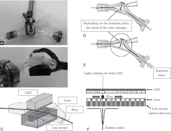
脳動脈瘤コイル塞栓術時のコイル挿入力を,デリバリ ーワイヤーの「たわみ」から光学的センサーによって測 定する装置が開発され,Matsubaraらによって報告され
ている1,3,7-9).この装置は,Yコネクター内部の構造に屈
曲を加えることで,コイル挿入力によって生じるデリバ リーワイヤーの「たわみ」を発生させる.そして,この ワイヤーの「たわみ」による位置の変化を,LEDから
Fig. 1 A and B:Photograph illustrating the newly developed sensor. C:Schematic showing the optical force sensor. D and E:Schematics showing the optical system. D:Without insertion force, the coil-delivery wire does not bend. E:With insertion force, the wire bends. F:The sensor optically detects the position of the bending wire.
Depending on the insertion force, the bend of the wire changes
Light coming out from LED Insertion
force
LED
Lens Line sensor
(optical detector)
Darkest point Wire
Line sensor Lens LED
Wire
C F
D
E A
B
Fig. 2 Photographs of the silicone aneurysm model. Dotted lines showing the inner lumen of the aneurysm.
A:The aneurysm is not filled with a coil,B:The aneurysm is filled with a coil.
発せられる光によってできたワイヤーの影から光学的セ ンサーで検出する.その「たわみ」の位置情報を基にコ イル挿入力を計算する仕組みである(Fig. 1).本装置 の最大の特徴は,手元のYコネクター部でコイル挿入力 が測定できることである.
本装置を用いて,1)通常の手技で術者がコイルを挿 入した場合,2)一定の挿入速度で機械によってコイル を挿入した場合について,シリコン動脈瘤モデルを用い て,塞栓中のコイル挿入力を測定した.
シリコン動脈瘤は分岐のないterminal typeの瘤で,直 径は4及び5mm,親血管の径は2mmのものを使用した
(Fig. 2).これに径3-6mmの各種コイル(GDC10 2D Soft 3mm-8cm,GDC10 3D 3mm-4cm,GDC10 3D 5mm-10cm,GDC10 3D 6mm-15cm; Boston Scientific Neurovascular,Natick,MA,USA)をfirst coilとして 挿入した.マイクロカテーテルはほぼ直線状に配置し,
その先端は瘤の正中に固定した.脳血管内治療専門医を 術者としてデータを収集した.また,この条件下で,コ イルが逸脱せずに瘤内に納まるスピードを検討した予備 実験の結果から,機械によるコイル挿入時のスピードは 0.5[mm/s], 1.0[mm/s]に設定した.コイル挿入力と コイルやカテーテルの挙動との関係をみるために,顕微 鏡にて塞栓中のコイルとカテーテル先端の動きを観察し た.
結 果
コイル挿入力の測定データはFig. 3の通りであった.
術者の場合,挿入力は手の往復運動に伴って鋸歯状のパ
ターンを示した.機械による挿入の場合,3Dコイルで は鋸歯状のパターンに近かったが,helicalコイルではコ イル挿入後半に1-2回のピークを示す所見がみとめら れた.
最大コイル挿入力についてまとめたものをFig. 4に示 す.術者の平均挿入スピードは,2.8-3.4[mm/s]であ った.術者,機械とも最大挿入力は,0.23-0.30[N]の 間であり,1.0[mm/s]では,0.25[N]に集中していた.
コイルの挙動やカテーテル先端の動きの観察から,その 発生はいずれもカテーテルのペインティング動作時であ った.
術者,挿入速度0.5[mm/s],1.0[mm/s]それぞれの,
挿入操作中の挿入力の時間割合をFig. 5に示す.術者の 場合,挿入力は0.20-0.25[N]に低いピークを1つ示した.
機械による挿入の場合,1.0[mm/s]のスピードでは 0.05-0.10[N]に強いピーク点が1つ存在し,一方0.5
[mm/s]ではピーク点が存在しなかった.コイルとカテ ーテル先端の挙動に関して,0.5[mm/s]に比べて1.0
[mm/s]で,カテーテルのペインティングの発生が少な かった.
考 察
脳動脈瘤コイル塞栓術時のコイル挿入操作は,トルク 操作を伴わず,単純にワイヤーを前進させる動きである.
しかし,その手技自体は決して容易なものではなく,そ の習得に訓練を要することはよく知られている5).1990 年代以降,脳動脈瘤コイル塞栓術は急速に広まったもの の12),コイル挿入操作とコイルやカテーテルの挙動やコ
A B
イル挿入力の発生パターンについて,客観的に分析され た報告は乏しく,熟練者の経験に基づいた意見がほとん
どである4,11).本研究では,術者によるコイル挿入と機
械による一定速度での挿入についてコイル挿入力を測定 し,コイル挿入力の発生パターンについて分析した.コ イルの種類,形状やサイズとコイル挿入力に関する研究 については,すでにMatsubaraらが報告しているため2), 本稿では最もストレスなくコイルが挿入できる挿入方法 や挿入速度の条件について,コイルと瘤壁との摩擦状態 に注目して考察を行った.
1.挿入力の発生パターン
術者の場合,3Dコイル,helicalコイルにかかわらず,
鋸歯状の発生パターンが認められた.術者の挿入速度は 速く,速い挿入速度自体が,コイル挿入力に影響を及ぼ した可能性も考えられるが,主には手の往復運動に伴う Fig. 3 The coil insertion force obtained with silicone aneurysms is demonstrated. A:Data with GDC10 3D 3mm-4cm in a 4-mm
aneurysm. B:Data with GDC10 3D 5mm-10cm in a 4-mm aneurysm. C:Data with GDC10 3D 6mm-15cm in a 5-mm aneurysm. Data obtained over 100 seconds are not described here to simply compare the force pattern. D:Data with GDC10 2D Soft 3mm-8cm in a 4-mm aneurysm. For manual insertion, the saw-like (repeated peak) force pattern is described. For mechanical insertion, the force pattern differs with the insertion speed.
0.35 0.30 0.25 0.20 0.15 0.10 0.05
0.000 20 40 60 80 100
Time[sec]
0 20 40 60 80 100
Time[sec]
0.35 0.30 0.25 0.20 0.15 0.10 0.05 0.00 Silicone aneurysm 4mm/GDC10 3D 3mm-4cm
Insertion Force[N]
Insertion Force[N]
Silicone aneurysm 4mm/GDC10 3D 5mm-10cm
0.35 0.30 0.25 0.20 0.15 0.10 0.05 0.00
Insertion Force[N]
0.35 0.30 0.25 0.20 0.15 0.10 0.05 0.00
Insertion Force[N]
Silicone aneurysm 4mm/GDC10 3D 6mm-15cm Silicone aneurysm 4mm/GDC10 Soft 3mm-8cm
0 10 20 30 40 50 60 70 80 90 100 Time[sec]
0 10 20 30 40 50 60 70 80 90 100 Time[sec]
Manual insertion:Surgeon Mechanical insertion:1.0[mm/s] Mechanical insertion:0.5[mm/s]
C D
B A
0.40 0.35 0.30 0.25 0.20 0.15 0.10 0.05
0.000 1 2 3 4
Average insertion speed[mm/s]
Surgeon 1.0[mm/s]
0.5[mm/s]
Maximum insertion force[N]
Fig. 4 The maximum coil insertion force is plotted according to the insertion speed, reaching the range of approximately 0.23-0.30[N]regardless of insertion speed.
前進と停止操作の繰り返しによって,挿入力のピークが 断続的に発生したものと考えられた.
機械による挿入の場合,3Dコイルでは鋸歯状のパタ ーンに近く,helicalコイルでは終盤に1-2回のピーク を示す所見が認められた.これはコイルの特性による影 響と考えられた2).
2.最大挿入力
シリコン動脈瘤モデルでは,術者,機械のスピードに かかわらず最大挿入力は,0.23-0.30[N]の間であり,
その発生はカテーテルのペインティング動作時であっ た.コイルが瘤壁とスムーズに滑るとき挿入力は上昇せ ず,瘤壁と滑らない時にカテーテルのペインティングが 発生していた.ペインティングが発生する時の挿入力は 大きく,最大挿入力となり,術者・機械いずれでもカテ ーテルのペインティングが最低1回は発生していたた め,最大挿入力はほぼ等しくなったと考えられる.なお,
ペインティングが発生せずに,コイルがそれ以上瘤内に 進入できない場合には,挿入力が蓄積し,コイルが瘤外 に出たり,kick backによってマイクロカテーテル先端 が瘤外へ逸脱したりする3).
3.コイル挿入力の時間割合
術者の場合,挿入力の分布には1つの低いピークが認 められた.コイルの挙動との関係を分析すると,停止か ら前進操作に移る時に,コイル挿入力が発生していた.
一度停止した後,再び前進する際に,動脈瘤壁とコイル の滑りがスムーズな場合は挿入力が上昇しないが,スム ーズに滑らない場合に,ペインティングが短時間で発生 し0.20-0.25[N]に分布のピークを形成したものと考え られた.
機械による一定速度での挿入では,挿入速度1.0[mm/s]
の場合,挿入力の分布に1つのピーク(弱い力)が認め られた.このスピードでは,瘤壁とコイルがスムーズに 滑っていることが多いため,ペインティングの発生が少 なかったことが主な要因と考えられた.
挿入速度0.5[mm/s]の場合は,挿入力の分布に明確 なピークが認められなかった.コイルの挙動の観察から,
コイルは瘤壁を滑らず,先端は静止した状態でゆっくり とコイルが充填されていき,ある瞬間にペインティング を起こしていた.1回のペインティングまでの時間が長 く,徐々に挿入力が上昇していく過程で,その分布が均 100
80 60 40 20
00.00 0.05 0.10 0.15 0.20 0.25 0.30 0.35 Insertion force[N]
0.00 0.05 0.10 0.15 0.20 0.25 0.30 0.35 Insertion force[N]
0.00 0.05 0.10 0.15 0.20 0.25 0.30 0.35 Insertion force[N]
100 80 60 40 20 0
100 80 60 40 20 0
Surgeon
Time distribution ratio[%]Time distribution ratio[%] Time distribution ratio[%]
0.5[mm/s]
1.0[mm/s]
Fig. 5
The time distribution ratio of the coil insertion force with silicone aneurysms is demonstrated. A:Data in the manual insertion showing one low peak. B and C:Data in the mechanical insertion showing one peak in 1.0 [mm/s] (B) and no peak in 0.5[mm/s](C).
A B
C
一化されたと考えられた.
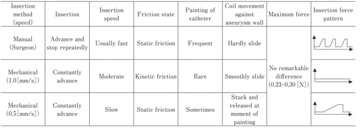
これらの実験結果から,動脈瘤壁に対してコイルが滑 るかどうかは,挿入方法と挿入速度によって決定される 摩擦状態に依存していることが推測された.それぞれの 特徴をまとめると以下のようになる.
術者の場合は,手の往復運動に伴いデリバリーワイヤ ーが停止から前進する際に,コイルと瘤壁との間に静止 摩擦が作用する.そして,静止摩擦によってコイルが滑 るのよりも先にペインティングが発生する傾向がみられ る.
挿入速度0.5[mm/s]の場合,挿入速度が遅いために 静止摩擦によって静止状態が維持されてコイルが滑らな い状態が続き,ある瞬間にペインティングを起こし力が 解放されることとなる.一方,1.0[mm/s]では,コイ ルが動き出すと動摩擦状態を維持して,コイルが動脈瘤 に対してスムーズに滑るような動きを示す(Table 1).
クローンの法則から,常に(最大)静止摩擦力は動摩 擦力より大きいことが知られており,コイルと瘤壁がス ムーズに滑る動摩擦状態を維持できるように適切な挿入 スピードを設定すれば,コイル挿入力の上昇を回避でき る可能性がある.ただし,3Dコイルのように,ペイン ティング動作に伴いコイル塊の形状が変化することによ って良好なframeを形成する場合があり,ある程度の静 止摩擦状態が発生するスピードの方が良い状況も想定さ れる.状況に応じた挿入スピードを的確に設定すること ができれば,機械による一定速度でのコイル自動挿入が,
臨床の場においても有望である可能性が示唆された6).
4.今後の研究課題
今後,ある程度コイルが瘤内に入った状態での評価や 動脈瘤の親動脈との位置関係や形状の違いによる挿入力 発生パターンの分析等,さまざまな条件下における研究 が必要と考える.その他,脳動脈瘤コイル塞栓術におい ては,コイルの挿入とともに,マイクロカテーテル操作 が必要となる場面も多い.そのため,コイル挿入力とマ イクロカテーテル操作との関連についての分析も必要と 考えている.また,先述の通り,コイル塞栓術において は,瘤のタイトパッキングとともに,いかにうまくコイ ルフレームを形成するかも重要なポイントである10).今 回の実験では,コイル挿入操作やコイル挿入力とコイル の展開形状についての十分な検討はされておらず,今後 の研究課題である.
結 論
コイル挿入操作と挿入速度によってコイルと瘤壁との 摩擦状態(静止摩擦状態か動摩擦状態)が決まり,コイ ル挿入力の発生パターンに影響することが判明した.コ イルと瘤壁の間の動摩擦状態を維持できるような挿入方 法が設定できれば,最もストレスなくコイルが挿入でき ると考えられた.
文 献
1) Matsubara N, Miyachi S, Nagano Y, et al: A novel pressure sensor with an optical system for coil embolization of intracranial aneurysms. J Neurosurg 111:41-47, 2009.
2) Matsubara N, Miyachi S, Nagano Y, et al: Evaluation of Insertion
method (speed)
Insertion Insertion
speed Friction state Painting of catheter
Coil movement against aneurysm wall
Maximum force Insertion force pattern
Manual (Surgeon)
Advance and
stop repeatedly Usually fast Static friction Frequent Hardly slide
No remarkable difference (0.23-0.30[N])
Mechanical (1.0[mm/s])
Constantly
advance Moderate Kinetic friction Rare Smoothly slide
Mechanical (0.5[mm/s])
Constantly
advance Slow Static friction Sometimes
Stuck and released at
moment of painting
Table 1 Summary of the differences between the coil insertion methods during the embolization with silicone aneurysms.
Friction state between the coil and the aneurysm wall is the most important factors affecting the coil insertion force.
the characteristics of various types of coils for the embolization of intracranial aneurysms with an optical pressure sensor system. Neuroradiology. Published online Jun 3, 2010. DOI: 10.007/s00234-010-0722-5.
3) 松原功明,宮地 茂,大島共貴,他:光学的センサーに よ る 脳 動 脈 瘤 塞 栓 用 coil の 挿 入 力 測 定 装 置 の 開 発.
JNET 2:113-118, 2008.
4) 松本康史,江面正幸:破裂動脈瘤.吉田 純,宮地 茂編,
脳血管内治療の Do's & Don'ts 第2版,東京,医学書院,
2006,60-62.
5) Miyachi S, Okamoto T, Kobayashi N, et al: Nagoya University training system for neuroendovascular therapists. Interventional Neuroradiol 10 (Suppl 1):
S103-106, 2004.
6) 永野佳孝,佐野明人,松原功明,他:脳動脈瘤コイル塞 栓術用ワンサージョン手術システムの開発.J JSCAS 11:244-245, 2009.
7) Nagano Y, Sano A, Sakaguchi M, et al: Development of force sensor for a linear object. Proceeding of Institute of Electrical and Electronics Engineers Conference on Sensors 2006:823-826, 2006.
8) 永野佳孝,佐野明人,坂口正道,他:極細かつ長尺の線 状体用力センサの開発-脳動脈瘤コイル塞栓術への適応
-.計測自動制御学会論文集.44:278-284, 2008.
9) 永野佳孝,佐野明人,坂口正道,他:脳動脈瘤コイル塞 栓術用センシングシステムの開発-血管モデルと In vivo の模擬瘤を用いた挿入力の評価- . J JSCAS 10:147-153, 2008.
10) 中澤和智,太田剛史,今村博敏,他:コイルの特性を考 慮した脳動脈瘤瘤内塞栓術-そのコンセプトと実験的
データより-第22回脳神経血管内治療学会総会講演集.
11-16, 2006.
11) 根本 繁:未破裂動脈瘤 . 吉田 純,宮地 茂編,脳血管 内治療の Do's & Don'ts 第2版,東京,医学書院,2006, 85-90.
12) Prestigiacomo CJ: Historical perspectives: the microsurgical and endovascular treatment of aneurysms. Neurosurgery 59 (Suppl 3): S39-47, 2006.
13) Richling B: History of endovascular surgery: personal accounts of the evolution. Neurosurgery 59 (Suppl 3):
S30-38, 2006.
JNET 4:84-90, 2010
要 旨
【目的】脳動脈瘤モデルにおいてコイル挿入力の測定を行い,コイルやカテーテル先端の挙動との関係を検討することで,
コイル挿入力の発生パターンを分析した.そして,よりストレスなくコイルが挿入できる挿入方法と挿入速度の条件につ
いて考察した.【方法】Yコネクター内のワイヤーの「たわみ」を光学的センサーで計測することでコイル挿入力を測定す
る装置が開発されている.本装置を用いて,術者が挿入した場合,一定の挿入速度で機械的に挿入した場合について,コ
イル挿入力を測定した.同時に,顕微鏡で塞栓中のコイルやカテーテルの動きを観察した.【結果】術者による挿入の場合,
手の往復運動による影響がみられ,停止から再び動き出す際に静止摩擦が作用していた.機械による挿入の場合,挿入速 度によってコイル挿入力の発生パターンに違いがみられ,瘤壁とコイルの間の摩擦状態(静止摩擦状態か動摩擦状態か)
が影響していた.【結論】挿入操作と挿入速度によってコイルと瘤壁との摩擦状態が決まり,挿入力の発生パターンに影響
することが判明した.コイルと瘤壁の間の動摩擦状態を維持できるような挿入条件が設定できれば,ストレスなくコイル が挿入できると考えられた.

![Fig. 4 The maximum coil insertion force is plotted according to the insertion speed, reaching the range of approximately 0.23-0.30[N]regardless of insertion speed.](https://thumb-ap.123doks.com/thumbv2/123deta/7474770.2486088/4.892.85.806.101.568/maximum-insertion-according-insertion-reaching-approximately-regardless-insertion.webp)