はじめに
脳血管内治療は,近年急速に発展してきた治療方法で あり,特に,脳動脈瘤に対するcoil塞栓術はその代表的 な治療法の一つである.
脳動脈瘤に対するcoil塞栓術において,最も避けるべ き合併症は動脈瘤の破裂であり,致命的な結果となる場 合が少なくない2, 4).術中破裂は,マイクロカテーテル
やマイクロガイドワイヤーの穿通による以外にも,coil の挿入に伴い瘤壁に過度の力がかかることによっても引 き起こされる3,8).一方,coil塞栓術の終盤でcoilに過度 の力を加えるとkick backによりマイクロカテーテルの 先端が瘤外へ逸脱してしまうことがあり,その結果,意 図せず不十分な塞栓に終わってしまう場合もある.
この瘤壁への過度の力による破裂やkick backによる カテーテル先端の瘤外への逸脱を予防するには,マイク
光学的センサーによる脳動脈瘤塞栓用coilの 挿入力測定装置の開発
松原功明1) 宮地 茂1) 大島共貴1) 細島 理1) 泉 孝嗣1) 靍見有史1) 錦古里武志1)
吉田 純1) 永野佳孝2,3) 佐野明人2) 坂口正道2) 藤本英雄2)
Development of optical force sensor system for cerebral aneurysm coil embolization
Noriaki MATSUBARA
1)Shigeru MIYACHI
1)Tomotaka OHSHIMA
1)Osamu HOSOSHIMA
1)Takashi IZUMI
1)Arihito TSURUMI
1)Takeshi KINKORI
1)Jun YOSHIDA
1)Yoshitaka NAGANO
2,3)Akihito SANO
2)Masamichi SAKAGUCHI
2)Hideo FUJIMOTO
2)1) Department of Neurosurgery, Nagoya University Graduate School of Medicine 2) Graduate School of Engineering, Nagoya Institute of Technology
3) New Product Development R & D Center, NTN Corporation
●Abstract●
Objective: In endovascular coil embolization for a cerebral aneurysm, excessive stress from coil insertion into an aneurysm may cause a catastrophic rupture or the microcatheter tip to dislocate from the aneurysm dome, resulting in insufficient embolization. Such mechanical stress caused by the coil insertion force can only be subjectively detected by the minor tactile feedback the surgeon receives. Therefore, a new sensor device, that adequately measures coil insertion force via an optical system was developed. This new sensor device is described here.
Method: This system is incorporated into a Y-connector (hemostatic valve which is connected to the proximal end of a microcatheter).
The sensor principle is based on an optical system consisting of an LED and line sensor. The line sensor measures how the coil delivery wire slightly bends according to the insertion force by detecting the wire shadow. This information is translated into a given force level. An experimental silicon aneurysm embolization was performed with this optical sensor. The sensor continuously observed the mechanical force during the insertion of coils into the dummy aneurysm.
Results: The sensor adequately recorded the coil insertion force during embolization. When the embolization was successfully performed, the maximum insertion force did not exceed 0.3[N], and the record showed a repeated peak pattern change in force, reflecting actual clinical experience. Presence of the sensor did not hinder performance of embolization in any way.
Conclusion: This new sensor device adequately measures coil insertion force. This system provides potential for safer and more certain aneurysm embolization.
●Key Words●
cerebral aneurysm, coil embolization, insertion force, sensor
(Recieved April 14, 2008:Accepted May 23, 2008)
1)名古屋大学大学院医学系研究科 脳神経外科 2)名古屋工業大学大学院 工学研究科 3)NTN株式会社 商品開発研究所
<連絡先:松原功明 〒466-8550 名古屋市昭和区鶴舞町65 E-mail:[email protected]>
ロカテーテルからcoilを挿入する時にかかる力の程度を うまく加減しなければならない.しかし,現在この力の 加減は,デリバリーワイヤーを持った術者の指にかかる 微細な力の感覚のみによってコントロールされている.
そのため,より安全で確実な治療のためには,動脈瘤壁 にかかるcoil挿入力の定量化を行い客観的に評価できる システムが必要であると考えた.
我々は,名古屋工業大学およびNTN株式会社との共 同研究により,脳動脈瘤coil塞栓術時に瘤壁にかかる挿 入力を,デリバリーワイヤーの「たわみ」から光学的セ ンサーによって測定する装置を開発したので,概要と基 礎的な実験の結果について報告する.
方 法
光学的センサーによるcoil挿入力測定装置(以降,本 装置と記載)の概観はFig. 1B, Cの通りである.市販さ れているY-コネクター(止血用ハブ)(Fig. 1A)の形を アレンジし内部にLEDと128列の光学的ラインセンサー を搭載した.模式図を示す(Fig. 1D).マイクロカテー テルとの接続部(出口部)とcoil/ワイヤー挿入部(入
従来のY-コネクターでは,ワイヤーの入口部と出口 部は直線的であるが,あえてこの間に少し屈曲をくわえ ることでデリバリーワイヤーの「たわみ」を発生させる
(Fig. 2).Y-コネクター内において,指先からのcoil挿 入力によって生じるこのデリバリーワイヤーの「たわみ」
による位置の変化を検出する.これはLEDから発せら れる光によってできたワイヤーの影を光学的センサーで 検出することにより行われる(Fig. 3).その「たわみ」
の位置情報が,制御ボックスに内蔵されたコンピュータ ーに送られ,自動的に挿入力に変換される原理である.
詳細な物理工学的な原理に関しては本論文では割合する が,共著者らの文献報告を参考にされたい5, 6).
結 果
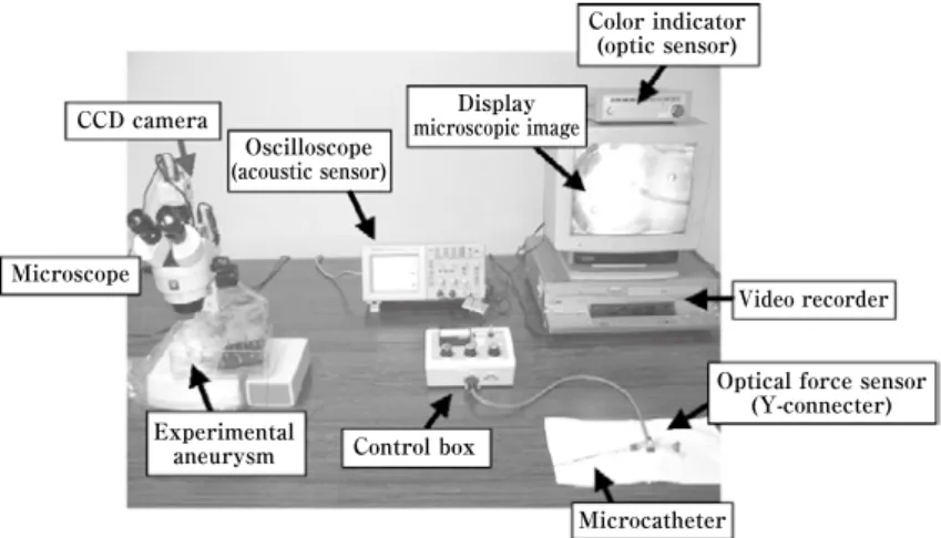
本装置を用いて,シリコン性の実験的動脈瘤に対して coil挿入を行った.実験システムはFig. 4の通りである.
coilやマイクロカテーテル先端の動きを顕微鏡で観察し ながら,同時に挿入力を測定した.代表的な挿入力の測 定結果をFig. 5に示す.
coilの挿入がスムーズに成功した場合には,カテーテ
3 4
2 1
Fig. 1
A:Y connector available commercially.
B, C: Y-connector with the optical force sensor system. Arrow indicates a part of the optical force sensor.
D: Schematic image of the optical force sensor. 1. Part connecting to the proximal end of the microcatheter. 2. Part connecting to the infusion line with normal saline. 3. Part to insert into the coil delivery wire. 4. Coil delivery wire.
A B
C D
Insertion force Depending on the insertion force,
the bend in the wire changes
Fig. 2 Schematic image of the optical system
A:Without insertion force, the coil delivery wire does not bend.
B: With insertion force, the wire bends. The sensor optically detects the position of the bending wire.
(optical detector) Line sensor
Wire Lens
Darkest point 63.5μm
LED
Optical sensor system
Line sensor Wire Lens LED
Fig. 3
A: The line sensor determines the wire position by measuring the shadow cast.
The darkest point is defined as the wire position, that is translated into insertion force. Line sensor resolution is 63.5μm.
B:Optical system for the force sensor.
A
B
A
B
挿入に伴って振り子状に動くこと)の前に挿入力がかか り,painting後に挿入力が低下するパルス状の力のパタ ーンが認められた.挿入力の最大値は0.3[N]を大きく越 えることはなかった.それと対照的に,coil挿入力が,
0.3[N]前後の強さで,一定のまま変化しない場合には,
coilがそれ以上動脈瘤内に進入できず挿入力がたまり,
その結果,coilが瘤外に出てしまったり,kick backによ ってマイクロカテーテル先端の瘤外への逸脱が認められ たりした.またcoilを順につめていくに従って,最大挿 入力が上昇しており,coilの性質によって挿入力が変化 していた.このように,これまでの臨床経験を反映する 結果が得られた.
また,センサー付きY-コネクターはcoilの操作性に支 障はなく,ほぼ従来通りのデバイスとしての使用が可能
考 察
現在,脳動脈瘤に対するcoil塞栓術において,coil挿 入時にかかる力の加減はデリバリーワイヤーを通じて術 者の指にかかる微細な力の感覚のみで調整されている.
そのため,力の強さについては,「かたい」・「やわらかい」
などの言葉で表現されるのみであった.我々は,この指 にかかる挿入力の測定および記録が可能となるよう,光 学的センサーによるcoil挿入力測定システムを開発し た.
本装置を用いると,客観的に挿入力を測定・表示する ことができるので,他の術者にもワイヤーにかかってい る力が認識可能となる.これにより,マイクロカテーテ ルの操作とcoil挿入を複数の術者で行う場合には協調し
Optical force sensor (Y-connecter) Color indicator
(optic sensor)
Experimental aneurysm
Display microscopic image Oscilloscope
(acoustic sensor)
Microcatheter Microscope
CCD camera
Control box
Video recorder
Fig. 4 Photograph showing the experimental system measuring coil insertion force by optical sensor during embolization of a dummy aneurysm.
No.1 Success: ED ExtraSoft 3-6 No.2 Success: ED ExtraSoft 3-6 No.3 Failure: GDC UltraSoft 3-6 No.4 Failure: GDC UltraSoft 3-6
Time [sec]
Force [N]
0-100 [sec]
Measuring condition (4mm aneurysm, insertion of third coil)
Fig. 5 Results for insertion force measured by the optical system. No. 1 and 2 resulted in successful embolization. No. 3 and 4 failed to embolize.
In a successful embolization case (No. 1 and 2), maximum insertion force did not exceed 0.3[N] and a repeated peak (short pulse) pattern of force was observed. In contrast, in a failed case (No. 3 and 4), force reached 0.3[N]and did not show the repeated peak pattern.
とができる.
このシステムのきわめて画期的な点は,手元のワイヤ ー挿入部でcoil挿入力の測定が可能なことである.従来 のcoil挿入力の研究では,実験的動脈瘤壁に装着したセ ンサーを用いていたため,生体内での測定は不可能であ った7).カテーテル先端に付けた圧力センサーや挿入部 でカテーテルからの反発力から力を測定する装置の報告 もみられるが,いずれも特殊な装置が必要であり臨床応 用は困難である1,9,10).この点,本システムではcoil挿入 部で力を測定でき,さらに従来どおりの使用が可能とな るため,in vivoでの使用や将来的には実際の臨床応用が 可能である.なお,各coilによりデリバリーワイヤーの 特性(たわみ具合/Young率)が違うため,coilごとに 違うプログラムを使用しコンピューターが挿入力を計算 する.また,coilのみならずマイクロガイドワイヤーの 先端にかかる力も計算式のプログラムを作成すれば測定 できる.今回の実験では,マイクロカテーテルが大きく 屈曲している状況ではないため,Y-コネクター部での 力の測定結果について,coilがマイクロカテーテル内を 通過する際に発生する摩擦力が問題になることはなかっ た.しかし,実際のcoil塞栓術においては,アクセス途 中にマイクロカテーテルが強く屈曲している場合も考え られ,本装置では正確に動脈瘤壁にかかる力を反映でき ない可能性がある.マイクロカテーテルの屈曲の程度と coil/デリバリーワイヤーとの間に生じる摩擦力につい ては今後の検討課題である.
Coil挿入力はデジタル・アナログ表示とも可能であ る.挿入力はオシロスコープに伝達され音声に変換する ことができ,カラーインジケーターによる視覚センサー にも変換することが可能である.さまざまな表示システ ムを試行した結果,塞栓術中の術者の視線は,通常モニ ターに集中しているため,挿入力の大きさを知らせ,デ リバリーワイヤーの操作にフィードバックさせるための 方法としては,音声センサーが最も現実的であると考え られた.挿入力が上昇するとアラームが鳴るように設定 し,coilの挿入停止やコントロールのための指標として 用いることが可能である.
他の大きな特徴として,本装置は光学的センサーであ り,デリバリーワイヤーの通るY-コネクター内部の空 間と光学的システムが直接,接することはない点が挙げ られる.これによりデリバリーワイヤーの操作にセンサ ーが影響を及ぼしたり,センサーが血液等に汚染された りすることがない.
また,実験の結果,Y-コネクター型センサーは,現 在用いられているY-コネクターとほぼ同様な形で使用 可能であり,これまでの塞栓術の手技の妨げとならない ことが確認された.このY-コネクター型光学的センサ ー自体は,特殊な原材料を要するものではないため,ニ ーズがあり製品化・実用化された場合には,大量生産可 能で安価に提供できる見込みである.そして,制御コン ピューターや挿入力表示モニター等の測定機器は別とし て,Y-コネクター部分は基本的にディスポーザブル使 用が可能である.
結 論
光学的センサーによる脳動脈瘤塞栓用coilの挿入力測 定装置の開発について報告した.本装置は,より安全・
確実な脳動脈瘤coil塞栓術を行うための術中モニターと して有用であると考えられた.
文 献
1)Katsumata T, Haga Y, Minami K, et al: Micromachined 125μm diameter ultra miniature fiber-optic pressure sensor for catheter. Trans. IEEE of Japan. 120-E-2:
58-63, 2000.
2)Li MH, Gao BL, Fang C, et al: Prevention and management of intraprocedural rupture of intracranial aneurysm with detachable coils during embolization.
Neuroradiology 48: 907-915, 2006.
3)松本康史 , 江面正幸:破裂動脈瘤.吉田 純,宮地 茂編 , 脳血管内治療の Do's & Don'ts.第2版,東京,医学書院,
2006,60-62.
4)Miyachi S, Negoro M, Sahara Y, et al: Treatment strategy for cerebral aneurysms based on the evidence of the efficacy of GDC embolization. Interventional Neuroradiol 9(Suppl 1):51-55, 2003.
5)Nagano Y, Sano A, Sakaguchi M, et al: Development of force sensor for a linear object. Proc. IEEE Sensors 2006, 823-826, 2006.
6)永野佳孝 , 佐野明人 , 坂口正道,他:極細かつ長尺の線 状体用力センサの開発−脳動脈瘤コイル塞栓術への適応
−.計測自動制御学会論文集.44:278-284,2008.
7)中澤和智,太田剛史,今村博敏,他:コイルの特性を考 慮した脳動脈瘤瘤内塞栓術−そのコンセプトと実験的 データより−第22回脳神経血管内治療学会総会講演集.
11-16,2006.
8)根本 繁:未破裂動脈瘤.吉田 純,宮地 茂編,脳血 管内治療の Do's & Don'ts.第2版,東京,医学書院,
2006,85-90.
9)Tanimoto M, Arai F, Fukuda T, et al: Micro force sensor for intravascular neurosurgery. Proc. 1997 IEEE
Int. Conf. on Robotics and Automation (ICRA'97), 1561-1566, 1997.
10)Tanimoto M, Arai F, Fukuda T, et al: Force display method to improve safety in teleoperation system for
intravascular neurosurgery. Proc. 1999 IEEE Int. Conf.
on Robotics and Automation (ICRA'99), 1728-1733, 1999.
JNET 2:113-118, 2008
要 旨
【目的】脳動脈瘤の塞栓術において,coil挿入時に過度の負荷が加わると瘤の破裂により重篤な合併症を起こしたり,マイク ロカテーテル先端が瘤内から逸脱し不十分な塞栓となったりする可能性がある.我々は,光学的システムを用いてcoilの挿 入に伴う力を測定できる装置を開発したので,概要と基礎的実験の結果について報告する.【方法】Y-コネクターを改良し,
内部に光学的センサーを組み込んだ.内部を通るコイルデリバリーワイヤーは挿入力により「たわみ」を生じる.LEDから 発せられる光によってできるワイヤーの影によりその「たわみ」を同定し,それが定量的な挿入力の値へと変換される.シ リコン動脈瘤に対してcoilの挿入を行い,本装置により挿入力を測定した.【結果】正確なcoil挿入中の力の測定が可能であ った.また,本装置の使用に際してcoil挿入操作の妨げにならなかった.挿入が成功した場合の最大の挿入力は0.3[N]を 超えることがなく,力の2峰性の分布が観察された.【結論】光学的センサーによるcoil挿入力測定装置を開発した.この装 置の臨床応用が広がれば,より安全・確実に脳動脈瘤塞栓術が施行できると期待された.