ORIGINAL ARTICLE
Long-term outcome of idiopathic steroid-resistant nephrotic syndrome in children
Aya Inaba1&Yuko Hamasaki2&Kenji Ishikura3,7&Riku Hamada3&Tomoyuki Sakai4&
Hiroshi Hataya3&Fumiyo Komaki5&Tetsuji Kaneko6,8&Masaaki Mori1&
Masataka Honda3
Received: 8 December 2014 / Revised: 13 July 2015 / Accepted: 15 July 2015 / Published online: 3 September 2015
#IPNA 2015
Abstract
Background Several recent studies have shown improved short-term outcome of steroid-resistant nephrotic syndrome (SRNS) in children; however, only a few studies have evalu- ated the long-term outcome. The aims of our study were to obtain detailed data and analyze the long-term outcome of children with SRNS.
Methods Sixty-nine children with idiopathic SRNS were en- rolled and divided into two groups based on initial histopath- ological patterns: focal segmental glomerulosclerosis (FSGS) and minimal change (MC)/diffuse mesangial proliferation ( D M P ) . T h e e ffe c t s of i n i t i a l t r e a t m e nt w i t h t h e
immunosuppressant of choice (cyclosporine or cyclophospha- mide) on renal survival, remission, and incidence of compli- cations were analyzed in both groups (4 subgroups).
Results The renal survival rate was significantly different among the four different subgroups based on different combi- nations of initial histopathological pattern (FSGS vs. MC/
DMP) and initial immunosuppressant used for treating SRNS (cyclosporine vs. cyclophosphamide) (P=0.013), with renal survival in the FSGS (cyclophosphamide) subgroup being especially low (54.6 %). Disease- and/or treatment- associated complications were relatively low; however, hyper- tension at last examination was observed in a considerable number of patients (31.9 %).
Conclusions Our results suggest that a recently developed therapeutic regimen with cyclosporine considerably improves both the initial remission rate and the long-term renal survival rate of children with idiopathic SRNS.
Keywords Children . Steroid-resistant nephrotic syndrome . Long-term outcome . Immunosuppressant . Minimal change . Diffuse mesangial proliferation . Focal segmental
glomerulosclerosis
Introduction
In general, 10 % of children with idiopathic nephrotic syn- drome (INS) show steroid resistance. Early studies reported that 30–40 % of children with steroid-resistant nephrotic syn- drome (SRNS) progress to end stage-kidney disease (ESKD) during follow-up of 10 years [1,2].
Methylprednisolone pulse therapy and/or immunosuppres- sant therapy, such as cyclophosphamide or cyclosporine, have been used for many years to treat children with SRNS. Cyclo- phosphamide has been used since the 1960s, and in the 1990s, Electronic supplementary materialThe online version of this article
(doi:10.1007/s00467-015-3174-7) contains supplementary material, which is available to authorized users.
* Yuko Hamasaki
1 Department of Pediatrics, Yokohama City University Medical Center, Kanagawa, Japan
2 Department of Pediatric Nephrology, Toho University Faculty of Medicine, 6-11-1, Omori-Nishi, Ota-ku, Tokyo 143-8541, Japan
3 Department of Nephrology, Tokyo Metropolitan Children’s Medical Center, Tokyo, Japan
4 Department of Pediatrics, Shiga University of Medical Science, Shiga, Japan
5 Community Health Welfare Division, Kawasaki Saiwai Ward Office Health and Welfare Center, Kanagawa, Japan
6 Department of Clinical Research, Tokyo Metropolitan Children’s Medical Center, Tokyo, Japan
7 Department of Nephrology and Rheumatology, National Center for Child Health and Development, Tokyo, Japan
8 Teikyo Academic Research Center, Teikyo University, Tokyo, Japan DOI 10.1007/s00467-015-3174-7
uncontrolled trials had found that up to 60 % of children treated with the combination of methylprednisolone pulse therapy and cyclophosphamide or chlorambucil achieved complete remission [3]. Cyclosporine was added to the battery of therapeutic strategies in 1987, and many recent studies have shown the efficacy of cyclosporine for SRNS [4–6]. Hamasaki et al. recently reported a high remission rate (88.6 %) and high renal survival rate (94.3 %) in a prospective 12-month proto- col treatment trial using a combined cyclosporine/
prednisolone therapeutic regimen for children with SRNS [7]. However, the long-term outcome of children with SRNS, including the renal survival rate, permanent remission rate, and incidence of treatment-related complications, as well as the impact of each therapeutic regimen on these rates have not yet been sufficiently evaluated.
The aims of this retrospective cohort study were to analyze the clinical and histopathological parameters of children with SRNS and to evaluate the remission rate of the initial SRNS episode, renal survival rate, permanent remission rate, and long-term complications. We also analyzed whether the remis- sion rate of the initial SRNS episode and the long-term out- come differ according to the choice of initial immunosuppres- sant used to treat the SRNS, namely, cyclosporine or cyclo- phosphamide. The incidence of complications related to SRNS and treatment for SRNS was also evaluated.
Methods Patients
This study was a retrospective analysis of children with SRNS who were followed in Tokyo Metropolitan Kiyose Children’s Hospital (predecessor of Tokyo Metropolitan Children’s Med- ical Center), a tertiary care center for children with kidney disease. Data were retrieved from the hospital’s database on children with nephrotic syndrome who fulfilled the following criteria: (1) INS initially diagnosed between 1 January 1990 and 1 January 2005; (2) SRNS, either initial non-responder or late non-responder; (3) followed up for≥4 years. Children were excluded if they (1) had underlying secondary causes [Henoch–Schönlein nephritis, systemic lupus erythematosus, immunoglobulin (Ig) A nephropathy, membranous nephropa- thy, membranoproliferative glomerulonephritis, among others]; (2) had congenital or inherited forms of nephrotic syndrome; (3) were younger than 1 year or older than 15 years when diagnosed with nephrotic syndrome; (4) had not under- gone renal biopsy.
The database and medical records were reviewed to collect relevant data over the period from diagnosis of INS to last examination, including the date on which each child was di- agnosed as having INS and SRNS, demographic characteris- tics (age, gender) at the time of diagnosis of SRNS, initial
histopathological pattern [minimal change (MC), diffuse mesangial proliferation (DMP), or focal segmental glomerulosclerosis (FSGS)], steroid response (primary or late non-responder), therapeutic strategies of immunosuppressant, the date on which each child achieved complete remission of the initial SRNS episode, the date of diagnosis of ESKD, the date on which each child achieved complete remission after last relapse episode, clinical aspects (height, weight, and blood pressure), the usage of anti-hypertensive agents, renal func- tion, the condition of nephrotic syndrome, the complications of nephrotic syndrome or treatment-related complications at the last examination, and the findings of chronic cyclosporine nephrotoxicity at the last renal biopsy.
Definitions
Nephrotic syndrome was diagnosed if the urinary protein/
creatinine ratio was≥1.8 mg/mg and the serum albumin level was≤2.5 g/dl [7]. SRNS was diagnosed if complete remission was not achieved after treatment with 2 mg/kg prednisolone daily for 4 weeks [8]. Complete remission was defined as negative or trace proteinuria (by the dipstick method or a uri- nary protein/creatinine ratio of ≤0.20 mg/mg) on urinalysis and a serum albumin level of>2.5 g/dl. Partial remission was defined as a serum albumin level of >2.5 g/dl, but persisting proteinuria on urinalysis (dipstick method +1 or greater, or urinary protein/creatinine ratio of>0.2 mg/mg). The state of remission included both complete remission and partial remis- sion. Non-remission was defined as persisting nephrotic syn- drome. Relapse of nephrotic syndrome was defined as in- creased proteinuria and a serum albumin level of ≤2.5 g/dl.
Permanent remission was defined as the relapse-free state without any immunosuppressant or steroid over the previous 2 years or more up to the last examination. Frequently relaps- ing nephrotic syndrome (FRNS) was defined as four or more relapses within any 12-month period or the condition in which any immunosuppressant was used to control relapse of steroid- sensitive nephrotic syndrome (SSNS). ESKD was defined as the requirement for dialysis or kidney transplantation. Late non-response to steroids was defined as an initial response to steroid therapy but none during a subsequent relapse.
The estimated glomerular filtration rate (eGFR) was cal- culated using the Schwartz formula for patients aged
≤17 years [9]: 194 × SCr−1.094× Age−0.278in male patients aged ≥18 years, and 194 × SCr−1.094 × Age−0.278 × 0.739 in female patients aged ≥18 years, where SCr is the serum creatinine level [10]. Hypertension was defined as the need for anti-hypertensive therapy, except when given for renoprotective purpose. The height measurements were expressed as the height standard deviation score (SDS) compared with normal stature values for age- and sex- matched healthy Japanese children. Short stature was de- fined as a height of less than −2.0 SDS.
Histopathology
Renal biopsy was performed after the diagnosis of SRNS.
Repeat biopsy was performed for the patients who were treat- ed with cyclosporine to evaluate cyclosporine-related nephro- toxicity. Renal pathologists at our institution evaluated the initial histopathological findings and the development of c h r o n i c c y c l o s p o r i n e n e p h r o t o x i c i t y, d e f i n e d a s cyclosporine-associated arteriolopathy and/or cyclosporine- induced tubulointerstitial lesions showing characteristic striped tubulointerstitial lesions.
Treatment
All patients were treated initially with prednisolone at a dose of 2 mg/kg per day administered in three separate doses for 4 weeks (maximum 80 mg/day) and diagnosed with SRNS if they did not achieve complete remission during this period.
Up until the 1990s, following a diagnosis of SRNS, oral cyclophosphamide, methylprednisolone pulse therapy, or a combination of these therapies were administered. Cyclophos- phamide was used at a dose 2.5 mg/kg per day (maximum 100 mg per day) orally for 12 weeks (with a total cumulative dose of 210 mg/kg). The basic protocol of methylprednisolone pulse therapy consisted of the administration of methylpred- nisolone 30 mg/kg per day (2-h infusion, maximum 1000 mg) every day for 3 days—considered to be one course; one course per week was given in weeks 1 and 2, then one course per month was given from week 4 to month 6, followed by one course per 3 months from month 7 to year 2 [11]. Beginning in the mid-1990s, the main immunosuppressant used to treat SRNS was changed to cyclosporine combined with predniso- lone, the dose of which was adjusted to maintain a whole- blood trough level of 120–150 ng/ml for the initial 3 months, followed by 80–100 ng/ml for months 4–12, and 60–80 ng/ml for months 13–24, with subsequent tapering of the cyclospor- ine dose after 2 years of therapy. Among those patients treated with cyclosporine, some additionally received methylprednis- olone pulse therapy consisting of methylprednisolone 30 mg/kg per day (2-h infusion, maximum 1000 mg) every day for 3 days—considered to be one course; one course per week was given in weeks 1, 2, 5, 9, and 13 [7]. Prednisolone was started at 1 mg/kg per day in three separate doses for 4 weeks and then was reduced to 1 mg/kg in a single dose every other day for generally 1 year in those patients diag- nosed with SRNS.
If a therapeutic regimen failed to induce remission, another regimen or a combination of regimens was considered. That is, if the regimen with cyclosporine failed to induce remission, we switched to the regimen with cyclophosphamide, and if the cyclophosphamide regimen failed to induce remission, we switched to one with cyclosporine. We considered a therapeu- tic regimen as having failed to induce remission if remission
was not induced within 4–6 months after commencement of administration.
Statistical analyses
Results were expressed in terms of median, range, and percent- age. The endpoints were the incidences of complete remission of the initial SRNS episode, ESKD, and permanent remission.
The duration from the date of diagnosis of SRNS to the date on which each respective endpoint was reached was measured and evaluated using Kaplan–Meier analysis according to sub- group analysis of the initial histopathological patterns (FSGS or MC/DMP) and initial immunosuppressant used for SRNS (cyclosporine or cyclophosphamide). Thus, there were four subgroups. Differences between subgroups were compared using the log-rank test. Regarding renal survival rate, variables, including gender, age at time of SRNS diagnosis, initial histo- pathological pattern (FSGS or MC/DMP), and initial immuno- suppressant used for SRNS were assessed by multivariate anal- ysis with Cox regression. A two-sidedP-value of <0.05 was considered to be statistically significant. All statistical analyses were performed using the SAS software package for Windows (release 9.3; SAS Institute Inc., Cary, NC, USA).
Results
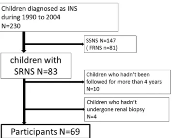
In total, 230 patients were diagnosed with INS between 1 January 1990 and 1 January 2005, of whom 147 showed SSNS. Among the remaining 83 SRNS patients, ten patients had been followed for <4 years and four patients had not undergone renal biopsy. Therefore, 69 children were ultimate- ly enrolled in the analysis (Fig. 1). Basic characteristics of these children are shown in Table1. The first renal biopsy showed MC in 39 children (57 %), FSGS in 22 (32 %), and DMP in eight (11 %). Among the eight patients who showed DMP, two showed mild IgM deposition on immunofluores- cence staining, one showed mild C1q deposition, one showed intense C3 deposition and mild IgM and C1q deposition, two showed no deposition, and two had no data about immuno- fluorescence staining. The median age at diagnosis of SRNS was 3.2 (range 1.1–15.3) years. In children with FSGS diag- nosed on the first renal biopsy, cyclophosphamide and cyclo- sporine were used as the initial immunosuppressant for the initial SRNS episodes in 11 and ten patients, respectively;
one patient did not receive any immunosuppressant. In chil- dren with MC/DMP on the first renal biopsy, cyclophospha- mide was used as the initial immunosuppressant for initial SRNS episodes in 13 patients, cyclosporine was used in 29 patients, and no immunosuppressant was used in five patients, two of whom underwent methylprednisolone pulse therapy.
Therapeutic strategies from the time of the initial diagnosis of SRNS to achievement of first complete remission or
progression to ESKD are shown in Table2. During the obser- vation period of this study we did not use rituximab, myco- phenolate mofetil, or tacrolimus. The overall median follow- up period was 10.1 (range 4.5–19.3) years.
No immunosuppressant was used in six patients (1 in FSGS, 5 in MC/DMP). Of these, three patients attained com- plete remission early and maintained that condition; one pa- tient was diagnosed with SRNS and had been treated with
prednisolone for 1 year only at another institution and did not require any immunosuppressant when she came to our hospital for the first time; one patient had relatively mild pro- teinuria and spontaneously attained complete remission after prednisolone was discontinued; one patient was treated with prednisolone only and achieved partial remission but not com- plete remission within 1 month of the start of prednisolone, prednisolone therapy was discontinued after 2 months, and complete remission was finally achieved 7 years after the ini- tial diagnosis of SRNS and maintained to date. Because the above six patients achieved complete remission without any immunosuppressant, they were excluded from the analysis, and the analyses of remission rate, renal survival rate, and permanent remission rate were conducted using only the data of the remaining 63 patients.
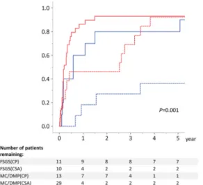
Kaplan–Meier analysis revealed that the complete re- mission rate of the initial SRNS episode at 4 months in those patients whose initial histopathological pattern was FSGS and initial immunosuppressant was cyclosporine [FSGS (cyclosporine) subgroup] was 40 %. In compari- son, that of the patients whose initial histopathological pattern was FSGS and initial immunosuppressant was cyclophosphamide [FSGS (cyclophosphamide) subgroup]
was 0 %. The complete remission rate of those patients whose initial histopathological pattern was MC/DMP and initial immunosuppressant was cyclosporine [MC/
DMP (cyclosporine) subgroup] was 69.0 %. In compar- ison, that of the patients whose initial histopathological pattern was MC/DMP and initial immunosuppressant was cyclophosphamide [MC/DMP (cyclophosphamide) Fig. 1 Overview of idiopathic nephrotic syndrome (INS) patients in this
study. Of the 230 children diagnosed with INS between 1 January 1990 and 1 January 2005, 69 patients with steroid-resistant nephritic syndrome (SRNS) met the entry criteria. SSNS, steroid-sensitive nephrotic syndrome;FRNS, frequently relapsing nephrotic syndrome
Table 1 Basic (background) characteristics of participants in the study Basic (background) characteristics of participants
First renal biopsy finding: FSGS MC/DMP Total
First immunosuppressant used to treat SRNS:
CP CSA None CP CSA None
Number of patients 11 10 1 13 29 5 69
Gender (n)
Male 6 7 0 8 21 3 45
Female 5 3 1 5 8 2 24
Age when diagnosed with SRNS (years)
<3 4 5 0 5 15 1 30
≥3, <7 5 1 0 5 7 2 20
≥7, <11 2 1 0 2 4 2 11
≥11 0 3 1 1 3 0 8
Median age (years) at last follow-up (range)
19.7 (13.6–26.9) 12 (10.0–28.1) 23.3 17.7 (8.2–23.6) 12.1 (6.9–24.9) 16.8 (15.5–27.6) 13.9 (6.9–28.1) Steroid response
Late non-responder 0 2 1 3 9 0 15
SRNS, Steroid-resistant nephritic syndrome; MC, minimal change; DMP, diffuse mesangial proliferation; FSGS, focal segmental glomerulosclerosis;
CP, cyclophosphamide; CSA, cyclosporine
subgroup] was 38.5 % (P= 0.001) (Fig. 2). After exclu- sion of patients with DMP from the analysis, the com- plete remission rate of the initial SRNS of the MC (cyclosporine) subgroup was 75.0 % and that of the MC (cyclophosphamide) subgroup was 45.4 % (not shown).
The actual renal survival rate at 10 years in the FSGS (cyclosporine) subgroup was 100 %, as calculated by Kaplan–Meier analysis (P=0.013). However, actual renal sur- vival rate in the FSGS (cyclophosphamide) subgroup, MC/
D M P ( c y c l o s p o r i n e ) s u b g r o u p , a n d M C / D M P (cyclophosphamide) subgroup was 54.6, 96.6, and 90.9 %, Table 2 Therapeutic strategies in each initial treatment from the initial diagnosis of steroid-resistant nephrotic syndrome to the first complete remission or progression to end-stage kidney disease
Therapeutic strategies FSGS MC/DMP All patients
Number of patients
CR Progressing to ESKD
Number of patients
CR Progressing to ESKD
Number of patients
CR Progressing to ESKD CSA (n=39)
CSA alone 1 0 1 20 20 0 21 20 1
CSA+MPT 7 7 0 8b 7 0 15b 14 0
CSA+MPT→CP→CSA+MZB 0 0 0 1 0 1 1 0 1
CSA→CP+MPT→CSA+MPT→CSA+MZB→ MPT→LDL apheresis→CSA
1 0 1 0 0 0 1 0 1
CSA→CP+MPT→MZB 1 1 0 0 0 0 1 1 0
Total 10 8 2 29b 27 1 39b 35 3
Median follow-up period (years) (range) 9.0 (6.3–16.1) 8.6 (4.5–16.4) 8.7 (4.5–16.4) Median duration (years) to CR (range) 0.4 (0.1–5.1) 0.2 (0.0–1.5) 0.2 (0.0–5.1) Median duration (years) to ESKD (range) 11.5 (10.1–12.8) 1.8 (–) 10.1 (1.8–12.8) CP (n=24)
CP alone 2 2 0 7 7 0 9 9 0
CP→CSA 3 2 1 0 0 0 3 2 1
CP→CSA→MZB 1 0 1 0 0 0 1 0 1
CP→CSA→CP 1 0 1 0 0 0 1 0 1
CP→CSA→CP+MPT 1 1 0 0 0 0 1 1 0
CP→CSA→MPT 0 0 0 1 1 0 1 1 0
CP→CSA→MPT→CSA 0 0 0 1 1 0 1 1 0
CP→CSA→MPT→CSA→LDL apheresis 0 0 0 1 0 1 1 0 1
CP+MPTa 1 0 1 2 2 0 3 2 1
CP+MPT→CSA→CPa 1 0 1 0 0 0 1 0 1
CP+MPT→CSA→MPTa 1 1 0 1 1 0 2 2 0
Total 11 6 5 13 12 1 24 18 6
Median follow-up period (years) (range) 14.6 (10.4–19.0) 11.5 (5.3–16.3) 12.1(5.3–19.0) Median duration (years) to CR (range) 2.5 (0.7–8.6) 1.5 (0.1–3.8) 2.0 (0.1–8.6) Median duration (years) to ESKD (range) 2.2 (1.0–3.7) 8.5 (−) 2.3(1.0–8.5) Others (n=6)
MPTa 0 0 0 2 2 0 2 2 0
PSL only 1 1 0 3 3 0 4 4 0
Total 1 1 0 5 5 0 6 6 0
Median follow-up period (years) (range) 19.3 (−) 12.8 (6.9–17.7) 15.1(6.9–19.3)
Median duration (years) to CR (range) 2.4 (−) 1.4 (0.0–7.4) 1.2 (0.0–7.4)
Median duration (years) to ESKD (range) – – –
Data are presented as numbers unless stated otherwise
MPT, methylprednisolone pulse therapy; MZB, mizoribine; PSL, prednisolone; CR, complete remission; ESKD, end-stage kidney disease; LDL apheresis, low-density lipoprotein apheresis
aThe maximum number and median number of courses of methylprednisolone pulse therapy were 16 and 3.5, respectively
bOf those, one patient did not achieve CR and remained as partial remission until the last examination
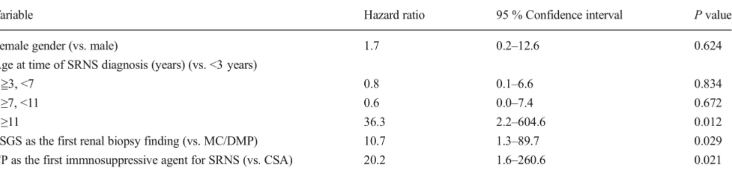
respectively (Fig.3). The risk of ESKD was assessed by mul- tivariate analysis, revealing that the initial immunosuppressant of cyclophosphamide [hazard rate (HR) 20.2; 95 % confi- d e n c e i n t e r v a l ( C I ) 1 . 6–2 6 0 . 6 ; P= 0 . 0 2 1 ] , i n i t i a l histopathogical pattern of FSGS (HR 10.7; 95 % CI 1.3–
89.7;P=0.029), and age of >11 years when diagnosed with SRNS (HR 36.3; 95 % CI 2.2–604.6;P=0.012) were signif- icant risk factors for progression to ESKD (Table3). After exclusion of patients with DMP from the analysis, the renal survival rate at 10 years was 100 % in both the MC
(cyclosporine) group and MC (cyclophosphamide) subgroup (not shown).
As calculated by Kaplan–Meier analysis, the permanent remission rate at 10 years in the FSGS (cyclosporine) group, FSGS (cyclophosphamide) group, MC/DMP (cyclosporine) group, and MC/DMP (cyclophosphamide) group was 10.0, 27.3, 24.1, and 23.1 %, respectively (Fig.4). Significant dif- ferences between groups were not found (P=0.747).
At the last follow-up examination, the disease was in re- mission in 57 (83 %) patients, was not in remission in three (4 %) patients, and had progressed to ESKD in nine (13 %; see Electronic Supplementary Material for the detailed clinical course of these 9 patients) patients (Fig.5). Of those patients who achieved remission at the last follow-up examination (n= 57), 48 (84 %) were in complete remission but only 18 (31 %) were in permanent remission.
Nephrotic syndrome- or treatment-related complications at the last examination are listed in Table 4. No patient died during the follow-up period. The median eGFR, excluding children with ESKD at the last examination, was 121.4 ml/
min/1.73 m2(81.0–180.8 ml/min/1.73 m2), that of FSGS group was 121.0 ml/min/1.73 m2 (82.5–180.8 ml/min/
1.73 m2), and that of the MC/DMP group was 123.0 ml/
min/1.73 m2(81.0–170.4 ml/min/1.73 m2).
In total, 55 patients underwent repeat renal biopsy, and six of these patients (10.9 %) exhibited significant cyclosporine- related nephrotoxicity in the last renal biopsy (FSGS group, 3/19; MC/DMP group, 3/36). Of these six patients, three showed tubulointerstitial fibrosis only, one showed arteriolar hyalinosis only, and two showed both arteriolar hyalinosis and tubulointerstitial fibrosis. The median duration from the date of starting of cyclosporine therapy to the date on which cyclo- sporine toxicity was found was 4.8 (range 2.0–5.8) years.
Discussion
In this retrospective cohort study, we found that children with SRNS treated with cyclosporine had better outcome than those treated with cyclophosphamide, particularly those with FSGS, in terms of both induction of complete remission and long-term renal survival. On the other hand, most of the pa- tients had been suffering frequent relapse for a long-term pe- riod, up to 10 years.
Among our study subjects, our analysis showed that the renal survival rates of those who received cyclosporine as the initial immunosuppressant were better than those of patients who received cyclophosphamide, regardless of the initial his- topathological patterns. Those who received cyclosporine as initial immunosuppressant showed both higher short-term re- mission rate and higher long-term renal survival rate, suggest- ing that early induction of complete remission by cyclosporine might lead to better renal survival. SRNS has been recognized Fig. 2 The complete remission rate of the initial SRNS episode, analyzed
b y K a p l a n - M e i e r a n a l y s i s a c c o r d i n g t o s u b g r o u p { i n i t i a l histopathological patterns [minimal change (MC)/diffuse mesangial proliferation (DMP) vs. focal segmental glomerulosclerosis (FSGS)]
and initial immunosuppressive agents used to treat for SRNS [cyclosporine (CSA) vs. cyclophosphamide (CP)]}. Number of children at risk at each time point is shownbelow the x-axis
Fig. 3 Renal survival rate in subgroups based on the initial histopathological patterns [minimal change (MC)/diffuse mesangial proliferation (DMP) vs. focal segmental glomerulosclerosis (FSGS)]
and initial immunosuppressive agents [cyclosporine (CSA) vs.
cyclophosphamide (CP)] used to treat SRNS. Number of children at risk at each time point is shownbelow the x-axis
as a disease associated with a high risk to progress to ESKD over the long term, although several recent studies have report- ed good short-term remission rate in patients with SRNS and, consequently, the effect of cyclosporine to induce remission in SRNS patients is beginning to be established [7,12–14]. How- ever, there have been only a few reports of children with SRNS in which the observation time was >10 years [15,16]. In our study, we assessed long-term renal survival rate in children with SRNS mainly with respect to the initial immunosuppres- sant and showed for the first time the higher effectiveness of cyclosporine.
Our results show that the immunosuppressant used to treat SRNS, the initial histopathological pattern, and the age of patient diagnosis of SRNS were significant predictive factors of renal survival. Children who received cyclosporine had a significantly higher renal survival, even after adjustment for the other factors. In previous studies, FSGS as the initial
histopathological pattern was found to be a predictive factor of progression to ESKD, particularly in those who could not attain remission [16–20]. In this regard, our results are com- patible with those reported previously. Moreover, our results demonstrate that those children with FSGS treated with oral cyclophosphamide had a much lower renal survival rate (Fig.3). Treatment regimens for patients with SRNS that in- clude oral cyclophosphamide have been tentatively advocated by the authors of a number of reports (including patients with FSGS) [16,21]; however, the results of our study suggest that the treatment regimen of oral cyclophosphamide can not be recommended—at least not to patients with SRNS with the FSGS histopathological pattern. Our institution has not adopted regimens with intravenous cyclophosphamide for the treatment of children with SRNS, but the results of other studies evaluating the efficacy of such therapeutic regimens are conflicting [12,22,23]. The older children in our study Table 3 Risk factor for end-stage kidney disease by multivariate analysis
Variable Hazard ratio 95 % Confidence interval Pvalue
Female gender (vs. male) 1.7 0.2–12.6 0.624
Age at time of SRNS diagnosis (years) (vs. <3 years)
≧3, <7 0.8 0.1–6.6 0.834
≥7, <11 0.6 0.0–7.4 0.672
≥11 36.3 2.2–604.6 0.012
FSGS as the first renal biopsy finding (vs. MC/DMP) 10.7 1.3–89.7 0.029
CP as the first immnosuppressive agent for SRNS (vs. CSA) 20.2 1.6–260.6 0.021
The data are presented as numbers unless stated otherwise
SRNS, Steroid-resistant nephritic syndrome; MC, minimal change; DMP, diffuse mesangial proliferation; FSGS, focal segmental glomerulosclerosis;
CP, cyclophosphamide; CSA, cyclosporine
Fig. 4 The permanent remission rate in subgroups based on the initial histopathological patterns [minimal change (MC)/diffuse mesangial proliferation (DMP) vs. focal segmental glomerulosclerosis (FSGS)]
and initial immunosuppressive agents [cyclosporine (CSA) vs.
cyclophosphamide (CP)] used to treat SRNS. Number of children at risk at each time point is shownbelow the x-axis
Fig. 5 Condition of patients at the last follow-up examination. Of 69 patients with SRNS, 57 patients had achieved remission by the last follow-up examination (right pie chart).SRNS, steroid-resistant nephritic syndrome;ESKD, end-stage kidney disease;PR, partial remission;FRNS, frequently relapsing nephrotic syndrome. The median duration of follow-up for patients with permanent remission was 10.3 (range, 5.2–15.9) years; with non-FRNS, 8.7 (5.5–12.5) years; with FRNS, 8.6 (4.9–15.8) years; with PR, 16.3 (4.5–19.3) years; with non- remission, 10.1 (6.3–16.4) years; with ESKD, 14.6 (9.2–19.0) years
cohort showed a significantly lower renal survival rate, which is a similar result to that reported previously [15]
On the other hand, the permanent remission rate among our study cohort was relatively low regardless of the initial immu- nosuppressant, and we found no evidence of any superiority of cyclosporine in achieving permanent remission. Moreover, many children in our study showed were in the state of FRNS at the last follow-up examination (Fig.5). These results sup- port those which we reported previously [24] and suggest that treatment strategies for children with SRNS to achieve long- term remission without immunosuppressant or steroid therapy still need to be improved.
Disease- and/or treatment-associated complications were relatively low among our study cohort and were comparable with those of the previous studies on long-term outcomes in patients with SSNS, except for hypertension [25,26]. Exclud- ing those who deteriorated to ESKD, only a few children showed mild kidney dysfunction. There were also only a few children who showed short stature or obesity, which might be attributable to the treatment, such as alternate-day administration of prednisolone. The rate of children who showed chronic cyclosporine nephrotoxicity in repeated biop- sy was 10.9 %, which is lower than that reported in previous studies [27–31], possibly due to the advantage of trough con- trol of cyclosporine. Close attention should be paid to cyclo- sporine nephrotoxicity. The rate of children who showed hy- pertension at last examination was relatively high, which might be attributed to the use of prednisolone or cyclosporine.
However, the frequency of hypertension in our children with SRNS and in children with SRNS in previously reported studies was higher than that of children with FRNS [24,32], which suggests that SRNS might itself be a significant risk factor for developing hypertension.
Our study has a number of strengths. First, a relatively large number of patients were included in the analysis, among whom 22 showed FSGS as the initial histopathological pat- tern. Second, we analyzed a rare refractory disease. Third, patient data for the analysis were collected over a relatively long term. These strengths should be balanced against this study’s limitations. First, there was a diversity with the patient study groups, with the analysis including patients with MC and those with DMP in the same group. Second, the study cohort consisted of a homogeneous population.
This study was confined to Japanese patients and did not include other races, such as Black Africans who typically show poorer long-term outcome. The homogeneity of our patient population might have contributed to the better outcome demonstrated in this study. Third, no genetic analyses were performed. Several recent papers have re- ported lack of efficacy of immunosuppression in SRNS secondary to genetic causes. We did not perform genetic analyses; however, none of our patients were indicated for this at the time of diagnosis. Fourth, the immunosuppres- sion treatment course (including methylprednisolone pulse therapy) was quite complex, making the analysis of any given variable impossible. Patients who used cyclosporine only as the first immunosuppressive agent for SRNS and those who used cyclosporine plus methylprednisolone pulse therapy were not analyzed separately; as such, it is very difficult to compare the results of this study with those having other protocols. Finally, our assessment of the side effects in this study relied mainly on medical records, and thus some side effects might not have been identified and the assessment might be insufficient.
In conclusion, our results suggest that the recently re- ported therapeutic regimen with cyclosporine could Table 4 Nephrotic syndrome or
treatment-related complications at the last examination
Variable Total Initial renal biopsy finding
FSGS MC/DMP
Number of patients 69 22 47
Short stature 5 (7.2) 1 (4.5) 4 (8.5)
Excessive body weighta 9 (13.0) 3 (13.6) 6 (12.8)
Obesityb 4 (5.8) 1 (4.5) 3 (6.4)
Hypertension 22 (31.9) 11 (50.0) 11 (23.4)
Number of patients with ESKD 9 7 2
Hypertension among patients with ESKD 5 (55.6) 3 (42.8) 2 (100)
Number of patients undergoing repeat renal biopsy 55 19 36
CSA-related nephrotoxicity 6 (10.9) 3 (15.8) 3 (8.3)
The data are presented as numbers with the percentage in parenthesis
ESKD, end-stage kidney disease; FSGS, focal segmental glomerulosclerosis; MC, minimal change; DMP, dif- fuse mesangial proliferation; CSA, cyclosporine
aDefined as the body mass index (BMI): weight (kg)/square of height (m2) of >25 in men and >24 in women
bDefined as a BMI of >30
considerably improve both the initial remission rate and the long-term renal survival rate of children with idiopath- ic SRNS, regardless of initial histopathological pattern (i.e., FSGS or MC/DMP). However, further studies are required to resolve remaining problems, such as the man- agement of SRNS in children who show frequent relapse after SRNS remission, and the establishment of a new therapeutic regimen for those who show resistance to the existing therapeutic regimens.
Acknowledgments The results presented in this paper have not been published previously in whole or part. The authors would like to thank Drs. Kentaro Ogata (Tokyo), Ryugo Hiramoto (Chiba), Takeshi Matsu- yama (Tokyo), Hitoshi Wakaki (Tokyo), and Kaori Kikunaga (Tokyo) for their contributions to this study.
Financial declaration Kenji Ishikura has received lecture fees from Novartis Pharma and Asahi Kasei Pharma. Yuko Hamasaki has received research grants from Novartis Pharma and lecture fees from Novartis Pharma, Astellas Pharma, and Pfizer Japan. Hiroshi Hataya has received lecture fees from Asahi Kasei Pharma, Astellas Pharma, Baxter, JMS, and Meiji Seika Pharma Co. Ltd. Masataka Honda has received lecture fees from Novartis Pharma, Takeda Pharmaceutical Co. Ltd., Chugai Pharma- ceutical Co. Ltd., Japan Blood Products Organization, JCR Pharmaceu- ticals Co. Ltd., and Asahi Kasei Pharma. Masaaki Mori has received lecture fees from Astellas Pharma, Pfizer Japan, Asahi Kasei Pharma, Meiji Seika Pharma Co. Ltd., and Chugai Pharmaceutical Co. Ltd.
Ethical approval The study was conducted in accordance with the ethical principles set out in the Declaration of Hesinki, and with the ethical guideline for epidemiological studies issued by the Minstry of Health, Labor and Welfare in Japan. The study was approved by the ethics committee of Tokyo Metropolitan Children's medical Center (H25-2).
Informed Consent Because data were reported retrospectively based on patient charts, informed consent was not obtained, in accordance with the above guidelines.
References
1. Mendoza SA, Reznik VM, Griswold WR, Krensky AM, Yorgin PD, Tune BM (1990) Treatment of steroid-resistant focal segmental glomerulosclerosis with pulse methylprednisolone and alkylating agents. Pediatr Nephrol 4:303–307
2. Cattran DC, Rao P (1998) Long-term outcome in children and adults with classic focal segmental glomerulosclerosis. Am J Kidney Dis 32:72–79
3. Tune BM, Kirpekar R, Sibley RK, Reznik VM, Griswold WR, Mendoza SA (1995) Intravenous methylprednisolone and oral alkylating agent therapy of prednisone-resistant pediatric focal seg- mental glomerulosclerosis: a long-term follow-up. Clin Nephrol 43:
84–88
4. Garin EH, Orak JK, Hiott KL, Sutherland SE (1988) Cyclosporine therapy for steroid-resistant nephrotic syndrome. A controlled study. Am J Dis Child 142:985–988
5. Ponticelli C, Rizzoni G, Edefonti A, Altieri P, Rivolta E, Rinaldi S, Ghio L, Lusvarghi E, Gusmano R, Locatelli F, Pasquali S, Castellani A, Casa-Alberighi OD (1993) A randomized trial of cyclosporine in steroid-resistant idiopathic nephrotic syndrome.
Kidney Int 43:1377–1384
6. Lieberman KV, Tejani A (1996) A randomized double-blind place- bo-controlled trial of cyclosporine in steroid-resistant idiopathic focal segmental glomerulosclerosis in children. J Am Soc Nephrol 7:56–63
7. Hamasaki Y, Yoshikawa N, Hattori S, Sasaki S, Iijima K, Nakanishi K, Matsuyama T, Ishikura K, Yata N, Kaneko T, Honda M (2009) Cyclosporine and steroid therapy in children with steroid-resistant nephrotic syndrome. Pediatr Nephrol 24:2177–2185
8. Ishikura K, Matsumoto S, Sako M, Tsuruga K, Nakanishi K, Kamei K, Saito H, Fujinaga S, Hamasaki Y, Chikamoto H, Ohtsuka Y, Komatsu Y, Ohta T, Nagai T, Kaito H, Kondo S, Ikezumi Y, Tanaka S, Kaku Y, Iijima K (2015) Clinical practice guideline for pediatric idiopathic nephrotic syndrome 2013: medical therapy.
Clin Exp Nephrol 19:6–33
9. Schwartz GJ, Haycock GB, Edelmann CM Jr, Spitzer A (1976) A simple estimate of glomerular filtration rate in children derived from body length and plasma creatinine. Pediatrics 58:259–263 10. Matsuo S, Imai E, Horio M, Yasuda Y, Tomita K, Nitta K,
Yamagata K, Tomino Y, Yokoyama H, Hishida A (2009) Revised equations for estimated GFR from serum creatinine in Japan. Am J Kidney Dis 53:982–992
11. Mori K, Honda M, Ikeda M (2004) Efficacy of methylprednisolone pulse therapy in steroid-resistant nephrotic syndrome. Pediatr Nephrol 19:1232–1236
12. Plank C, Kalb V, Hinkes B, Hildebrandt F, Gefeller O, Rascher W (2008) Cyclosporin A is superior to cyclophosphamide in children with steroid-resistant nephrotic syndrome-a randomized controlled multicentre trial by the Arbeitsgemeinschaft fur Padiatrische Nephrologie. Pediatr Nephrol 23:1483–1493
13. Ehrich JH, Geerlings C, Zivicnjak M, Franke D, Geerlings H, Gellermann J (2007) Steroid-resistant idiopathic childhood nephro- sis: overdiagnosed and undertreated. Nephrol Dial Transplant 22:
2183–2193
14. Choudhry S, Bagga A, Hari P, Sharma S, Kalaivani M, Dinda A (2009) Efficacy and safety of tacrolimus versus cyclosporine in children with steroid-resistant nephrotic syndrome: a randomized controlled trial. Am J Kidney Dis 53:760–769
15. Mekahli D, Liutkus A, Ranchin B, Yu A, Bessenay L, Girardin E, Van Damme-Lombaerts R, Palcoux JB, Cachat F, Lavocat MP, Bourdat-Michel G, Nobili F, Cochat P (2009) Long-term outcome of idiopathic steroid-resistant nephrotic syndrome: a multicenter study. Pediatr Nephrol 24:1525–1532
16. Kirpekar R, Yorgin PD, Tune BM, Kim MK, Sibley RK (2002) Clinicopathologic correlates predict the outcome in children with steroid-resistant idiopathic nephrotic syndrome treated with pulse methylprednisolone therapy. Am J Kidney Dis 39:1143–1152 17. Abrantes MM, Cardoso LS, Lima EM, Penido Silva JM, Diniz JS,
Bambirra EA, Oliveira EA (2006) Predictive factors of chronic kidney disease in primary focal segmental glomerulosclerosis.
Pediatr Nephrol 21:1003–1012
18. Paik KH, Lee BH, Cho HY, Kang HG, Ha IS, Cheong HI, Jin DK, Moon KC, Choi Y (2007) Primary focal segmental glomerular sclerosis in children: clinical course and prognosis. Pediatr Nephrol 22:389–395
19. Gipson DS, Chin H, Presler TP, Jennette C, Ferris ME, Massengill S, Gibson K, Thomas DB (2006) Differential risk of remission and ESRD in childhood FSGS. Pediatr Nephrol 21:344–349
20. Martinelli R, Okumura AS, Pereira LJ, Rocha H (2001) Primary focal segmental glomerulosclerosis in children: prognostic factors.
Pediatr Nephrol 16:658–661
21. Mantan M, Sriram CS, Hari P, Dinda A, Bagga A (2008) Efficacy of intravenous pulse cyclophosphamide treatment versus combina- tion of intravenous dexamethasone and oral cyclophosphamide treatment in steroid-resistant nephrotic syndrome. Pediatr Nephrol 23:1495–1502
22. Hari P, Bagga A, Jindal N, Srivastava RN (2001) Treatment of focal glomerulosclerosis with pulse steroids and oral cyclophosphamide.
Pediatr Nephrol 16:901–905
23. Bajpai A, Bagga A, Hari P, Dinda A, Srivastava RN (2003) Intravenous cyclophosphamide in steroid-resistant nephrotic syn- drome. Pediatr Nephrol 18:351–356
24. Hamasaki Y, Yoshikawa N, Nakazato H, Sasaki S, Iijima K, Nakanishi K, Matsuyama T, Ishikura K, Ito S, Kaneko T, Honda M (2013) Prospective 5-year follow-up of cyclosporine treatment in children with steroid-resistant nephrosis. Pediatr Nephrol 28:
765–771
25. Fakhouri F, Bocquet N, Taupin P, Presne C, Gagnadoux MF, Landais P, Lesavre P, Chauveau D, Knebelmann B, Broyer M, Grunfeld JP, Niaudet P (2003) Steroid-sensitive nephrotic syn- drome: from childhood to adulthood. Am J Kidney Dis 41:550–557 26. Ruth EM, Kemper MJ, Leumann EP, Laube GF, Neuhaus TJ (2005) Children with steroid-sensitive nephrotic syndrome come of age:
long-term outcome. J Pediatr 147:202–207
27. Niaudet P, Fuchshuber A, Gagnadoux MF, Habib R, Broyer M (1997) Cyclosporine in the therapy of steroid-resistant idiopathic nephrotic syndrome. Kidney Int Suppl 58:S85–S90
28. Iijima K, Hamahira K, Tanaka R, Kobayashi A, Nozu K, N a k a m u r a H , Yo s h i k a w a N ( 2 0 0 2 ) R i s k f a c t o r f o r cyclosporine-induced tubulointerstitial lesions in children with minimal change nephrotic syndrome. Kidney Int 61:
1801–1805
29. Kengne-Wafo S, Massella L, Diomedi-Camassei F, Gianviti A, Vivarelli M, Greco M, Stringini GR, Emma F (2009) Risk factors for cyclosporin A nephrotoxicity in children with steroid-dependent nephrotic syndrome. Clin J Am Soc Nephrol 4:1409–1416 30. Sinha A, Sharma A, Mehta A, Gupta R, Gulati A, Hari P, Dinda
AK, Bagga A (2013) Calcineurin inhibitor induced nephrotox- icity in steroid resistant nephrotic syndrome. Indian J Nephrol 23:41–46
31. Fujinaga S, Shimizu T (2013) Chronic cyclosporine-induced neph- rotoxicity in children with steroid-resistant nephrotic syndrome.
Pediatr Nephrol 28:2065–2066
32. Ishikura K, Yoshikawa N, Nakazato H, Sasaki S, Iijima K, Nakanishi K, Matsuyama T, Ito S, Yata N, Ando T, Honda M (2012) Two-year follow-up of a prospective clinical trial of cyclo- sporine for frequently relapsing nephrotic syndrome in children.
Clin J Am Soc Nephrol 7:1576–1583
O R I G I N A L A R T I C L E
Clinical and genetic characteristics of Japanese nephronophthisis patients
Keisuke Sugimoto1• Tomoki Miyazawa1•Takuji Enya1•Hitomi Nishi1• Kohei Miyazaki1• Mitsuru Okada1•Tsukasa Takemura1
Received: 30 August 2015 / Accepted: 4 October 2015 ÓJapanese Society of Nephrology 2015
Abstract
Background Nephronophthisis (NPH) accounts for 4–5 % of end-stage renal disease occurring in childhood.
Method We investigated the clinical context and char- acteristics of renal and extrarenal symptoms, as well as the NPHP genes, in 35 Japanese patients with clinical and histologic features suggesting NPH.
Results NPH occurred fairly uniformly throughout Japan irrespective of region or gender. In three families, NPH affected siblings. The median age of patients was 12.5 years. Renal abnormalities attributable to NPH dis- covered through mass screening, such as urine tests in school. However, NPH accounted for less than 50 % of children with abnormal findings, including incidentally discovered renal dysfunction during evaluation of extra- renal symptoms or during routine check-ups. Typical extrarenal manifestations leaded to discovery including anemia and delayed physical development. The urine often showed low gravity specific density and low molecular weight proteinuria. Frequent renal histologic findings included cystic dilation of tubules, mainly in the medulla, and irregularity of tubular basement membranes. Geneti- cally abnormalities of NPHP1 were not common, with large deletions frequently noted. Compound heterozygotes showing single abnormalities in each ofNPHP1,NPHP3, andNPHP4were observed.
Conclusions Our findings resemble those reported in Western populations.
Keywords End-stage renal diseaseRenal cystsNPHP genesChildrenRenal tubules
Introduction
Nephronophthisis (NPH) is a disease characterized by renal medullary cyst formation. Additional histologic findings include tubulointerstitial nephritis accompanied by pro- gressive sclerosis and hyaline glomeruli. Although NPH characteristically shows autosomal recessive inheritance, it may occur sporadically [1]. NPH accounts for approximately 4–5 % of end-stage renal disease (ESRD) in childhood.
Disease subtypes include: infantile NPH (NPH2), which progresses to ESRD around the age of 5 years; juvenile NPH (NPH1), which develops from early childhood to school age and usually progresses to ESRD by an age of about 13 or 14 years; and adolescent NPH (NPH3), with development of ESRD at an average age of 19 years. Juvenile NPH is reported to be the most common subtype [1].
NPHP1, the gene most often responsible for juvenile nephronophthisis, encodes the nephrocystin-1 molecule.
This gene has an extent of approximately 11 kbp, and is located on chromosome 2q12-13 [2]. The nephrocystin-1 protein consists of 677 amino acids and includes three coiled domains; two highly acidic negatively charged glutamic acid-rich domains; and an Src-homology 3 domain. Nephrocystin-1 has a molecular weight of 83 kD.
As this protein is located in the transition zone of primary cilia of renal tubular epithelial cells, its abnormalities typically cause dysfunction of these primary cilia (cil- iopathy) [1,2].
NPHP4, whose abnormalities cause a second form of NPH1, is located on chromosome 1p36 and encodes the nephrocystin-4 (nephroretinin) molecule. Nephrocystin-4
& Keisuke Sugimoto
1 Department of Pediatrics, Kinki University Faculty of Medicine, 377-2 Ohno-higashi, Osaka-Sayama 589-8511, Japan
DOI 10.1007/s10157-015-1180-5
has been shown to carry out signal transmission between renal tubular epithelial cells, in cooperation with nephro- cystin-1 [3].
NPHP2, the gene responsible for infantile NPH (NPH2), is located on 9q22-31 [4].NPHP2encodes a protein termed inversin (INVS). An abnormality in INVS can cause situs inversus, pancreatic islet-cell dysplasia, cardiovascular
abnormalities, and hepato-biliary disorders. In addition, INVS abnormalities can cause cyst formation resembling that in juvenile nephronophthisis. However, the renal prognosis is worse progression to ESRD in early childhood.
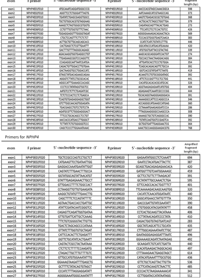
The gene responsible for adolescent NPH (NPH3), NPHP3is located on chromosome 3q21-22 [5].NPHP3is believed to encode a protein involved in signal Fig. 1 Genomic DNA extraction, PCR, and determination ofNPHP1, 2, 3 and 4gene sequence. PCR primers were prepared to amplify approximately 200–300 bp fragments based onNPHP 1–4gene sequences registered in GenBank, the following primers were used as shown
Fig. 1 continued
transmission in renal tubular epithelial cells, such as sig- naling involving diacylglycerol kinase-zeta and receptor- like tyrosine kinase. Abnormalities of the protein disrupt urinary concentrating ability and the structure of cilia of renal tubules, as in the other types of NPH.
Previous reports describe occurrence of NPHP1 muta- tions in approximately 30–50 % of juvenile nephronoph- thisis patients in Western countries [1, 6], where genetic analysis of NPHP1 is performed initially when juvenile NPH is suspected. If mutation is detected, kidney biopsy usually is deferred [7]. Genetic diagnosis is made less frequently in Japan; so kidney biopsy often is performed to obtain a definitive diagnosis. Not infrequently, NPH is discovered in the advanced or end stage in many Japanese patients, in whom treatment no longer can slow progres- sion. Unfortunately, symptoms typically seen in early stages are incompletely characterized.
In the present study, we investigated clinical, histologic, and genetic features in 35 Japanese patients clinically and histologically suspected to have NPH, aiming to promote early diagnosis. We studied many exons as many as 13 NPHPgenes. Since such genetic analysis involves signif- icant cost and time, we also screened biopsy specimens by immunohistologic methods employing antibodies against relevant peptides.
Methods
Patient registration and informed consent
Our subjects included 35 patients with clinicopathologic findings suggestive of NPH who were referred to our department from various regions of Japan. The study was performed following approval by the Ethics Committee of Kinki University Faculty of Medicine and acquisition of
written informed consent from patients or their parents (Actual state of Japanese juvenile nephronophthisis patients and identification of gene aberrations; approval number 20–99).
Genomic DNA extraction, polymerase-chain reaction (PCR), and determination ofNPHPgene sequence.
After approximately 5 mL of peripheral blood was col- lected from patients into tubes containing Na-EDTA, genomic DNA was extracted using NucleoSpin for Blood (TaKaRa Bio Inc, Shiga, Japan). Human genomic DNA (TaKaRa Clontech, code 636401; Shiga, Japan) was used as a control. Patient samples and control genomic DNA were diluted with sterile water to prepare 10 ng/lL solu- tions. PCR was performed using these as templates and TaKaRa PCR Thermal Cycler Dice Gradient (TaKaRa Bio Inc, Shiga, Japan). To determine extent of deletions and identify break points, PCR primers were prepared to amplify approximately 200–300 bp fragments based on NPHPgene sequences registered in GenBank (Fig.1). For PCR, annealing temperatures and times were 63°C and 15 s forNPHP1andNPHP3; 60°C and 15 s for NPHP2;
and 60°C and 20 s forNPHP4, respectively. For sequence analysis, PCR products were purified by an enzyme reac- tion, and templates for sequencing were prepared. The sequencing reaction was carried out using the prepared template DNA and a BigDye Terminator v.3.1 Cycle Sequencing Kit (Applied Biosystems, CA, USA), employing the dye terminator method. Reaction products were purified by gel filtration, and sequence analysis was performed using a capillary-type sequencer, ABI3730xl (Applied Biosystems, CA, USA). The algorithm estab- lished by Salomon et al. [8]. was adapted for use in our analytical procedure. In children with renal dysfunction Fig. 2 Percentage of NPH
patients withNPHPgene mutation.NPHPgene mutation was detected in 19 patients. No NPHPgene aberration detected within the sequences analyzed in the other 16 patients with suspicion of NPH
clinicopathologically

![Fig. 2 Histopathological findings of the kidney biopsy taken at day 37. (A) Almost all glomeruli had evidence of global sclerosis or were collapsed (periodic acid Schiff stain [PAS]; × 100)](https://thumb-ap.123doks.com/thumbv2/123deta/7455976.2477288/35.891.85.837.110.493/histopathological-findings-glomeruli-evidence-sclerosis-collapsed-periodic-schiff.webp)