A Novel Treatment for Arrhythmias via the Control of the Degradation of Ion
Channel Proteins
Junichiro Miake
Division of Pharmacology, Department of Pathophysiological and Therapeutic Science, School of Medicine, Faculty of Medicine, Tottori University, Yonago 683-8503, Japan
ABSTRACT
Although there are many reports on the regulation of ion channel expression in transcription and translation, few drugs have been studied to influence post-translational modification of ion channel proteins. The Kv1.5 channel is a potassium ion channel expressed in atrial muscle, belongs to the voltage-gated K+ channel superfamily, and forms an ultrarapid delayed rectifier potassium ion current. It is important to understand the fate of these channel proteins, as cardiac Kv1.5 mutations can cause arrhythmias. Disruption of quantitative and qualitative control mechanisms of channels leads to stagnation and degradation of intracellular channel proteins. As a result, ion channel proteins are not transported to the cell membrane and are involved in the development of atrial fibrillation. This review takes the Kv1.5 channel as an example and focuses on the degradation mechanism of ion channel proteins, and discusses its application to the treatment of arrhythmia by drugs that control the mechanism of ion channel protein degradation.
Key words arrhythmia; chaperon; degradation; ion channel; trafficking
Ion channels are membrane proteins responsible for the formation of resting membrane potential and action potential of the myocardium. The mRNA transcribed from the nuclear DNA is spliced when passing through the nuclear pore and translocating to the cytoplasm, and then an ion channel protein is formed on the ribo-some as a protein having a primary structure. The ion channel protein having a primary structure undergoes post-translational modification such as folding and
sugar chain modification via the endoplasmic reticu-lum and Golgi apparatus, and is then transported to the cell membrane via microtubules. In addition, the transported ion channel proteins function by binding to anchor proteins present on the membrane.1, 2 To date, there have been many reports on the regulation of ion channel expression in transcription and translation, but few studies on drugs that affect post-translational modification of ion channel proteins. The Kv1.5 channel is a potassium ion channel expressed in atrial muscle, belongs to the voltage-gated K+ channel superfamily and forms ultrarapid delayed rectifier K+ currents (IK
ur) in the myocardium.3 Since mutations in the cardiac Kv1.5 can cause arrhythmias,4 it is important from a cell biol-ogy perspective to understand the fate of these channel proteins. Disruption of the quantitative and qualitative control mechanisms of the channel due to innate and acquired factors leads to stagnation and degradation of the channel protein in the cell. As a result, ion channel proteins are not transported to the cell membrane and are involved in the development of atrial fibrillation.5 This review takes the Kv1.5 channel as an example and focuses on the degradation mechanism of ion channel proteins, and describes its application to the treatment of arrhythmias by drugs that control the mechanism of ion channel protein degradation.
MOLECULAR MECHANISM THAT REGULATES THE DEGRADATION RATE OF ION CHANNEL PROTEINS
As previously mentioned, the quantity and quality of proteins is determined by transcription and translational synthesis, and degradation by lysosomes and protea-somes. In the process of post-translational modification of cell membrane proteins, proteins translated by cytoplasmic ribosomes are first transported to the endoplasmic reticulum based on mRNA transcribed in the nucleus. After cleavage of the signal sequence and transfer of the core sugar chain, they undergo quality control by molecular chaperones, followed by complete sugar chain modification, and are transported to the cell membrane via the Golgi apparatus (Fig. 1).1, 2
The channel protein functions after being trans-ported to the cell membrane following post-translational
Review Article: Special Contribution Yonago Acta Medica 2020;63(3):146–153 doi: 10.33160/yam.2020.08.002
Corresponding author: Junichiro Miake, MD, PhD jmiake@tottori-u.ac.jp
Received 2020 May 25 Accepted 2020 June 16 Online published 2020 July 13
Abbreviations: CHIP, carboxyl-terminus of Hsc70 interacting pro-tein; EPA, eicosapentaenoic acid; 4-AP, 4-aminopyridine; GGA, geranylgeranylacetone; HSC, heat shock cognate; HSF, heat shock factor; HSP, heat shock protein; IKur, ultrarapid delayed rectifier K+ current; PDZ, PSD95/Dlg/ZO-1; SAP97, synapse-associated protein 97; siRNA, small interfering RNA; TPR, tetratricopeptide repeat; UPS, ubiquitin-proteasome system
modification, bound to the anchor protein existing in the cell membrane, and localized in the cell membrane. In these processes, if the protein undergoes complete fold-ing and sugar chain modification, the protein becomes stable, escapes from the protein degradation system described below and reaches the cell membrane, and the amount of ion channel protein increases. On the other hand, some of the proteins recognized as incomplete in the endoplasmic reticulum are retrogradely transported from the endoplasmic reticulum to the cytoplasm, ubiquitinated, and then degraded by the proteasome (ubiquitin-proteasome system, UPS). In addition, some defective proteins are transferred from the Golgi or cell membrane to the lysosomal/endosome system and de-graded. When these degradative systems are activated, the ion channel protein cannot reach the cell membrane and the ion channel activity is reduced. Target proteins that are degraded by UPS are ubiquitin-activating E1
enzyme, ubiquitin-conjugating E2 enzyme, and ubiq-uitin E3 ligase, which are ubiqubiq-uitin molecules that are covalently linked to their lysine residues. The ubiquiti-nated proteins are degraded by the threonine protease 26S proteasome.6, 7 Increased expression of channel proteins at cell membranes due to slower protein deg-radation is referred to as protein stabilization, and as a result of protein stabilization, expression of channel proteins at cell membranes is increased (Fig. 1).
The degradation of channel proteins is defined by (1) degradation by proteasome, (2) ubiquitination in cy-toplasm, and (3) folding and sugar chain modification in endoplasmic reticulum (Fig. 1). Therefore, degradation of Kv1.5 channel proteins is delayed by the proteasome inhibitor, and as a result, expression of Kv1.5 channel proteins on the membrane is stabilized (Fig. 1). The deg-radation of channel proteins and stabilization of channel protein expression in the membrane could be achieved
Fig. 1. Overall view of the anterograde trafficking of the normal Kv1.5 proteins. The degradation of channel proteins is defined by (1)
ubiquitination, (2) degradation by proteasome in cytoplasm, and (3) folding and sugar chain modification in endoplasmic reticulum.
The degradation of channel proteins and stabilization of channel protein expression in the membrane could be achieved by one of the following methods: promotion of protein folding and glycosylation at endoplasmic reticulum level; control of protein ubiquitination at cytoplasmic level; inhibition of protein degradation in the proteasome at cytoplasmic level.
J. Miake by one of the following methods: promotion of protein folding and glycosylation (control at endoplasmic reticu-lum level); control of protein ubiquitination (control at cytoplasmic level); inhibition of protein degradation in the proteasome (control of final degradation apparatus). INHIBITION OF PROTEASOME AND STABILI-ZATION OF KV1.5 CHANNEL
Kv1.5 channels are degraded by UPS and classified into proteins with short half-lives. Our in vitro studies using cultured cells and forced expression of the Kv1.5 chan-nel gene showed a half-life of Kv1.5 chanchan-nel protein of 9.2 hours.8 Pretreatment of MG132, a proteasome inhibitor, extended the half-life of Kv1.5 channel. Furthermore, the expression of Kv1.5 channel proteins was stabilized at the membrane with an increase in ubiquitination after treatment with MG132.
Then, inhibition of proteasome increases the ex-pression of Kv1.5 channel protein, but how does it affect the intracellular trafficking of Kv1.5? To clarify this, we investigated the intracellular localization of Kv1.5. As a result, the localization of Kv1.5 channel proteins was consistent with the distribution of endoplasmic reticulum (reticulated distribution in the cytoplasm) and Golgi apparatus (distribution around the nucleus).8 We observed that proteasome inhibition increased the localization of Kv1.5 channel proteins in both the endo-plasmic reticulum and the Golgi apparatus.8
In electrophysiological studies, proteasome inhibi-tion significantly increased 4-AP (4-aminopyridine) -sensitive IKur (ultrarapid delayed rectifier K+ current) compared to controls. This increase in IKur due to proteasome inhibition was abolished by brefeldine A, a selective inhibitor of endoplasmic reticulum-to-Golgi protein transport, and colchicine, a microtubule-blocking drug.8 From the above, it was shown that when the proteasome system was suppressed, the Kv1.5 chan-nel protein increased its expression at the cell membrane through the endoplasmic reticulum-Golgi apparatus, and thereby its channel activity increased (Fig. 1). KV1.5 CHANNEL PROTEIN STABILIZATION VIA PROTEASOME INHIBITION BY NA+ CHAN-NEL INHIBITORS
Proteasome is a huge protein with a molecular weight of 26S consisting of 19S of regulatory subunit and 20S subunit having an active center. The 20S proteasome of eukaryotic cells has three types of protease activities: caspase-like activity (cleaving acidic amino acids), trypsin-like activity (cleaving basic amino acids), and chymotrypsin-like activity (cleaving hydrophobic or aromatic amino acids).9 These three types of activities
are localized in β1, β2, and β5 subunits, respectively. The mammalian β5 subunit shares 67% homology with yeast and has chymotrypsin-like activity.10 Structural biology has shown that classical proteasome inhibitors such as MG132 inhibit their activity by covalently binding to the chymotrypsin-like active site of the 20S proteasome β subunit.10
Recently, we reported that aprindine, a Na+ channel inhibitor belonging to class 1b, stabilizes Kir6.2 proteins that are degraded in proteasome, and its mechanism of action may be the inhibitory effect of 20S proteasome
Since Na+ channel blockers such as pilsicainide and lidocaine have an aromatic ring and an amide bond, and this structure is similar to that of proteasome inhibi-tors, it was hypothesized that these drugs may have an inhibitory effect on the proteasome. According to our hypothesis, the administration of pilsicainide markedly extended the half-life of Kv1.5 channel proteins and significantly increased its expression.8 Administration of pilsicainide significantly increased ubiquitination of Kv1.5, and markedly increased intracellular localization of Kv1.5 channel proteins in Golgi and endoplasmic reticulum. Furthermore, administration of pilsicainide significantly increased IKur current in COS7 cells expressing Kv1.5 genes. Next, the effect of ion channel inhibitors on 20S proteasome activity was measured.8 As a result, the Na+ channel inhibitors pilsicainide and lidocaine dose-dependently suppressed 20S proteasome activity, and their IC50 were 1.2 μM and 75 μM, respec-tively; on the other hand, K+ channel inhibitors and Ca2+ channel antagonists did not suppress their activity.
Next, we hypothesized that the difference in IC50 between pilsicainide and lidocaine was due to the difference in proteasome inhibitory action mode. The structure of bovine 20S proteasome is known at the crystallographic level. Therefore, for the purpose of ver-ifying this hypothesis, we examined the binding mode of pilsicainide and lidocaine to the active site of bovine proteasome using the docking test.11–13 Interestingly, it was revealed that the binding sites of pilsicainide and lidocaine are different from the nucleophilic reaction site of β5 subunit, Thr1, (Fig. 2). That is, in general, a binding site of proteasome inhibitor is an S1 site containing Thr1 which is a nucleophilic reaction site of β5 subunit.11–13 However, in the results of model experi-ments, the binding site of pilsicainide and lidocaine was an S1’ site near the S1 site, but not the S1 site.14 It was suggested that both drugs stably occupy the S1’ site by binding to the S1’ site and further by hydrogen-bonding the amide groups of pilsicainide and lidocaine between Gly129 and Tyr113 of the proteasome.14 Since this S1’ site is the binding site of the target protein that is cleaved
by the chymotrypsin-like activity, it was thought that inhibition of this binding site would suppress the degra-dation of the protein and stabilize the expression. From the structure of this S1’ site, it was considered possible to explain the difference in the strength of the protea-some inhibitory action of pilsicainide and lidocaine (Fig. 2). That is, in pilsicainide, its pyrrolidine ring can sufficiently fit in the recess before the S1’ site, while li-docaine does not have a pyrrolidine group so that the fit in the recess in the S1’ site is insufficient (Fig. 2). Based on this difference, the potency of proteasome inhibi-tion of both drugs was considered to be different. This inhibition of proteasome activity and the stabilizing effect of Kv1.5 channel proteins were also confirmed by other Na+ channel inhibitors mexiletine and flecainide (unpublished data). The inhibitory effect of Na+ channel inhibitors on proteasome was considered to be a class effect (Fig. 2). Moreover, since it was suggested that the S1’ site could be a target site for the development of pro-teasome inhibitors, future research in the development of new proteasome inhibitors is expected.
STABILIZATION OF KV1.5 CHANNEL PROTEIN BY MOLECULAR CHAPERONE
We searched for a protein that binds to Kv1.5 and found that one of the heat shock proteins (HSP), HSP70, binds to Kv1.5. The cytoplasmic molecular chaperones HSP70, HSC70 (heat shock cognate 70), and HSP90 play important roles in protein folding and degradation of proteins with abnormal folding.15, 16 In fact, overexpres-sion of the HSP70 gene significantly increased the half-life of Kv1.5 channel proteins degradation and increased protein expression at the cell membrane without altering the level of ubiquitinated Kv1.5 channel proteins.17 The results of co-immunoprecipitation experiments showed that Kv1.5 interacts with HSP70. Furthermore, the re-sults of immunostaining experiments on cells revealed that Kv1.5 and HSP70 colocalized with the endoplasmic reticulum and the Golgi apparatus. And, overexpres-sion of HSP70 gene markedly enhanced Kv1.5 channel proteins expression in the endoplasmic reticulum, Golgi apparatus and cell membrane from the results of immunostaining experiments, and the results of patch-clamp experiments revealed that Kv1.5 currents were significantly increased in the test pulse range of +20 mV to +80 mV. The increase in Kv1.5 currents by HSP70
Fig. 2. Molecular mechanism of proteasome inhibition by Na+ channel inhibitors and Kv1.5 protein stabilization. Pilsicainide occupies the peptide binding site and exerts a proteasome inhibitory effect, which is different from the chymotrypsin site where MG132 acts. 10 μM lidocaine and 10 μM pilsicainide inhibited proteasome activity by 20% and 50%, respectively. Pilsicainide suppresses proteasome activity below clinical use levels by occupying the S1’ pocket of the proteasome (Kd = 1.99 mM).
J. Miake
was abolished by brefeldin A and colchicine which are protein transport inhibitors. In addition, from the cell fraction extraction experiment, the expression level of Kv1.5 channel proteins in the endoplasmic reticulum fraction was increased by forced expression of the HSP70 gene. In summary, it was concluded that the molecular chaperone HSP70 acts on the endoplasmic reticulum to stabilize the structure of the Kv1.5 channel proteins (Fig. 3).17
STABILAIZION OF KV1.5 CHANNEL PROTEIN BY CONTROLLING UBIQUTIN LIGASE
Kv1.5 channel proteins are ubiquitinated and degraded by endoplasmic reticulum-related UPS, but the specific E3 ligase that ubiquitinates Kv1.5 channel proteins is unknown at present. The carboxyl-terminus of HSC70 interacting protein (CHIP) was found as a co-chaperone that inhibits folding by HSP70, but was later shown to be an ubiquitin ligase dependent on cytoplasmic chaper-one. CHIP has three N-terminal tetratricopeptide repeat
(TPR) domains, which bind to the C-terminus of HSP70 or HSP90, and a Ubox domain required for interaction with ubiquitin-conjugating E2 enzyme.18, 19 Therefore, we examined the possibility that CHIP acts as an E3 ligase of Kv1.5.18, 19 When the wild-type CHIP gene was introduced, the degradation of Kv1.5 was promoted in a CHIP expression-dependent manner. On the other hand, the amount of Kv1.5 did not change when the Ubox domain of CHIP, which is the binding site of ubiquitin conjugating E2 enzyme, or the TPR domain, which is the binding site of CHIP to HSP70, was deleted. Furthermore, to confirm that CHIP regulates the degra-dation of Kv1.5 channel proteins, we created inhibitory small interfering RNA (siRNA) against CHIP and introduced this into cells to suppress the endogenous CHIP in COS7 cells. As a result, Kv1.5 channel proteins increased depending on the decrease of CHIP expres-sion. These results indicated that CHIP acts as an E3 ligase for UPS degradation of Kv1.5 channel proteins. Taken together, it was considered that the binding of
Fig. 3. The anterograde trafficking of the normal Kv1.5 proteins, and the degradation of misfolded Kv1.5 channel proteins dependent on
an E3 ligase, CHIP. Left: Normal Kv1.5 proteins traffic to the membrane. Right: the misfolded Kv1.5 channel proteins undergo degrada-tion by proteasome after poly-ubiquitinadegrada-tion by an E3 ligase (CHIP) and an E2 ligase. Mutant CHIP proteins (E2 binding site defect, CHIPΔUbox or HSP70 binding site defect, CHIPΔTPR) fail to poly-ubiquitination of the Kv1.5 channel proteins. Novel treatments for arrhythmias suggested in the text are depicted.
Kv1.5 and HSP70 stabilized the protein structure, CHIP could not ubiquitinate Kv1.5 channel proteins, and Kv1.5 channel proteins expression was increased (Fig. 3). STABILIZATION OF KV1.5 CHANNEL PRO-TEINS BY A CHEMICAL CHAPERONE IN THE ENDOPLASMIC RETICULUM
It has been suggested that chronic administration of eicosapentaenoic acid (EPA), an n-3 polyunsaturated fatty acid, may influence the biosynthesis and intracel-lular transport of Kv1.5 channels.20 We hypothesized that the antiarrhythmic effects of EPA are explained by modifications of myocardial ion channels. To prove the hypothesis, the effects of n-3 polyunsaturated fatty acids EPA and docosahexaenoic acid on Kv1.5 channel protein and channel activity were examined.20 As a result, chronic administration of EPA dose-dependently increased Kv1.5 channel proteins in the range of 0.1 to 10 μM. From the study of intracellular distribution of Kv1.5 channel proteins, Kv1.5 channel proteins expres-sion was markedly increased in endoplasmic reticulum, Golgi apparatus and cell membrane under chronic administration of 1 μM EPA. Consistent with this result, the Kv1.5 channel current IKur was also significantly increased. Examination of the mechanism of Kv1.5 channel increasing action by this low concentration EPA revealed that EPA significantly extended the half-life of Kv1.5 channel proteins. In addition, pretreatment with protein transport inhibitors abolished the effect of EPA on increasing Kv1.5 currents, suggesting that EPA acts mainly on Kv1.5 channel proteins localized on the endoplasmic reticulum membrane. Interestingly, acute treatment with 1 mM 4-AP increased Kv1.5 channel proteins, whereas 12-hour treatment with 4-AP abol-ished the EPA-induced increase in Kv1.5 proteins. These facts suggest that EPA acts as a chemical chaperone by altering the structure of Kv1.5 channel proteins in the endoplasmic reticulum and stabilizing it by acting on the 4-AP binding site of Kv1.5 channel proteins (Fig. 3). STABILAIZAION OF KV1.5 CHANNELS PRO-TEINS BY ION CHANNERL ANCHOR PTOTEIN Propagation of electrical excitation in myocardial tissue requires the formation of action potentials of cardio-myocytes, and expression of voltage-gated K+ channels in the cell membrane is important for the formation of action potentials. The anchor protein containing the PSD95/Dlg/ZO-1 (PDZ) domain plays an important role in the organization and intracellular localization of voltage-gated K+ channels in the plasma membrane.21–24 Synapse-associated protein 97 (SAP97) is an anchor protein that belongs to the membrane-associated
guanylate kinase protein family that has a PDZ domain, and is an anchor protein that colocalizes with Kv1.5 at the intercalated disc of atrial and ventricular muscles (Fig. 1).25, 26 SAP97 stabilizes the Kv1.5 channel pro-teins by binding via the amino acid on the carboxyl-terminal side of the Kv1.5 channel proteins. From the results of immunostaining and immunoprecipitation experiments using mouse atrial myocytes, HL-1 cells, it was confirmed that SAP97 and Kv1.5 co-localize.26 Furthermore, it was observed that overexpression of the SAP97 gene increased the expression level of Kv1.5 channel proteins. From these results, it was speculated that the Kv1.5 channel proteins and the anchor protein SAP97 were post-translationally modified and stabilized on the endoplasmic reticulum and that SAP97 and Kv1.5 interact with each other to stabilize the proteins (Fig. 1). Recently, we revealed that geranylgeranylacetone (GGA) that induces heat shock factor (HSF) -1 and HSP70 di-rectly stabilize the Kv1.5 channel proteins by increasing the transcriptional activity of SAP97.26 From the results of the reporter assay, it was confirmed that the HSF-1 binding domain was present in the transcriptional regulatory region of the SAP97 gene. From the above, it is considered that regulation of SAP97 protein expres-sion by heat shock proteins may be applicable as a novel arrhythmia treatment via a mechanism that regulates expression in an intervening version of Kv1.5 channel proteins (Fig. 3).25–27
CONCLUSION
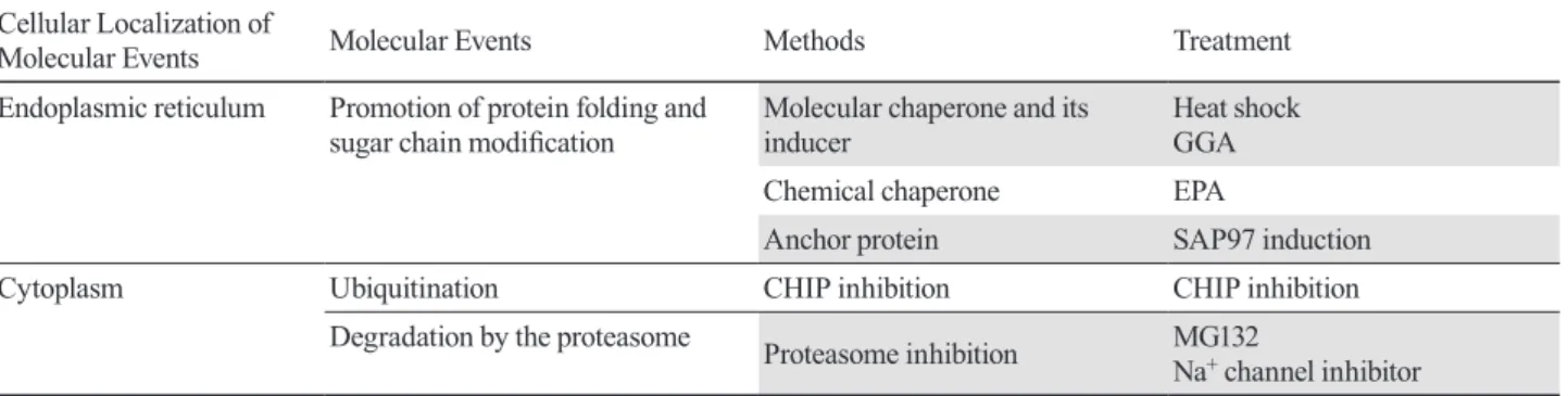
In this paper, I reviewed the possibilities of a novel treat-ment for arrhythmias via the control of the degradation of ion channel proteins (Fig. 3). In order to stabilize the expression of ion channel proteins on the cell membrane and maintain channel activity by controlling protein degradation, several strategies such as those discussed in this review can be considered (Table 1): Proteasome inhibition (e.g., MG132 and Na+ channel inhibitor); Delayed degradation by suppressing ubiquitination (e.g., CHIP inhibition); Acceleration of protein folding and sugar chain modification in the endoplasmic reticulum (e.g., chemical or molecular chaperones such as EPA and SAP97). Further development of research in this field is expected in the future.
Acknowledgments: I would like to thank Ichiro Hisatome, Haruaki Ninomiya, Udin Bahrudin, Hiroshi Hirota, Toshiya Koshida, Peili Li, Yakuang Ting, Yasutaka Yamamoto, Yasuaki Shirayoshi, Masaru Kato, Hiroaki Tanaka, Kazuhiko Iizuka, Yasutaka Kawada, Yasutaka Kurata, Tatsuo Matsuoka and Akira Nakai for their cooperation.
J. Miake
REFERENCES
1 Hurtley SM, Bole DG, Hoover-Litty H, Helenius A, Copeland CS. Interactions of misfolded influenza virus hemagglutinin with binding protein (BiP). J Cell Biol. 1989;108:2117-26.
DOI: 10.1083/jcb.108.6.2117, PMID: 2738090
2 Hammond C, Helenius A. Quality control in the secre-tory pathway. Curr Opin Cell Biol. 1995;7:523-9. DOI: 10.1016/0955-0674(95)80009-3, PMID: 7495572
3 Nattel S, Yue L, Wang Z. Cardiac ultrarapid delayed rectifi-ers: a novel potassium current family o f functional similarity and molecular diversity. Cell Physiol Biochem. 1999;9:217-26.
DOI: 10.1159/000016318, PMID: 10575199
4 Snyders J, Knoth KM, Roberds SL, Tamkun MM. Time-, voltage-Time-, and state-dependent block by quinidine of a cloned human cardiac potassium channel. Mol Pharmacol. 1992;41:322-30. PMID: 1538710
5 Brundel B, Ausma J, van Gelder IC, Van der Want JJ, van Gilst WH, Crijns HJ, et al. Activation of proteolysis by calpains and structural changes in human paroxysmal and persistent atrial fibrillation. Cardiovasc Res. 2002;54:380-9.
DOI: 10.1016/S0008-6363(02)00289-4, PMID: 12062342
6 Ciechanover A. The ubiquitin-proteasome proteolytic path-way. Cell. 1994;79:13-21. DOI: 10.1016/0092-8674(94)90396-4, PMID: 7923371
7 Jentsch S, Schlenker S. Selective protein degradation: A journey’s end within the proteasome. Cell. 1995;82:881-4.
DOI: 10.1016/0092-8674(95)90021-7, PMID: 7553848
8 Kato M, Ogura K, Miake J, Sasaki N, Taniguchi S, Igawa O, et al. Evidence for proteasomal degradation of Kv1.5 channel protein. Biochem Biophys Res Commun. 2005;337:343-8.
DOI: 10.1016/j.bbrc.2005.09.053, PMID: 16185660
9 Groll M, Huber R. Substrate access and processing by the 20S proteasome core particle. Int J Biochem Cell Biol. 2003;35:606-16. DOI: 10.1016/S1357-2725(02)00390-4,
PMID: 12672453
10 Cardozo C, Eleuteri AM, Orlowski M. Differences in cata-lytic activities and subunit pattern of multicatacata-lytic proteinase complexes (proteasomes) isolated from bovine pituitary, lung, and liver. Changes in LMP7 and the component necessary for expression of the chymotrypsin-like activity. J Biol Chem. 1995;270:22645-51. DOI: 10.1074/jbc.270.38.22645, PMID: 7673255
11 Groll M, Ditzel L, Löwe J, Stock D, Bochtler M, Bartunik HD, et al. Structure of 20S proteasome from yeast at 2.4Å resolution. Nature. 1997;386:463-71. DOI: 10.1038/386463a0,
PMID: 9087403
12 Groll M, Koguchi Y, Huber R, Kohno J. Crystal structure of the 20 S proteasome:TMC-95A complex: a non-covalent proteasome inhibitor 1 1Edited by I. A. Wilson. J Mol Biol. 2001;311:543-8. DOI: 10.1006/jmbi.2001.4869, PMID: 11493007
13 Smith DM, Daniel KG, Wang Z, Guida WC, Chan TH, Dou QP. Docking studies and model development of tea poly-phenol proteasome inhibitors: applications to rational drug design. Proteins. 2004;54:58-70. DOI: 10.1002/prot.10504,
PMID: 14705024
14. litsuka K, Kato M, Ogura K, Miake J, Igawa O, Ninomiya H, et al. Molecular mechanism on augmentation of ion channel expression by sodium channel blocker, Pilsicainide: compari-son with lidocaine. JPN J Electrocardiology. 2005;25:502-16. Japanese.
15 Ficker E, Dennis AT, Wang L, Brown AM. Role of the cytosolic chaperones Hsp70 and Hsp90 in maturation of the cardiac potassium channel HERG. Circ Res. 2003;92:e87-100. DOI: 10.1161/01.RES.0000079028.31393.15, PMID: 12775586
16 Meacham GC, Patterson C, Zhang W, Younger JM, Cyr DM. The Hsc70 co-chaperone CHIP targets immature CFTR for proteasomal degradation. Nat Cell Biol. 2001;3:100-5. DOI: 10.1038/35050509, PMID: 11146634
17 Hirota Y, Kurata Y, Kato M, Notsu T, Koshida S, Inoue T, et al. Functional stabilization of Kv1.5 protein by Hsp70 in mammalian cell lines. Biochem Biophys Res Commun. 2008;372:469-74. DOI: 10.1016/j.bbrc.2008.05.068, PMID: 18502196
18 Scheufler C, Brinker A, Bourenkov G, Pegoraro S, Moroder L, Bartunik H, et al. Structure of TPR domain-peptide complexes: critical elements in the assembly of the Hsp70-Hsp90 multichaperone machine. Cell. 2000;101:199-210. DOI: 10.1016/S0092-8674(00)80830-2, PMID: 10786835
19 Ballinger CA, Connell P, Wu Y, Hu Z, Thompson LJ, Yin LY, et al. Identification of CHIP, a novel tetratricopeptide repeat-containing protein that interacts with heat shock proteins and negatively regulates chaperone functions. Mol Cell Biol. 1999;19:4535-45. DOI: 10.1128/MCB.19.6.4535, PMID: 10330192
20 Koshida S, Kurata Y, Notsu T, Hirota Y, Kuang TY, Li P, et al. Stabilizing effects of eicosapentaenoic acid on Kv1.5 chan-nel protein expressed in mammalian cells. Eur J Pharmacol. 2009;604:93-102. DOI: 10.1016/j.ejphar.2008.12.016, PMID: 19121632
Table 1. Overall view of degradation and stabilization of Kv1.5 channel proteins
Cellular Localization of
Molecular Events Molecular Events Methods Treatment
Endoplasmic reticulum Promotion of protein folding and
sugar chain modification Molecular chaperone and its inducer Heat shock GGA
Chemical chaperone EPA
Anchor protein SAP97 induction
Cytoplasm Ubiquitination CHIP inhibition CHIP inhibition
Degradation by the proteasome Proteasome inhibition MG132
21 Garner CC, Nash J, Huganir RL. PDZ domains in synapse assembly and signalling. Trends Cell Biol. 2000;10:274-80.
DOI: 10.1016/S0962-8924(00)01783-9, PMID: 10856930
22 Kim E, Niethammer M, Rothschild A, Nung Jan Y, Sheng M. Clustering of Shaker-type K+ channels by interaction with a family of membrane-associated guanylate kinases. Nature. 1995;378:85-8. DOI: 10.1038/378085a0, PMID: 7477295
23 Niethammer M, Kim E, Sheng M. Interaction between the C terminus of NMDA receptor subunits and multiple members of the PSD-95 family of membrane-associated guanylate kinases. J Neurosci. 1996;16:2157-63. DOI: 10.1523/JNEU-ROSCI.16-07-02157.1996, PMID: 8601796
24 Wyszynski M, Lin J, Rao A, Nigh E, Beggs AH, Craig AM, et al. Competitive binding of α-actinin and calmodulin to the NMDA receptor. Nature. 1997;385:439-42. DOI: 10.1038/385439a0, PMID: 9009191
25 Godreau D, Vranckx R, Maguy A, Rücker-Martin C, Goyenvalle C, Abdelshafy S, et al. Expression, regulation and role of the MAGUK protein SAP-97 in human atrial myocardium. Cardiovasc Res. 2002;56:433-42. DOI: 10.1016/ S0008-6363(02)00602-8, PMID: 12445884
26 Murata M, Buckett PD, Zhou J, Brunner M, Folco E, Koren G. SAP97 interacts with Kv1.5 in heterologous expression sys-tems. Am J Physiol Heart Circ Physiol. 2001;281:H2575-84.
DOI: 10.1152/ajpheart.2001.281.6.H2575, PMID: 11709425
27 Ting YK, Morikawa K, Kurata Y, Li P, Bahrudin U, Mizuta E, et al. Transcriptional activation of the anchoring protein SAP97 by heat shock factor (HSF)-1 stabilizes Kv1.5 chan-nels in HL-1 cells. Br J Pharmacol. 2011;162:1832-42. DOI: 10.1111/j.1476-5381.2011.01204.x, PMID: 21232033