A Case Report of CD56-Positive Aggressive
Lymphoma Involving Skin and Subcutaneous
Tissue
著者
YAMABE Hirohiko, KOBASHI Yoichiro, YUBA
Yoshiaki, HAGA Hironori, KASHU Ippei
journal or
publication title
鹿児島大学医学雑誌=Medical journal of
Kagoshima University
volume
47
number
Suppl. 2
page range
167-170
URL
http://hdl.handle.net/10232/18338
Med. J. Kagoshima Univ., Vol. 47, Suppl. 2. 167-170, November, 1995
Case Report
A Case Report of CD56-Positive Aggressive Lymphoma
Involving Skin and Subcutaneous Tissue
Hirohiko YAMABE1,
Yoichiro KOBASHI2, Yoshiaki YUBA2, Hironori HAGA1, and Ippei KASHU1
'Laboratory of Anatomic Pathology, Kyoto University Hospital, Kyoto, Japan
department of Pathology, Tenri Hospital, Nara, Japan
Abstract
We report the clinical and pathologic features of a CD56-positive aggressive lymphoma occurring in the skin and subcutaneous tissue of a 44-year-old female. The disease started with skin pigmentation of the left flank, pancytopenia and a high serum LDH level. A skin and subcutaneous biopsy revealed diffuse lymphoblastic-like lymphoma of mainly medium-sized cells with a small area of necrosis. Lymphoma cells showed cytoplasmic azurophilic granules. These cells were positive for CD2, CD56 and HLA-DR but negative for CD3 on flow cytometry, positive for CD2, CD7 and CD56 on frozen sections, and positive for cytoplasmic CD3 and Epstein-Barr virus EBER RNA on paraffin sections. Despite extensive chemotherapy, the patient had a downhill course and died 111 days after admission.
Key words: CD56-positive lymphoma, skin and subcu taneous tissue, Epstein-Barr virus
Introduction
It has been noted that CD56 (NKH 1), a natural killer (NK) cell-associated antigen, is often expressed in sinonasal lymphomas including polymorphic
reticulosis1'2'. However, CD56-positive lymphomas
occurring outside the sinonasal and upper aerodigestive
tract are very rare3"6'.
We report herein a case of CD56-positive aggressive
lymphoma occurring in the skin and subcutaneous tissue of a 44-year-old woman.
Address for correspondence: Hirohiko YAMABE, M.D. Laboratory of Anatomic Pathology, Kyoto University Hos
pital, 54 Shogoin Kawahara-cho, Sakyo-ku, Kyoto 606-01,
Japan
Case Report
The patient, a 44-year-old female, had been well
until November 1992, when she noticed pigmentation in the skin of the left flank. She had past history of chronic pyelonephritis at age 40. Her family history was
unremarkable. In January 1993, periphiral edema
developed in the feet and spread over the thighs. Intermittent fever as high as 39°C developed. The patient consulted a physician who discovered pancy
topenia and an elevated LDH level. She was admitted to Tenri Hospital, Nara, on May 1, 1993.
Examination on admission revealed high fever
(39°C), anemia, erythema with pigmentation in the
trunk (Fig. 1) and extremities, peripheral edema, and
[168] Med. J. Kagoshima Univ.. Vol. 47. Suppl. 2. November. 1995
tenderness in the legs. A small bean-sized lymph node was palpable in the right inguinal region. The liver was not felt. The spleen extended 2 cm below the left costal margin. On laboratory data, pancytopenia and elevated
GOT and LDH levels were noted. (RBC 347 x 104/,u\,
Hb 8.1g/dl, Hct 25.2%, PLT 11.1 x 10⁴/,«1, WBC
1,800/ ft I, LDH 1,448 IU/L, GOT 63 IU/L). Anti-HTLV-1 antibody was negative. A biopsy was taken from the cutaneous-subcutaneous tissue in the right shoulder region, and was diagnosed as CD56-positive aggressive lymphoma. Chemotherapy was started on June 1, 1993 with VEPA-B(ADM, VCR, CPM, PDN)
which was not effective. Then, on June 29, it was
switched to FEPP-A (ADM, VP-16, VDS, PDN, PCZ). which worked well. The skin lesions disappeared and the LDH level decreased. This regimen had to be
discontinued, however, due to severe bone marrow
suppression. Then, skin lesions recurred. Acute renal failure appeared on July 24. On July 28, CRP and LDH levels increased abruptly. Repeated chemotherapy was not effective. Pancytopenia, renal failure and bleeding from the gastrointestinal tract appeared. The patient eventually died on August 19, the 111th hospital day. Autopsy was not permitted.
#
Iff*1 '••
n£fy
Ultrastructure of lymphoma cells with cytoplasmic granules. (Electron microscopy, x5000)
Pathology
The biopsy specimen measured 1.2 x 0.5 cm. Section showed extensive infiltration of atypical lymphoid cells in the dermis and subcutaneous adipose tissue. These were composed mainly of monotonous medium-sized cells with frequent mitotic figures (Figs. 2 and 3). The histology mimicked a high-grade lymphoma such as lymphoblastic lymphoma of the T-cell type. The tumor cells infiltrated around the skin appendages and blood
vessels in the dernis. Necrosis was not a main feature
but a small ischemic necrosis was seen in one area (Fig. 2). Giemsa stained imprint of the tissue showed lymphoid cells with oval or irregular nuclei and abundant cytoplasm containing azurophilic granules. Electron microscopic analysis showed atypical lympho cytes of 6 to 10 ft m in diameter with dense core granules similar to those of large granular lymphocytes
(Fig. 4). According to flow cytometric analysis, the
tumor cells were positive for CD2, CD56 and HLA-DR, and negative for most of T-cell markers like CD3, CD4, CD5 and CDS (Table 1). Immunohistochemical
analysis on frozen sections (Table 1) showed positivity
for CD2, CD7 and CD56 (Fig. 5), but CD1, CD4, CD5
Fig. 3. Lymphoma
. yy *** <&%
fit w1 & \
.... , « v
cells in high power. (H&E, x700)
* 7 v
•&••"-•:,
,••'': ."Vv y*
*>''•
IS 4 .%y- y
hi:'"'
"*?>•:'y
} ,..*»
y
~<-J y -j^yv.
ri&> ,-> '-i* :•-•Fig. 5. CD56 positivity in lymphoma cells. (ABC method,
CD56-Positive Aggressive Lymphoma [169]
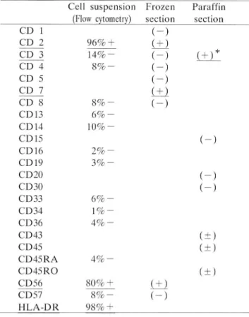
Table 1. Immunophenotyping of the lymphoma cells
Cell suspension Frozen Paraffin
(Flow cytometry) section section CD 1 CD 2 96% CD 3 14%-CD 4 8%-CD 5 CD 7 CD 8 8 % -CD13 6%-CD14 i o % -CD 15 CD 16 2%-CD19 3 % -CD20 CD30 CD33 6% -CD34 1%-CD36 4 % -CD43 CD45 CD45RA 4%-CD45RO CD56 80% + CD57 8%-HLA-DR 98% + Cytoplasmic CD 3. (-) (±1 (") (-) (-) i+1 (-) (±1 (-) i+y (-) (-) (-) (+) (±) (+) • H r y /.
Fig. 6. EBER RNA positivity in nuclei of lymphoma cells. (In situ hybridization, x 175)
and CDS were negative. Immunohistochemistry and in situ hybridization on paraffin sections showed positivity for cytoplasmic CD3 and Epstein-Barr virus EBER oligonucleotide (Fig. 6), respectively. Genotypic studies revealed a germlinc configuration in Jn and TCR genes. From these findings, we conclude that this tumor was a CD56-positivc aggressive lymphoma featuring large granular lymphocytes.
Discussion
NK cell marker(CD56)-positive aggressive lympho
ma has recently been described mostly in the sinonasal
and upper aerodigestive tract,l,2) but rarely in other
sites such as skin and subcutaneous tissue, intestines,
spleen, lymph node, bone marrow and so on.1'3'6'
Histologically, it is a diffuse lymphoma often with an angiocentric growth pattern and ischemic necrosis.
Cytologically, these neoplastic cells are mainly com
prised of large cells, but, there are some instances of
medium-sized cells, with azurophilic graunles in the
cytoplasm. Neoplastic cells in these tumors were usually positive for CD2, CD56 and HLA-DR, but negative for CD3 and B-cell markers. Expression of
CD4, CD7, CDS, CDll, CD!6, CD57 and other
markers was variable. In terms of markers,
CD56-positivc aggressive lymphomas are divided into CD3-negative and CD3-positive groups. The former group may be called CD56-positive cell lymphoma and the latter CD56-positive NK cell-like T-cell lymphoma. Regardless of differences in expression of these markers, the clinical course of CD56-positive lympho mas is very aggressive.
In the present case, the tumor was composed mainly of medium-sized lymphoblastic cells with frequent mitoses and intracytoplasmic azurophilic granules. Although cytoplasmic CD3 was positive on paraffin sections, surface CD3 was negative and TCR genes showed a germlinc configuration. This type of discre pancy in CD3 expression is also found in NK cells, since polyclonal CD3 stain may detect cytoplasmic CD3- e chain which is found in NK cells. The present case also showed EBER oligonucleotide of Epstein-Barr virus, which is consistent with the observations by Chan et al. of EBER positivity in non-nasal/nasopharyngeal
CD3 negative and CD56 positive lymphomas.S)
References
1) Ng CS, Chan JKC, Lo STH. Expression of natural killer cell markers in non-Hodgkin's lymphomas.
Hum Pathol 19S7; 18: 1257-62.
2) Ho FCS, Choy D, Loke SL. Kung ITM, Fu KH, Liang R, et al. Polymorphic reticulosis and conven tional lymphomas of the nose and upper aerodiges tive tract: A clinicopathologic study of 70 cases, and immunophenotypic studies of 16 cases. Hum Pathol
1990; 21: 1041-50.
3) Kern WF, Spier CM, Hanneman EH, Miller TP. Matzner M, Grogan TM. Neural cell adhesion
molecule-positive peripheral T-cell lymphoma: a
rare variant with a propensity for unusual sites of
involvement. Blood 1992; 79: 2432-7.
4) Nakamura S. Suchi T, Kosihkawa T, Takagi N, Hayashi K, Koike K, et al. Aggressive rectal lymphoma of large granular lymphocytes with the histologic feature of an angiocentric growth pattern.
[170] Med. J. Kagoshima Univ., Vol. 47, Suppl. 2, November, 1995
Cancer 1993; 71: 249-56.
5) Sun T, Brody J, Susin M, Marino J, Teichberg S, Koduru P, et al. Aggressive natural killer cell lymphoma/leukemia. A recently recognized clinico pathologic entity. Am J Surg Pathol 1993; 17: 1289-99.
6) Wong KF, Chan JKC, Ng CS, Lee KC, Tsang WYW, Cheung MMC. CD56(NKHl)-positive hematolymphoid malignancies: an aggressive neo plasm featuring frequent cutaneous/mucosal in volvement, cytoplasmic azurophilic granules and
angiocentricity. Hum Pathol 1992; 23: 798-804. 7) Biassoni R, Ferrini S, Prigione I, Moretta A, Long
EO. CD3 negative lymphokine-activated cytotoxic cell express the CD3 gene. J Immunol 1988; 140:
1685-9.
8) Tsang WYW, Chan JKC, Yip T, Ng CS, Wong KF. Demonstration of Epstein-Barr virus early RNA (EBER) in non-nasal/nasopharyngeal CD56-posi-tive lymphomas. Mod Pathol 1994; 7: 122A( Abstract).