RESEARCH ARTICLE
Lack of collagen
α6(IV) chain in mice does not
cause severe-to-profound hearing loss or
cochlear malformation, a distinct phenotype
from nonsyndromic hearing loss with COL4A6
missense mutation
Shaoying Tang1, Tomoko YonezawaID1*, Yukihide Maeda2, Mitsuaki Ono1,
Takahiro Maeba1¤, Toru Miyoshi3, Ryusuke Momota4, Yasuko Tomono5, Toshitaka Oohashi1
1 Department of Molecular Biology and Biochemistry, Okayama University Graduate School of Medicine,
Dentistry and Pharmaceutical Sciences, Okayama, Japan, 2 Department of Otolaryngology-Head and Neck Surgery, Okayama University Graduate School of Medicine, Dentistry and Pharmaceutical Sciences, Okayama, Japan, 3 Department of Cardiovascular Medicine, Okayama University Graduate School of Medicine, Dentistry and Pharmaceutical Sciences, Okayama, Japan, 4 Department of Human Morphology, Okayama University Graduate School of Medicine, Dentistry and Pharmaceutical Sciences, Okayama, Japan, 5 Division of Molecular and Cell Biology, Shigei Medical Research Institute, Okayama, Japan
¤ Current address: Department of Plastic Surgery, Japanese Red Cross Fukuoka Hospital, Fukuoka, Japan
Abstract
Congenital hearing loss affects 1 in every 1000 births, with genetic mutations contributing to more than 50% of all cases. X-linked nonsyndromic hereditary hearing loss is associated with six loci (DFNX1-6) and five genes. Recently, the missense mutation (c.1771G>A, p. Gly591Ser) in COL4A6, encoding the basement membrane (BM) collagenα6(IV) chain, was shown to be associated with X-linked congenital nonsyndromic hearing loss with cochlear malformation. However, the mechanism by which the COL4A6 mutation impacts hereditary hearing loss has not yet been elucidated. Herein, we investigated Col4a6 knock-out (KO) effects on hearing function and cochlear formation in mice. Immunohistochemistry showed that the collagenα6(IV) chain was distributed throughout the mouse cochlea within subepithelial BMs underlying the interdental cells, inner sulcus cells, basilar membrane, outer sulcus cells, root cells, Reissner’s membrane, and perivascular BMs in the spiral lim-bus, spiral ligament, and stria vascularis. However, the click-evoked auditory brainstem response analysis did not show significant changes in the hearing threshold of Col4a6 KO mice compared with wild-type (WT) mice with the same genetic background. In addition, the cochlear structures of Col4a6 KO mice did not exhibit morphological alterations, according to the results of high-resolution micro-computed tomography and histology. Hence, loss of Col4a6 gene expression in mice showed normal click ABR thresholds and normal cochlear formation, which differs from humans with the COL4A6 missense mutation c.1771G>A, p. Gly591Ser. Therefore, the deleterious effects in the auditory system caused by the mis-sense mutation in COL4A6 are likely due to the dominant-negative effects of theα6(IV) a1111111111 a1111111111 a1111111111 a1111111111 a1111111111 OPEN ACCESS
Citation: Tang S, Yonezawa T, Maeda Y, Ono M,
Maeba T, Miyoshi T, et al. (2021) Lack of collagen α6(IV) chain in mice does not cause severe-to-profound hearing loss or cochlear malformation, a distinct phenotype from nonsyndromic hearing loss with COL4A6 missense mutation. PLoS ONE 16(4): e0249909.https://doi.org/10.1371/journal. pone.0249909
Editor: Olivia Bermingham-McDonogh, University
of Washington, UNITED STATES
Received: December 5, 2020 Accepted: March 26, 2021 Published: April 13, 2021
Copyright:© 2021 Tang et al. This is an open access article distributed under the terms of the
Creative Commons Attribution License, which permits unrestricted use, distribution, and reproduction in any medium, provided the original author and source are credited.
Data Availability Statement: All relevant data are
within the manuscript and itsSupporting Informationfiles.
Funding: This work was supported in part by
grants from the JSPS KAKENHI (Grant Numbers: JP20K09867 to T.Y., and JP19H04754 to T.O.).
Competing interests: The authors have declared
chain and/orα5α6α5(IV) heterotrimer with an aberrant structure that would not occur in cases with loss of gene expression.
Introduction
The ear, a precise organ composed of the external, middle, and inner ear, that is heavily involved in the auditory system and balance. Auditory function plays an important role in communication and learning abilities [1]. The process of hearing begins when sound is con-ducted by air or bone. During air conduction, sound waves are collected, causing vibration of the tympanic membrane and ossicular chain. Sound waves also transfer through the skull, which is referred to as bone conduction. Vibrations within the endolymph through stapedial vibrations stimulate auditory receptors in the cochlea.
The cochlea, comprising the modiolus and osseous cochlear duct, is a spiral-shaped cavity in the bony labyrinth. The membranous cochlear duct is located in the osseous cochlear duct and includes the spiral limbus, basilar membrane, organ of Corti, and stria vascularis in the spiral ligament, which contribute to the mechanical-electrical signal conversion and ion trans-portation during sound conduction [1,2]. The extracellular matrix (ECM) also contributes to the auditory system; collagen and cochlin are the most abundant ECM components in the cochlea. Fibrillar collagen primarily provides tissue stability and strength, while cochlin inter-acts with collagen molecules [3]. Interestingly, a recent study has shown that cochlin also plays a role in innate immune responses within the inner ear [4]. Histologically, basement mem-branes (BMs) are present in the membranous labyrinth of the cochlea. The BM is a cell-adher-ent and sheet-like ECM found beneath the epithelium and endothelium, and surrounding smooth muscle cells, and adipocytes. The primary role of BMs is to provide tissue structure, divide the tissue into compartments, and influence cell behavior [5]. Collagen IV, laminin, per-lecan, and nidogen are major components of BMs. Collagen IV comprises six genetically dis-tinctα chains from α1(IV) to α6(IV) encoded by COL4A1 to COL4A6. Three unique triple-helical forms have been identified:α1α2α1, α3α4α5, and α5α6α5 (Fig 1). Furthermore, these protomers are extracellularly assembled into three hexamers, namely,α1α2α1-α1α2α1, α1α2α1-α5α6α5, and α3α4α5-α3α4α5 [6–8]. Previous reports showed thatα1(IV) and α2(IV) chains are abundant in all BMs, whereasα3(IV) to α6(IV) chains have a tissue-specific distri-bution. It is believed that the differential molecular composition of BM contributes to its spe-cific biological roles in tissues. Spespe-cifically, theα6(IV) chain is preferentially found in subepithelial- and smooth muscle cell-BMs in various organs [9–15].
Various genes encoding ECM or ECM-related proteins reportedly cause hereditary hearing loss, includingCOL4A6, which encodes the sixth alpha chain of collagen IV [16,17]. Previ-ously, we establishedCol4a6 knockout (KO) mice, which are apparently healthy and fertile,
but show a developmental delay in keratinization of the oral mucosal epithelium [18–20]. In zebrafish,col4a5 and col4a6 are essential for BM integrity, supporting the axogenesis of
gran-ule cells and retinal ganglion cells [21]. Thus, the collagenα6(IV) chain is a crucial element of BMs; however, its physiological role remains elusive.
Recently, the missense mutation (c.1771G>A, p.Gly591Ser) inCOL4A6 was reported to be
associated with X-linked congenital nonsyndromic hearing loss with cochlear malformation (OMIM: #300914); indeed, all the male subjects in this family experienced severe-to-profound hearing loss at all frequencies tested (0.125 to 8 kHz). Bioinformatic analysis predicted that the p.Gly591Ser missense mutation reduces the triple-helical conformational stability and triggers quaternary structure disassembly [22]. However, the impact of this mutation in the collagen α6(IV) chain in vivo remains unclear.
In this study, we investigate the collagenα6(IV) chain distribution in mouse cochlea and its effects on the cochlear formation and auditory function using aCol4a6 KO mouse model.
Overall, our study demonstrated the detailed distribution of collagenα6(IV) chain in mouse cochlea and showed that loss of expression ofCol4a6 in mice does not cause abnormalities in
the cochlear structure or severe-to-profound hearing loss, which occurs in humans with mis-sense mutation (c.1771G>A, p.Gly591Ser) ofCOL4A6.
Materials and methods
Animals
MaleCol4a6 KO and WT mice (8-week-old) were used in this study. Col4a6 KO mice were
generated by replacing part of exon 2 and intron 2 with a neomycin cassette in theCol4a6
Fig 1. Schematic view of the gene structure and assembly of collagen IV. TheCOL4A6/Col4a6 gene is located on the X chromosome and
paired withCOL4A5/Col4a5 in a head-to-head manner. COL4A1/Col4a1 and COL4A2/Col4a2, and COL4A3/Col4a3 and COL4A4/Col4a4
pairs are located in a head-to-head manner on their respective chromosomes (not shown). Each chain comprises a 400 nm long triple-helical domain, which contains many interruptions in the Gly-X-Y repeated sequence. The globular noncollagenous domain (NC1) is located at the C-terminal end, while the 7S domain is at the N-terminal end. Threeα chains assemble the triple-helical molecule (triple-helix). The heterotrimers extracellularly form a hexamer.α1α2α1(IV) and α5α6α5(IV) bind to each other through the NC1 domain [α1α2α1-α5α6α5 (IV)] and the 7S domains (not shown), which facilitates their assembly to a higher-ordered supra structure.α1α2α1(IV) is widely distributed, whereasα3α4α5(IV) and α5α6α5(IV) distribution is tissue specific [7–9,12]. InCol4a6 KO mice, the loss of α6(IV) chain expression inhibits
the formation of theα5α6α5(IV) heterotrimer. This study showed that the phenotypes of the auditory system in Col4a6 KO mice were distinct from those of individuals with theCOL4A6 missense mutation c.1771G>A, p.Gly591Ser. The presence of the mutant protein α6(IV)
chain and/orα5α6α5(IV) may induce deleterious effects on the cochlea. The pathogenesis involved in congenital hearing loss warrants further investigations.
gene in the 129 SV/J background, as previously described, and subsequently backcrossed with C57BL/6J (Charles River) over ten generations [18,19].
This study was conducted in strict accordance with the Policy on the Care and Use of Labo-ratory Animals, Okayama University. The protocol was approved by the Animal Care and Use Committee of the Okayama University (Protocol Number: OKU-2020035).
Immunohistochemistry
The temporal bone was dissected from the mice (8-week-old) anesthetized with intraperitoneal xylazine (8 mg/kg) and ketamine (80 mg/kg), and snap-frozen in super Cryoembedding medium (SECTION-LAB Co. Ltd., Hiroshima, Japan). Then, 6μm serial cryosections were prepared using Kawamoto’s film methods, as previously described with some modifications [20]. Briefly, the sections were fixed with acetone for 20 min followed by treatment with 6 M urea in 0.1 M glycine-HCl buffer (pH 3.5) to expose the epitope. Subsequently, 1% bovine serum albumin in phosphate buffered saline was used to block non-specific binding of anti-bodies for 1 h. The following rat monoclonal antianti-bodies were used: H11 (1:100); H22 (1:100); fluorescein isothiocyanate labeled-M26 (1:10); 129 (1:100); b42 (1:100); H53 (1:300); and B66 (1:10), recognizing collagenα1(IV), α2(IV), α2(IV), α3(IV), α4(IV), α5(IV), and α6(IV) chains (generated by Dr. Y Tomono and Y Sado in Shigei Medical Research Institute, Oka-yama, Japan) [9,11,23]. Antibodies against CD31 (1:50, ab28364; Abcam, UK), perlecan (1:1,000, A7L6; Millipore, Burlington, MA, USA), lamininα1 (1:200, AL-1; Chemicon, Temec-ula, CA, USA), lamininα2 (1:1,000, 4H8-2; Sigma, Saint Louis, MO, USA), laminin γ1
(1:2,000, A5; Chemicon), and nidogen-1 (1:2,000; kindly gift by Dr. Takako Sasaki, Oita Uni-versity) were used [24]. For the secondary antibody, Alexa Fluor1 488-conjugated goat anti-rat IgG (1:1,500; Invitrogen, Carlsbad, CA, USA), Alexa Fluor1 594-conjugated goat anti-anti-rat IgG (1:1,500; Invitrogen), and Alexa Fluor1 594-conjugated donkey anti-rabbit IgG (1:1,500; Invitrogen) were used. Nuclei were stained with Hoechst 33258 (2μM; Polysciences, Warring-ton, PA, USA). All images were obtained with a KEYENCE BZ-X710 microscope (Keyence, Osaka, Japan), and processed using BZ-X Analyzer software (Keyence). The experiment was repeated using three WT and threeCol4a6 KO mice.
Auditory function
Auditory function was assessed by click-evoked auditory brainstem response (ABR) as previ-ously described with minor modifications [25,26].Col4a6 KO mice (n = 5) and WT mice
(n = 5) were anesthetized with intraperitoneal xylazine (8 mg/kg) and ketamine (80 mg/kg). The generation of acoustic stimuli and simultaneous recording of evoked potentials were per-formed using Tucker Davis Technologies (TDT) ABR system (RA16, PA5, RP2.1, TDT, Gaines-ville, FL, USA). ABRs were evoked with clicks and the sound stimuli were applied in 5-dB SPL steps from 90 to 0 dB SPL. The click-ABR threshold was determined as the minimum sound pressure level at which the click-ABR waveform was visible on the recording monitor.
Statistical analysis
Statistical analysis was performed using Student’s unpairedt-test. The data are presented as
the mean± standard deviation (SD). P < 0.05 was considered statistically significant.
Micro-tomographic analysis (micro-CT)
The temporal bone was dissected fromCol4a6 KO and WT mice (8-week-old) anesthetized
in neutral buffer at 4˚C for 24 h. The samples were analyzed by micro-CT (SkyScan 1174 com-pact micro-CT, Bruker, Belgium) as described previously with some modifications [27]. Scans were performed at a resolution of 6.5μm, after which 1,004 sections were reconstructed to pro-duce the 3-D image of the petrous portion using SkyScan software (NRecon, CTAn, CTvol, and CTvox, SkyScan). In addition, to compare the shape and size of the cochlea between WT andCol4a6 KO mice, DataViewer software was used according to the manufacturer’s protocol
(Bruker). Briefly, we selected the volume of interest (VOI) in a 2-dimensional (2-D) plane with a clear view of the cochlea using WT scan data as a reference. The software automatically gen-erated three orthogonal views of the best-matched 2-D image from the scan data ofCol4a6 KO
as a target to the reference image. In the 2-D overlay image of WT andCol4a6 KO, the
match-ing degree was automatically represented by a color map based on the intensity of the radiog-raphy using DataViewer. The analysis was repeated using six cochleae from three mice of each genotype (WT, n = 3; KO, n = 3).
Hematoxylin and eosin (HE) staining
The temporal bone was dissected fromCol4a6 KO and WT mice (8-week-old) anesthetized
with intraperitoneal xylazine (8 mg/kg) and ketamine (80 mg/kg) and fixed in 4% paraformal-dehyde in 0.1 M phosphate buffer (pH 7.2). After decalcification with 0.5 M EDTA (pH 7.5; Wako, Japan) for 7 days at room temperature, the cochleae were dehydrated and embedded in paraffin and 5μm serial sections were prepared using a microtome (MICROM, HM 335E, Leica, Wetzlar, Germany). A new hematoxylin solution (Muto Pure Chemicals, Japan) and pure eosin solution (Muto Pure Chemicals) were used. All images were obtained with a KEY-ENCE BZ-X710 microscope (Keyence) and processed using BZ-X Analyzer software (Key-ence). The experiment was repeated using three WT and threeCol4a6 KO mice.
Results
Distribution of collagen
α6(IV) chain in the mouse cochlea
To examine the distribution of the collagenα6(IV) chain in the mouse cochlea, we performed immunohistochemistry. Several BMs were found in the cochlea, including the subepithelial BMs of interdental cells, inner sulcus cells, basilar membrane, outer sulcus cells, root cells, and Reissner’s membrane; perivascular BMs in the spiral limbus, spiral ligament, and stria vascu-laris; and perineural BMs in the nerve fasciculus and spiral ganglion [28,29]. We confirmed that both collagenα1(IV) and α2(IV) chains were present in all BMs in the mouse cochlea, as described previously (Fig 2AandS1 Fig) [30]. Results show that the collagenα6(IV) chain was observed in the subepithelial (Fig 2B–2E) and perivascular BMs (Fig 2B, 2D and 2G–2I), but not in the perineural BMs (Fig 2B and 2F).
For further characterization, we compared the localization of the collagenα6(IV) chain with that of the collagenα2(IV) chain using immunohistochemistry. The interdental cells are oval or round in shape and connect to form a comb-shaped network. The surface of the spiral limbus beneath the tectorial membrane is covered by flat processes of the interdental cells [31]. Although both theα2(IV) and α6(IV) chains were detected as comb-shaped networks, the immunoreactivity ofα6(IV) chain was higher in the invaginated region than in the superficial plate of the network (Fig 2I). In the spiral ligament, theα6(IV) chain appeared conspicuously in the radiated epithelial BMs surrounding the root cells and beneath the outer sulcus cells compared with theα2(IV) chain (Fig 2G and 2JandS1 Fig). In the stria vascularis, the signal of theα6(IV) chain colocalized at the outer side of α2(IV) chain-containing BMs, which sur-round the endothelium and pericytes (Fig 2H and 2K).
Differential composition of collagen IV networks in the mouse cochlea
We performed immunohistochemistry to investigate the molecular composition of cochlear BMs usingCol4a6 KO and WT mice. In WT mice, collagen α5(IV) chain was distributed inthe subepithelial BMs of interdental cells, inner sulcus cells, basilar membrane, outer sulcus cells, root cells, and Reissner’s membranes; perivascular BMs in the spiral limbus, spiral liga-ment, and stria vascularis; and the perineural BMs of the neural fasciculus and spiral ganglion (Fig 3A and 3D–3F). Based on the results for the collagenα6(IV) chain, α5α6α5 was suggested to be present in all subepithelial BMs and perivascular BMs in the mouse cochlea (Figs2and
3).
Notably, the collagenα5(IV) chain was observed in the subepithelial BMs and perineural BMs, however, it was scarcely detected in the perivascular BMs ofCol4a6 KO mice (Fig 3B and
Fig 2. Distribution of collagenα6(IV) chain in the adult mouse cochlea. Sections were immunostained using
antibodies against collagenα2(IV) (A, I–K: green) and α6(IV) (B–H: green; I–K: red) chains, and CD31 (G, H: red) for the detection of endothelial cells. Subepithelial BMs and perivascular BMs are indicated by open arrowheads and solid arrowheads in the insets, respectively (G, J, K). (I) The superficial plate and invaginated region of the interdental cells network are indicated by open arrows and solid arrows in insets, respectively. (L) Schematic diagram of the distribution of collagenα6(IV) chain [yellow line: subepithelial BMs; pink line: perivascular BMs]. The negative control was established by immunostaining with only the secondary antibody and no specific reaction was observed (S2 Fig). Nuclei were counterstained with Hoechst 33258 (blue). SLim: spiral limbus; B, basilar membrane; SLig, spiral ligament; SV, stria vascularis; RM, Reissner’s membrane; NF, neural fasciculus; SG, spiral ganglion; IDC, interdental cells; ISC, inner sulcus cells; OSC, outer sulcus cells; RC, root cells; Cap, capillary. Scale bars = 50μm. Scale bars in inset = 10μm.
3G–3I). Moreover, images with longer exposure showed weakα5(IV) chain immunostaining in perivascular BMs fromCol4a6 KO mice (S2 Fig). Similarly,α3(IV) and α4(IV) chains were weakly immunopositive in the perivascular BMs (S2 Fig). Since collagen IV protomer assembly requires threeα(IV) chains, the lack of any one of these chains would result in a failure to assemble a triple-helical form by the remaining two chains [8]. Collectively, our results suggest thatα1α2α1, α3α4α5, and α5α6α5 in the subepithelial and perivascular BMs, and α1α2α1 and α3α4α5 in the perineural BMs contribute to the assembly of the collagen IV network. α5α6α5, in addition toα1α2α1, is a quantitatively major component of the collagen IV network in the perivascular BMs.Table 1presents the tissue distribution of collagen IV in mouse cochlea.
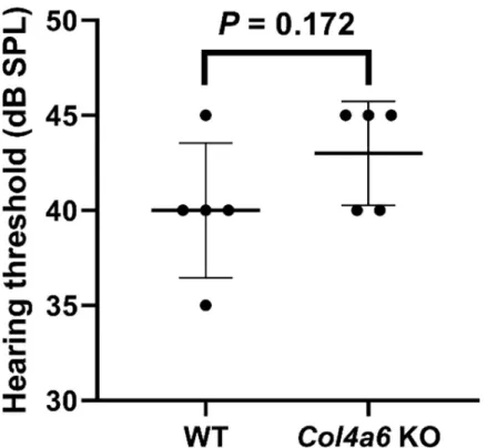
Assessment of hearing threshold by click-evoked ABR
To assess the hearing function inCol4a6 KO mice, we compared the click-ABR thresholds of
WT mice to those of ourCol4a6 KO mice (8-week-old male WT mice, C57BL/6J background).
The average threshold of the WT group andCol4a6 KO group were 40 ± 3.5 dB SPL and
43± 2.7 dB SPL, respectively (means ± SD, n = 5). The hearing threshold was not significantly different between WT andCol4a6 KO mice (P = 0.172,Fig 4).
Fig 3. Distribution of collagenα5(IV) chain in the cochlea of WT and Col4a6 KO mice. (A, D–F) The collagen α5(IV) chain located in the
subepithelial BMs, perivascular BMs, and perineural BMs in WT mice (green). (B, G–I) InCol4a6 KO mice, the α5(IV) chain is widely
distributed (green). However, weak signals were detected in the perivascular BMs of the spiral limbus (SLim), spiral ligament (SLig), and stria vascularis (SV) (dashed arrows). (C) No immunopositive signal of the collagenα6(IV) chain was present in Col4a6 KO mouse cochlea (green). Nuclei were counterstained with Hoechst 33258 (blue). B, basilar membrane; RM, Reissner’s membrane; NF, neural fasciculus; SG, spiral ganglion; IDC, interdental cells; ISC, inner sulcus cells; OSC, outer sulcus cells; RC, root cells; Cap, capillary. Scale bars = 50μm.
Micro-CT analysis
Patients with hearing loss caused by the X-linked hereditary mutation ofCOL4A6 were
reported to have malformed cochlea, as determined using high-resolution CT [22]. Therefore,
Table 1. Qualitative assessment using immunohistochemistry: Distribution of collagenα1(IV), α2(IV), α5(IV), and α6(IV) chains and the predicted molecular composition of the collagen IV network in mouse cochlea.
Classification¤ Location of BMs¤ α(IV) ¤ Predicted heterotrimer¤
α1a
¤ α2a¤ α5a¤ α6b¤ α1α2α1a¤ α3α4α5a¤ α5α6α5b¤
Subepithelial BMs¤ Interdental cells¤ +¤ +¤ +¤ +¤ +¤ +¤ +¤
Inner sulcus cells¤ +¤ +¤ +¤ +¤ +¤ +¤ +¤
Basilar membrane¤ +¤ +¤ +¤ +¤ +¤ +¤ +¤
Outer sulcus cells¤ +¤ +¤ +¤ +¤ +¤ +¤ +¤
Root cells¤ +¤ +¤ +¤ +¤ +¤ +¤ +¤
Reissner’s membrane¤ +¤ +¤ +¤ +¤ +¤ +¤ +¤
Perivascular BMs¤ Spiral limbus¤ +¤ +¤ +¤ +¤ +¤ +b¤ +¤
Spiral ligament¤ +¤ +¤ +¤ +¤ +¤ +b¤ +¤
Stria vascularis¤ +¤ +¤ +¤ +¤ +¤ +b¤ +¤
Perineural BMs¤ Neural fasciculus¤ +¤ +¤ +¤ -¤ +¤ +¤ -¤
Spiral ganglion¤ +¤ +¤ +¤ -¤ +¤ +¤ -¤
BM, Basement membrane; +, positive; -, negative.
a
Its presence was previously identified [28,30].
b
Data shown for the first time in the current study.α3α4α5(IV) protomer in the perivascular BMs was a quantitatively minor component of the collagen IV network.
https://doi.org/10.1371/journal.pone.0249909.t001
Fig 4. Hearing threshold assessed by click-evoked auditory brainstem responses in WT andCol4a6 KO mice. No
significant difference observed between WT mice (mean 40± 3.5 dB SPL, n = 5) and Col4a6 KO mice (mean 43 ± 2.7 dB SPL, n = 5).P = 0.172 by Student’s unpaired t-tests. The values are presented as mean ± SD.
to investigate the structural abnormalities inCol4a6 KO mouse cochlea, we performed a
micro-CT analysis. No differences were observed in the three-dimensional images of the petrous portion of the left temporal bone between the two groups of mice (Fig 5A). Moreover, three kinds of orthogonal 2-D cross-sections, including the transverse plane (X-Y), sagittal plane (Z-Y), and coronal plane (X-Z), were compared using DataViewer software. No abnor-malities were found in theCol4a6 KO cochlea structure (Fig 5B–5G). Furthermore, the overlay images based on the color map showed no differences in the size or shape of the cochlea between WT andCol4a6 KO mice (Fig 5H–5K). Taken together, our results demonstrate that
Col4a6 KO mice have no gross malformation of the osseous labyrinth.
Fig 5. Comparison of the cochlea bone structure of WT andCol4a6 KO mice by micro-CT analysis. (A) Representative
three-dimensional images of the petrous portion of the left temporal bone in WT andCol4a6 KO mice. The top shows the apex of petrous in the
superior view. (B–G) The three orthogonal plane images were automatically obtained as a best-matched plane from theCol4a6 KO data set
compared to the images manually selected from the cochlea structure of the WT mice data set. (H–J) The overlay images demonstrated that no differences in the size or shape are apparent in the cochlear bone between WT andCol4a6 KO mice. Blue squares represent the Volume of
Interest. (K) Light blue color indicates a highly matched degree based on the color map.
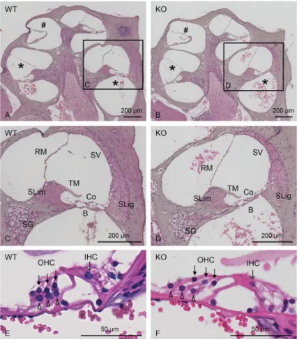
Histological analysis
To achieve a more detailed analysis of cochlear formation, both WT andCol4a6 KO
(8-week-old) mouse cochlea were compared histologically using HE-stained midmodiolar sections (Fig 6).Col4a6 KO mice exhibited regular morphological structures through the basal to apical
turn, as those observed in WT mice, comprising spiral limbus followed by inner sulcus, spiral ganglion, and neural fasciculus extended to the organ of Corti, which adheres to the basilar membrane (Fig 6A and 6B). Moreover, the tectorial membrane, spiral ligaments, stria vascu-laris, and Reissner’s membrane were similar in structure between the two groups (Fig 6C and 6D). The outer and inner hair cell rows, accompanied by Dieter’s cells, also showed regular morphology inCol4a6 KO and WT mice (Fig 6E and 6F). Hence, no significant differences in any structure were detected inCol4a6 KO mice compared to WT mice (n = 3).
No change in the distribution of major BM components
To examine the changes in the expression of the other major BM components, we performed immunohistochemistry. No apparent differences in the staining pattern of collagenα1(IV) chain, perlecan, lamininα1, laminin α2, laminin γ1, or nidogen-1 were detected in the cochlea ofCol4a6 KO and WT mice (Fig 7).
Fig 6. Representative images of HE-stained midmodiolar cochlear sections. Histological comparison between WT
(A, C, E) andCol4a6 KO (B, D, F) mice. (C-F) Images with higher magnification of cochlear basal turn are shown. No
differences are observed between WT andCol4a6 KO mice. , basal turn; #, apical turn; SG, spiral ganglion; SLim, spiral
limbus; TM, tectorial membrane; B, Basilar membrane; Co, organ of Corti; SLig, spiral ligament; SV, stria vascularis; RM, Reissner’s membrane; closed arrows, OHC (outer hair cells); arrows, IHC (inner hair cells); open arrowheads, Dieter’s cells.
Discussion
In this study, we characterized the localization of the collagenα6(IV) chain in mouse cochlea and demonstrated that the loss of collagenα6(IV) chain expression showed normal click ABR thresholds and normal cochlear formation, which differs from humans with theCOL4A6
mis-sense mutation c.1771G>A, p.Gly591Ser.
Our results showed that the collagenα6(IV) chain is expressed in both subepithelial BMs in the interdental cells, inner sulcus cells, basilar membrane, outer sulcus cells, root cells, and Reissner’s membrane, as well as the perivascular BMs in the spiral limbus, spiral ligament, and stria vascularis. A previous study reported that theα6(IV) chain exists intracellularly in a sub-group of ganglia cells, while our results, using monoclonal antibody B66, did not detect signals in these cells [22]. B66 has been established as a monoclonal antibody that specifically reacts with theα6(IV) chain of human, bovine, and mice [9,11,32]. We reconfirmed that B66 had no immunopositive signals in the tissue derived fromCol4a6 KO mice (Fig 3C). In addition, B66 showed clear immunopositive signals corresponding to the presence of BMs in the mouse cochlea. On the other hand, the distribution ofα3α4α5 in the cochlea of WT and Col4a3 KO mice by immunohistochemistry using antibodies against collagenα3(IV), α4(IV), and α5(IV) chains has been shown in previous reports [28,30]. In this study, we presented immunohis-tochemistry data using specific antibodies against each collagenα(IV) chain; hence, compari-son of the distribution of collagenα1(IV) to α6(IV) chains represented the tissue-specific collagen IV network of the mouse cochlea (Table 1). In particular, we demonstrated the pres-ence ofα1α2α1, α3α4α5, and α5α6α5 in subepithelial BMs, α1α2α1, α3α4α5, and α5α6α5 in perivascular BMs, andα1α2α1 and α3α4α5 in perineural BMs.
Fig 7. Distribution of major basement membrane components in the cochlea of WT andCol4a6 KO mice. Cryosections were
immunostained by antibodies against collagenα1(IV) chain (A, B), perlecan (C, D), laminin α1 (E, F), laminin α2 (G, H), laminin γ1 (I, J), and nidogen-1 (K, L). Nuclei were counterstained with Hoechst 33258 (blue). No differences are apparent between WT andCol4a6 KO mice.
Scale bars = 50μm.
The stria vascularis is responsible for supporting the endocochlear potential and high potas-sium content of the endolymph in the cochlear duct, which is necessary for sensory hair cell conduction [33]. The structure comprises marginal cells, intermediate cells, basal cells, and a dense capillary network. The capillaries and intermediate cells are located between the mar-ginal cell layer and the basal cell layer. Moreover, the perivascular BMs surround the endothe-lial cells and pericytes in the capillary network, which is contacted by the marginal cells and intermediate cells [34]. BMs regulate various cell functions, such as stable anchoring, prolifera-tion, and migraprolifera-tion, primarily mediated by integrin, a major cell surface receptor for the extra-cellular matrix. In addition, BMs serve as a filter for electrically charged molecules [5]. In fact, perivascular BMs may participate in the blood-labyrinth barrier, which is essential for prevent-ing the influx of harmful substances into the intrastrial space, similar to the blood-brain barrier [35,36]. Moreover, double-layered BMs are detected in the stria vascularis, which are derived from endothelial cells, pericytes, and marginal and intermediate epithelial cells [34,37]. Hear-ing impairment that is age-related, drug-induced, or results from acute injury or genetic dis-ease, such as Alport syndrome (AS), is associated with changes in the structure and
components of the perivascular BMs in the stria vascularis [28,38–41]. Interestingly, the colla-genα6(IV) chain was detected in the outer layer of BMs surrounding the capillary network of the stria vascularis, suggesting that theα6(IV) chain-containing BMs may functionally be related to marginal and intermediate epithelial cells.
Recently, a Hungarian family was reported to have congenital X-linked nonsyndromic hearing loss caused by a mutation inCOL4A6 (c.1771G>A, p.Gly591Ser), which was
accom-panied by cochlear malformation. All male members of this family with theCOL4A6 mutation
showed severe-to-profound hearing loss at all frequencies tested (0.125 to 8 kHz), which began during early childhood. Bioinformatic analysis showed that this mutation produces an atypical large side-chain in the interchain of the heterotrimer, which reduces the stability of the triple-helix and likely triggers disarrangement of the quaternary structure [22]. However, the current study demonstrates that the click ABR thresholds, histology, and micro-CT scanning images ofCol4a6 KO mice did not differ compared to those of WT mice. We also examined the
possi-bility that other BM components compensated for the deleterious effect induced by the absence of the collagenα6(IV) chain in the cochlea. However, immunohistochemistry staining showed that the expression of major BM components did not differ betweenCol4a6 KO and
WT mice. Taken together, our results suggest that a null-type mutation ofCol4a6 in mice had
no deleterious impact on cochlear formation or on click ABR thresholds. Our characterization of theCol4a6 KO mice differs dramatically from the phenotype observed in humans with the COL4A6 missense mutation c.1771G>A, p.Gly591Ser.
Collagens constitute a superfamily of extracellular matrix proteins that function primarily as structural proteins. Several collagen genes (COL1A1, COL1A2, COL2A1, COL4A3, COL4A4, COL4A5, COL11A1, and COL11A2) are associated with hereditary syndromic hearing loss.
Mutations inCOL11A1 and COL11A2 have been shown to cause nonsyndromic hereditary
hearing loss [42,43]. Osteogenesis imperfecta (OI) is commonly caused by autosomal domi-nant mutations in the genes encoding collagen I and is characterized by growth deficiency and vulnerability to fractures from minimal trauma [44]. The molecular defect in type I OI, the mildest form, is a nullCOL1A1 allele caused by frameshifts or a premature stop codon,
result-ing in reduced synthesis of structurally normal collagen, whereas types II–IV OI, the more severe forms, are caused by defects in the collagen I fibril structure, most commonly glycine substitutions (80%). The mutated collagenα(I) chain delays the folding of the heterotrimer and results in over-post-translational modification. Misfolded heterotrimers then induce the unfolded protein response (UPR) in the endoplasmic reticulum, associated with a pathogenic defect in the differentiation and maturation of osteoblasts. In cases where abnormal
procollagen can escape from the intracellular degradation pathway and are secreted, the colla-gen fiber with a misfolded heterotrimer has lower integrity and affects the mechanical strength and various cell-matrix interactions [45]. Moreover, AS is a hereditary disorder characterized by progressive renal failure, sensorineural hearing loss, and ocular defects caused by mutations inCOL4A3, COL4A4, and COL4A5. To date, over 500 mutations have been identified,
includ-ing point mutations, deletions, and insertions of a large or small nucleotide sequence [46–49]. Most mutations result in failure of assembly and/or secretion of collagenα3α4α5(IV) hetero-trimers, and consequently, all threeα chains are missing from the tissue. While collagen α3α4α5(IV) heterotrimers with subtle missense mutations can be assembled, secreted, and form the network in some cases; however, it still results in milder disease severity in patients due to the functionally impaired BMs [48]. For instance, Naitoet al. showed sparse or normal
immunostained signals of the collagenα5(IV) chains harboring the glycine substituted mis-sense mutation in the glomerular BM of patients [50]. Of note, a missense mutation,
COL4A3-G1334E, leads to collagenα3α4α5(IV) heterotrimer misfolding, which causes podocyte UPR [51]. Besides, chemical chaperones have recently been reported to have therapeutic potential for AS by normalizing the mutated collagenα3α4α5(IV) heterotrimer [52].
Collectively, the missense mutation ofCOL4A6 (c.1771G>A, p.Gly591Ser) may affect
hear-ing impairment as a consequence of the aberrant function associated with the collagenα6(IV) chain due to a misfolded collagen heterotrimer (Fig 1). Although the present study was not designed to show the direct effect of the mutated collagenα6(IV) chain on hearing function, our findings might provide a basis for developing advanced applications of aCol4a6 knock-in
mouse model carrying the missense mutation. Further studies are necessary to elucidate the pathogenesis of hereditary hearing loss and cochlear malformation associated withCOL4A6
missense mutations.
The primary limitation of this study is that the analysis of hearing levels in mice was exclu-sively based on the click-ABR method, which does not provide information on the frequency-specific hearing levels. Hearing assessment in our future studies will include ABR using pure tones to analyze hearing levels in low, middle, and high frequencies. Moreover, although the present study detected no histological differences in the organ of Corti between WT and
Col4a6 KO mice, via light microscopy, Col4a6 KO mice may have BM functional impairment
of the organ of Corti along the tonotopic axis of the apical, middle, and basal cochlear turns, which are involved in sound transduction at each frequency. In addition, startle response test-ing will provide insight into behavioral responses to brief, intense sound and otoacoustic emis-sions testing will further evaluate the function of outer hair cells along the tonotopic axis of the cochlea.
Supporting information
S1 Fig. Distribution of collagenα1(IV) and α2(IV) chains in the mouse cochlea.
Cryosec-tions were immunostained with antibodies againstα1(IV) (A–F: green) and α2(IV) chains (G–L: green). Nuclei were counterstained with Hoechst 33258 (blue). SLim, spiral limbus; NF, neural fasciculus; SG, spiral ganglion; B, basilar membrane; SLig, spiral ligament; SV, stria vas-cularis; RM, Reissner’s membrane. Scale bars = 50μm.
(TIF)
S2 Fig. Distribution of collagenα3(IV), α4(IV), and α5(IV) chains in the perivascular basement membranes of WT and Col4a6 KO mice. Cryosections were immunostained with
antibodies againstα3(IV) (A: green), α4(IV) (B: green), α5(IV) chains (C–F: green), and CD31 (D–F: red). The results suggest thatα3α4α5(IV) heterotrimer is present in the perivascular BMs of the mouse cochlea. (A–C) The subepithelial BMs in the spiral ligament (SLig) were
used as positive controls for each antibody in the inset. (G–I) The negative control was estab-lished by immunostaining with only secondary antibody; no specific reaction was observed. Nuclei were counterstained with Hoechst 33258 (blue). Arrows, immunopositive perivascular BMs in the spiral ligament (SLig); open arrowheads, immunopositive perivascular BMs in the stria vascularis (SV); SLim, spiral limbus. Scale bars = 50μm. Scale bars in inset = 20 μm. (TIF)
Acknowledgments
The authors would like to thank Ms. Junko Takahara (Okayama University Graduate School of Medicine, Dentistry, and Pharmaceutical Sciences) and the staff of the Central Research Laboratory at Okayama University Medical School for technical assistance. The authors also thank Dr. Takako Sasaki (Oita University) for providing antibody against nidogen-1, as well as Dr. Reinhard Fa¨ssler (Max Planck Institute of Biochemistry) for the support in generating knockout mice.
Author Contributions
Conceptualization: Tomoko Yonezawa, Toshitaka Oohashi.
Formal analysis: Shaoying Tang, Tomoko Yonezawa, Yukihide Maeda, Mitsuaki Ono, Toru
Miyoshi, Ryusuke Momota.
Funding acquisition: Tomoko Yonezawa, Toshitaka Oohashi.
Investigation: Shaoying Tang, Tomoko Yonezawa, Yukihide Maeda, Takahiro Maeba. Methodology: Shaoying Tang, Tomoko Yonezawa, Yukihide Maeda, Mitsuaki Ono. Project administration: Tomoko Yonezawa.
Resources: Tomoko Yonezawa, Yukihide Maeda, Mitsuaki Ono, Toru Miyoshi, Yasuko
Tomono, Toshitaka Oohashi.
Supervision: Toru Miyoshi, Ryusuke Momota, Yasuko Tomono, Toshitaka Oohashi. Validation: Shaoying Tang, Tomoko Yonezawa, Yukihide Maeda, Mitsuaki Ono, Takahiro
Maeba, Toru Miyoshi, Ryusuke Momota, Yasuko Tomono, Toshitaka Oohashi.
Writing – original draft: Shaoying Tang, Tomoko Yonezawa.
Writing – review & editing: Shaoying Tang, Tomoko Yonezawa, Yukihide Maeda, Toru
Miyoshi, Ryusuke Momota, Toshitaka Oohashi.
References
1. Fuchs JC, Tucker AS. Development and Integration of the Ear. Curr Top Dev Biol. 2015; 115: 213–32. https://doi.org/10.1016/bs.ctdb.2015.07.007PMID:26589927
2. Ekdale EG. Form and function of the mammalian inner ear. J Anat. 2016; 228(2): 324–37.https://doi. org/10.1111/joa.12308PMID:25911945
3. Peeleman N, Verdoodt D, Ponsaerts P, Van Rompaey V. On the Role of Fibrocytes and the Extracellu-lar Matrix in the Physiology and Pathophysiology of the Spiral Ligament. Front Neurol. 2020; 11: 580639.https://doi.org/10.3389/fneur.2020.580639PMID:33193034
4. Jung J, Yoo JE, Choe YH, Park SC, Lee HJ, Lee HJ, et al. Cleaved Cochlin Sequesters Pseudomonas aeruginosa and Activates Innate Immunity in the Inner Ear. Cell Host Microbe. 2019; 25(4): 513–25.e6. https://doi.org/10.1016/j.chom.2019.02.001PMID:30905438
5. Pozzi A, Yurchenco PD, Iozzo RV. The nature and biology of basement membranes. Matrix Biol. 2017;57–58: 1–11.https://doi.org/10.1016/j.matbio.2016.10.006PMID:27751945
6. Sado Y, Kagawa M, Naito I, Ueki Y, Seki T, Momota R, et al. Organization and expression of basement membrane collagen IV genes and their roles in human disorders. J Biochem. 1998; 123(5): 767–76. https://doi.org/10.1093/oxfordjournals.jbchem.a022003PMID:9562604
7. Khoshnoodi J, Pedchenko V, Hudson BG. Mammalian collagen IV. Microsc Res Tech. 2008; 71(5): 357–70.https://doi.org/10.1002/jemt.20564PMID:18219669
8. Brown KL, Cummings CF, Vanacore RM, Hudson BG. Building collagen IV smart scaffolds on the out-side of cells. Protein Sci. 2017; 26(11): 2151–61.https://doi.org/10.1002/pro.3283PMID:28845540
9. Ninomiya Y, Kagawa M, Iyama K, Naito I, Kishiro Y, Seyer JM, et al. Differential expression of two base-ment membrane collagen genes, COL4A6 and COL4A5, demonstrated by immunofluorescence stain-ing usstain-ing peptide-specific monoclonal antibodies. J Cell Biol. 1995; 130(5): 1219–29.https://doi.org/10. 1083/jcb.130.5.1219PMID:7657706
10. Seki T, Naito I, Oohashi T, Sado Y, Ninomiya Y. Differential expression of type IV collagen isoforms, alpha5(IV) and alpha6(IV) chains, in basement membranes surrounding smooth muscle cells. Histo-chem Cell Biol. 1998; 110(4): 359–66.https://doi.org/10.1007/s004180050296PMID:9792414
11. Saito K, Naito I, Seki T, Oohashi T, Kimura E, Momota R, et al. Differential expression of mouse alpha5 (IV) and alpha6(IV) collagen genes in epithelial basement membranes. Journal of biochemistry. 2000; 128(3): 427–34.https://doi.org/10.1093/oxfordjournals.jbchem.a022770PMID:10965041
12. Borza DB, Bondar O, Ninomiya Y, Sado Y, Naito I, Todd P, et al. The NC1 domain of collagen IV encodes a novel network composed of the alpha 1, alpha 2, alpha 5, and alpha 6 chains in smooth mus-cle basement membranes. J Biol Chem. 2001; 276(30): 28532–40.https://doi.org/10.1074/jbc. M103690200PMID:11375996
13. Urabe N, Naito I, Saito K, Yonezawa T, Sado Y, Yoshioka H, et al. Basement membrane type IV colla-gen molecules in the choroid plexus, pia mater and capillaries in the mouse brain. Arch Histol Cytol. 2002; 65(2): 133–43.https://doi.org/10.1679/aohc.65.133PMID:12164337
14. Hasegawa H, Naito I, Nakano K, Momota R, Nishida K, Taguchi T, et al. The distributions of type IV col-lagen alpha chains in basement membranes of human epidermis and skin appendages. Arch Histol Cytol. 2007; 70(4): 255–65.https://doi.org/10.1679/aohc.70.255PMID:18296826
15. Saito K, Yonezawa T, Minaguchi J, Kurosaki M, Suetsugu S, Nakajima A, et al. Distribution of alpha(IV) collagen chains in the ocular anterior segments of adult mice. Connective tissue research. 2011; 52(2): 147–56.https://doi.org/10.3109/03008207.2010.492062PMID:20672978
16. Oohashi T, Sugimoto M, Mattei MG, Ninomiya Y. Identification of a new collagen IV chain, alpha 6(IV), by cDNA isolation and assignment of the gene to chromosome Xq22, which is the same locus for COL4A5. J Biol Chem. 1994; 269(10): 7520–6. PMID:8125972
17. Zhou J, Ding M, Zhao Z, Reeders ST. Complete primary structure of the sixth chain of human basement membrane collagen, alpha 6(IV). Isolation of the cDNAs for alpha 6(IV) and comparison with five other type IV collagen chains. J Biol Chem. 1994; 269(18): 13193–9. PMID:8175748
18. Fox MA, Sanes JR, Borza DB, Eswarakumar VP, Fassler R, Hudson BG, et al. Distinct target-derived signals organize formation, maturation, and maintenance of motor nerve terminals. Cell. 2007; 129(1): 179–93.https://doi.org/10.1016/j.cell.2007.02.035PMID:17418794
19. Murata T, Katayama K, Oohashi T, Jahnukainen T, Yonezawa T, Sado Y, et al. COL4A6 is dispensable for autosomal recessive Alport syndrome. Sci Rep. 2016; 6: 29450.https://doi.org/10.1038/srep29450 PMID:27377778
20. Komori T, Ono M, Hara ES, Ueda J, Nguyen HTT, Nguyen HT, et al. Type IV collagen alpha6 chain is a regulator of keratin 10 in keratinization of oral mucosal epithelium. Sci Rep. 2018; 8(1): 2612.https:// doi.org/10.1038/s41598-018-21000-0PMID:29422532
21. Takeuchi M, Yamaguchi S, Yonemura S, Kakiguchi K, Sato Y, Higashiyama T, et al. Type IV Collagen Controls the Axogenesis of Cerebellar Granule Cells by Regulating Basement Membrane Integrity in Zebrafish. PLoS Genet. 2015; 11(10): e1005587.https://doi.org/10.1371/journal.pgen.1005587PMID: 26451951
22. Rost S, Bach E, Neuner C, Nanda I, Dysek S, Bittner RE, et al. Novel form of X-linked nonsyndromic hearing loss with cochlear malformation caused by a mutation in the type IV collagen gene COL4A6. Eur J Hum Genet. 2014; 22(2): 208–15.https://doi.org/10.1038/ejhg.2013.108PMID:23714752
23. Kohda T, Okada S, Hayashi A, Kanzaki S, Ninomiya Y, Taki M, et al. High nephritogenicity of monoclo-nal antibodies belonging to IgG2a and IgG2b subclasses in rat anti-GBM nephritis. Kidney Int. 2004; 66 (1): 177–86.https://doi.org/10.1111/j.1523-1755.2004.00719.xPMID:15200424
24. Fox JW, Mayer U, Nischt R, Aumailley M, Reinhardt D, Wiedemann H, et al. Recombinant nidogen con-sists of three globular domains and mediates binding of laminin to collagen type IV. Embo j. 1991; 10 (11): 3137–46. PMID:1717261
25. Maeda Y, Fukushima K, Omichi R, Kariya S, Nishizaki K. Time courses of changes in phospho- and total- MAP kinases in the cochlea after intense noise exposure. PLoS One. 2013; 8(3): e58775.https:// doi.org/10.1371/journal.pone.0058775PMID:23484051
26. Maeda Y, Kariya S, Omichi R, Noda Y, Sugaya A, Fujimoto S, et al. Targeted PCR Array Analysis of Genes in Innate Immunity and Glucocorticoid Signaling Pathways in Mice Cochleae Following Acoustic Trauma. Otol Neurotol. 2018; 39(7): e593–e600.https://doi.org/10.1097/MAO.0000000000001874 PMID:29995014
27. Khattab HM, Ono M, Sonoyama W, Oida Y, Shinkawa S, Yoshioka Y, et al. The BMP2 antagonist inhibi-tor L51P enhances the osteogenic potential of BMP2 by simultaneous and delayed synergism. Bone. 2014; 69: 165–73.https://doi.org/10.1016/j.bone.2014.09.011PMID:25240457
28. Cosgrove D, Samuelson G, Meehan DT, Miller C, McGee J, Walsh EJ, et al. Ultrastructural, physiologi-cal, and molecular defects in the inner ear of a gene-knockout mouse model for autosomal Alport syn-drome. Hear Res. 1998; 121(1–2): 84–98.https://doi.org/10.1016/s0378-5955(98)00069-0PMID: 9682811
29. Tsuprun V, Santi P. Proteoglycan arrays in the cochlear basement membrane. Hear Res. 2001; 157(1– 2): 65–76.https://doi.org/10.1016/s0378-5955(01)00278-7PMID:11470186
30. Cosgrove D, Samuelson G, Pinnt J. Immunohistochemical localization of basement membrane colla-gens and associated proteins in the murine cochlea. Hear Res. 1996; 97(1–2): 54–65. PMID:8844186
31. Shodo R, Hayatsu M, Koga D, Horii A, Ushiki T. Three-dimensional reconstruction of root cells and interdental cells in the rat inner ear by serial section scanning electron microscopy. Biomed Res. 2017; 38(4): 239–48.https://doi.org/10.2220/biomedres.38.239PMID:28794401
32. Sado Y, Kagawa M, Kishiro Y, Sugihara K, Naito I, Seyer JM, et al. Establishment by the rat lymph node method of epitope-defined monoclonal antibodies recognizing the six different alpha chains of human type IV collagen. Histochem Cell Biol. 1995; 104(4): 267–75.https://doi.org/10.1007/BF01464322 PMID:8548560
33. Hibino H, Nin F, Tsuzuki C, Kurachi Y. How is the highly positive endocochlear potential formed? The specific architecture of the stria vascularis and the roles of the ion-transport apparatus. Pflugers Arch. 2010; 459(4): 521–33.https://doi.org/10.1007/s00424-009-0754-zPMID:20012478
34. Takeuchi S, Ando M, Sato T, Kakigi A. Three-dimensional and ultrastructural relationships between intermediate cells and capillaries in the gerbil stria vascularis. Hear Res. 2001; 155(1–2): 103–12. https://doi.org/10.1016/s0378-5955(01)00252-0PMID:11335080
35. Yonezawa T, Ohtsuka A, Yoshitaka T, Hirano S, Nomoto H, Yamamoto K, et al. Limitrin, a novel immu-noglobulin superfamily protein localized to glia limitans formed by astrocyte endfeet. Glia. 2003; 44(3): 190–204.https://doi.org/10.1002/glia.10279PMID:14603461
36. Yonezawa T, Hattori S, Inagaki J, Kurosaki M, Takigawa T, Hirohata S, et al. Type IV collagen induces expression of thrombospondin-1 that is mediated by integrin alpha1beta1 in astrocytes. Glia. 2010; 58 (7): 755–67.https://doi.org/10.1002/glia.20959PMID:20091789
37. Suzuki M, Sakamoto T, Kashio A, Yamasoba T. Age-related morphological changes in the basement membrane in the stria vascularis of C57BL/6 mice. Eur Arch Otorhinolaryngol. 2016; 273(1): 57–62. https://doi.org/10.1007/s00405-014-3478-4PMID:25555607
38. Meehan DT, Delimont D, Dufek B, Zallocchi M, Phillips G, Gratton MA, et al. Endothelin-1 mediated induction of extracellular matrix genes in strial marginal cells underlies strial pathology in Alport mice. Hear Res. 2016; 341: 100–8.https://doi.org/10.1016/j.heares.2016.08.003PMID:27553900
39. Pakdaman MN, Ishiyama G, Ishiyama A, Peng KA, Kim HJ, Pope WB, et al. Blood-Labyrinth Barrier Permeability in Menière Disease and Idiopathic Sudden Sensorineural Hearing Loss: Findings on Delayed Postcontrast 3D-FLAIR MRI. AJNR Am J Neuroradiol. 2016; 37(10): 1903–8.https://doi.org/ 10.3174/ajnr.A4822PMID:27256854
40. Shi X. Pathophysiology of the cochlear intrastrial fluid-blood barrier (review). Hear Res. 2016; 338: 52– 63.https://doi.org/10.1016/j.heares.2016.01.010PMID:26802581
41. Ishiyama G, Lopez IA, Ishiyama P, Vinters HV, Ishiyama A. The blood labyrinthine barrier in the human normal and Meniere’s disease macula utricle. Sci Rep. 2017; 7(1): 253. https://doi.org/10.1038/s41598-017-00330-5PMID:28325925
42. McGuirt WT, Prasad SD, Griffith AJ, Kunst HP, Green GE, Shpargel KB, et al. Mutations in COL11A2 cause non-syndromic hearing loss (DFNA13). Nat Genet. 1999; 23(4): 413–9.https://doi.org/10.1038/ 70516PMID:10581026
43. Booth KT, Askew JW, Talebizadeh Z, Huygen PLM, Eudy J, Kenyon J, et al. Splice-altering variant in COL11A1 as a cause of nonsyndromic hearing loss DFNA37. Genet Med. 2019; 21(4): 948–54.https:// doi.org/10.1038/s41436-018-0285-0PMID:30245514
44. Marini JC, Forlino A, Cabral WA, Barnes AM, San Antonio JD, Milgrom S, et al. Consortium for osteo-genesis imperfecta mutations in the helical domain of type I collagen: regions rich in lethal mutations align with collagen binding sites for integrins and proteoglycans. Hum Mutat. 2007; 28(3): 209–21. https://doi.org/10.1002/humu.20429PMID:17078022
45. Forlino A, Cabral WA, Barnes AM, Marini JC. New perspectives on osteogenesis imperfecta. Nat Rev Endocrinol. 2011; 7(9): 540–57.https://doi.org/10.1038/nrendo.2011.81PMID:21670757
46. Gross O, Netzer KO, Lambrecht R, Seibold S, Weber M. Meta-analysis of genotype-phenotype correla-tion in X-linked Alport syndrome: impact on clinical counselling. Nephrol Dial Transplant. 2002; 17(7): 1218–27.https://doi.org/10.1093/ndt/17.7.1218PMID:12105244
47. Hashimura Y, Nozu K, Kaito H, Nakanishi K, Fu XJ, Ohtsubo H, et al. Milder clinical aspects of X-linked Alport syndrome in men positive for the collagen IV alpha5 chain. Kidney Int. 2014; 85(5): 1208–13. https://doi.org/10.1038/ki.2013.479PMID:24304881
48. Funk SD, Lin MH, Miner JH. Alport syndrome and Pierson syndrome: Diseases of the glomerular base-ment membrane. Matrix Biol. 2018.https://doi.org/10.1016/j.matbio.2018.04.008PMID:29673759
49. Rheault MN, Savige J, Randles MJ, Weinstock A, Stepney M, Turner AN, et al. The importance of clini-cian, patient and researcher collaborations in Alport syndrome. Pediatr Nephrol. 2020; 35(5): 733–42. https://doi.org/10.1007/s00467-019-04241-7PMID:31044288
50. Naito I, Kawai S, Nomura S, Sado Y, Osawa G. Relationship between COL4A5 gene mutation and dis-tribution of type IV collagen in male X-linked Alport syndrome. Japanese Alport Network. Kidney Int. 1996; 50(1): 304–11.https://doi.org/10.1038/ki.1996.316PMID:8807602
51. Pieri M, Stefanou C, Zaravinos A, Erguler K, Stylianou K, Lapathitis G, et al. Evidence for activation of the unfolded protein response in collagen IV nephropathies. J Am Soc Nephrol. 2014; 25(2): 260–75. https://doi.org/10.1681/ASN.2012121217PMID:24262798
52. Omachi K, Kamura M, Teramoto K, Kojima H, Yokota T, Kaseda S, et al. A Split-Luciferase-Based Tri-mer Formation Assay as a High-throughput Screening Platform for Therapeutics in Alport Syndrome. Cell Chem Biol. 2018; 25(5): 634–43.e4.https://doi.org/10.1016/j.chembiol.2018.02.003PMID: 29526710