Title
Clinical Significance of Adenosine Deaminase in Veterinary
Medicine( 本文(Fulltext) )
Author(s)
HANKANGA, Careen
Report No.(Doctoral
Degree)
博士(獣医学) 甲第239号
Issue Date
2007-09-14
Type
博士論文
Version
publisher
URL
http://hdl.handle.net/20.500.12099/23184
※この資料の著作権は、各資料の著者・学協会・出版社等に帰属します。
Clinical
SignirICanCe
Of Adenosine
I)eaminase
in Veterinary
Medicine
(獣医学領域におけるアデノシンデアミナーゼの
臨床診断的意義に関する研究)
2007
The
United
Graduate
School
of Veterinary
Science?
Gifu
University
(Iwate
University)
TABLE
OF
CONTENTS
ABBREVIATIONS
CIIAPTER 1 GENERAL INTRODUCTION INTRODUCTION
ADENOSINE DEAMINASE GENERAL OI∋JECTIVES
CHAPTER 2 PLASMA ADENOSINE DEAMINASE IN NORMAL AND DISEASED DOGS AND CATS
INTRODUCTION OBJECTIVE
MATERIALS AND METHODS ANIMAL S
MEASUREMENT OF P-ADA ACTIVITY RESULTS
DISCUSSION
CHAPTER 3 INVESTIGATION OF THE CLINICAL VALUE OF ADA IN CANINE LYMPHOMA
INTRODUCTION OBJECTIVE
MATERIALS AND METHODS RESULTS 4 6 6 8 ll 12 12 13 13 13 13 14 15 24 24 25 25 26
CIIAPTER 4 INVESTIGATION OF ADA ACTIVITY
IN LYMPHOCYTES AND NEUTROPHILS IN NORMAL AND DISEASED ANIMALS
INTRODUCTION OBJECTIVE
MATERIALS AND METHODS ANIMAL S
CELL SEPARATION
MEASUREMENT OF L-ADA AND N-ADA ACTIVITY RESULTS
CANINE LYMPHOCYTE/NEUTROPHIL ADA ACTIVITY FELINE LYMPHOCYTE/NEUTROPHIL ADA ACTIVITY DISCUSSION
CHAPTER 5 INVESTIGATION OF ADA IN T AND B LYMPHOCYTES
INTRODUCTION
EXPERIMENT 5A INVESTIGATION OF T AND B LYMPHOCYTE AI)A ACTIVITY IN DOGS AND CATS OBJECTIVE
MATERIALS AND METHODS ANIMAL S
P-ADA ACTIVITY MEASUREMENT
T AND B LYMPHOCYTES SEPARATION USING NYLON
33 33 34 35 35 35 36 36 36 37 38 48 48 48 48 49
WOOL RESULTS DISCUSSION
CHAPTER 5B PROLIFERATION ASSAY ANDAI)A mRNA MEASUREMENT IN CANINE LYMPHOCYTES AND TUMOR CELLS OBJEC TIVE
MATERIALS AND METHODS ANIMAL S
TUMOR CELLS CELL PROLIFERATION MTT MEASUREMENT
RNA EXTRACTION AND CDNA SYNTHESIS P RIMERS
SEMI-QUANTITATIVE
RT PCR CELLULAR ADA MEASUREMENT RESULTSDISCUSSION
CHAPTER 6 GENERAL CONCLUSION ACKNOWLEDGEMENTS REFERENCES 65 65 65 65 65 66 66 67 67 68 68 68 70 81 86 87
AC ACP Ado ADA AIDS ALT ALP AMV ARC CAMP CDNA ConA CPA CRF dAdo DXR D-PBS EHNA FAIDS FBS FIV GGT GLDH Hl.077 ABBREVIATIONS Asymptomatic carrier Acid phospbatase Adenosine Adenosine deaminase
Acquired immune de員ciency syndrome Alanine aminotransferase
Alkaline pbosphatase Avian myoblastosis virus AIDS-related complex
Cyclic adenosine monophospbate
Complementary deoxyribonucleic acid Concanavarin A
Cyc lopbospbamide Chronic renal failure 2、 deoxyadenosine Doxorubicin Dulbeccos'PBS
Erythro-9-
(hydroxyl-3-nonyl)
adenine Feline AIDSPetal bovine semm
Feline immunodeficiency virus Gamma glutamyl transferase Glutamate dehydrogenase Histopaque l・077
Hl.119 HIV L-ADA L-ASP LDH LPS mRNA MTT N-ADA P-ADA PBMC PBS PCR PGL PMNC Pred. RNA RT-PCR SCID SI T-ADA Tm VRC Histopaque 1.1 19
Human immunodeficiency virus Lymphocyte ADA
しAsparaglnaSe
Lactate dehydrogenase Lipopolysaccharide Messenger RNA
2-
[4,5-dimethylthiazol-2-y]-2,5-diphenyl
tetrazolium bromide Neutrophil ADAPlasma ADA
Peripheral blood mononuclear cells Phosphate-buffered saline
Polymerase chain reaction
Persistent generalized lympbadenopatby Polymorpbonuclear neutrophil cells Prednisolone
Ribonucleic acid
Reverse transcrlptaSe PCR
Severe Combined Immunodeficiency Disease Stimulation indexes
T cell ADA
Melting temperature Vincristine
CHAPTER 1
GENERAL
INTRODUCTION
1. 1 INTRODUCTION
Enzymes are proteins that have catalytlC Properties that include specific activation of their respective substrates・ Much emphasis is placed on the application
of plasma enzymes as markers of
organ damage,with many enzymes used in toxicologlCal studies to measure cellular Injury, enZyme induction and activation or
inhibition of enzymes・ The distribution of enzymes in different tissues varies between tissues, and therefore influences their diagnostic valueinparticular species・ The tissue distribution of an enzyme can be affected by age and sex and may vary ln the different
celltypeswithin an organ
(Braun
etal,1983).
The intracellular distribution of enzymes also varies and the proportions may be such that an enzyme can be regarded as relatively specific to a particulartype of
organelle・ Several enzymes are cytosolic, for example LDH, whilst other enzymes are
located in organelles such as GLDH in mitochondria or ACP in lysosomes・
Cytoplasmic enzymes are usually soluble, easily released, and readily pass血ough
the cell membrane
(even
when it appears microscopicallyintact).
This propertymakes them sensitive diagnostic皿arkers・ Some enzymes occur both in mitocbondria and inthe cytosol, whereas other enzymes may be largely membrane bound e・g・ GGT・ Membrane-bound enzymes are not soluble and are firmly attached to the cell
membrane and may be shed a鮎r severe damage・
When there is tissue injury there is increased release of some enzymes.
Injury
fromalterations without microscoplCally visible cell changes。 Hence, cells need not die to
release their enzymes・ A short period of hypoxia is enough to disrupt the integrityof the cell membrane and potentially allow soluble cytosolic enzymes to escape or leak into their surrounding matrix to be drained away in lymph
(Lemasters
et al,1983).
Hypoxic or toxic lnJury results in exocytosis or formation of membrane blebs wheretheir cytosoiic enzymes are released into the surrounding plasma
(Gores
et al,1990).
Plasma enzymes may be classified as
(i)
Plasma specificenzymes,
(ii)
other secreted enzymes, e・g・ amylase, and(iii)
intracellular enzymes. Plasma enzymes include those enzymes that are secreted by some organs and have a direct action in theplasma, for example coagulation enzymes・
For several enzyme measurements, 1t is preferable to use plasma rather than
serum because of the release of erythrocytlC enZymeS during the clottlng Process
(Korsrud
and Trick,1973)・
Some enzymes are present at relatively highconcentrations in erythrocytes compared to plasma and therefore may
Interfere with
the measurements
(Czerwek
and Bleuel, 1981).
There are a number of enzymes that are established as diagnostic enzymes in small animal medicine including'ALP which has been as an indicator
of hepatic Injury Since the twenties・ This enzyme is found pnmarily ln intestine, kidney, liver
and bone・ ALT is a cytoplasmic enzyme that is of great importance in diagnosis of
liver disease in small animals. LDH is contained in various tissues and is elevated during tissue damage・ Desplte the existence of many enzymes that are available fわr
clinical diagnosis in small animals, investlgations into new enzymes of clinical
significance is
justified
in order to improve or compliment the diagnostic abilityof1.2. ADENOSINE DEAMINASE
ADA
(EC 3・5・4・4)
isan important enzyme of purine catabolism. It catalysesthe deamination of Ado and dAdo toproduce inosine and 2、deoxylnOSine respectively. The importance of the enzyme for vertebrate organisms stems in part &om the physiologlCal impact of its substrates. ADA has in the past been thought to be purely cytosolic but has been also fourl-do_n_ the cell surfTace of lymphocytes
(Aran
et ai,1991)・
There isrecerl-t evidence about a specific role of ecto-ADA, which is differentfrom that of intracellular ADA. Apart from degrading extracellular Ado or dAdo,
ecto-ADA has an extraenzymatic function via its interaction with CD26. CD26 is a
sialoglycoprotein whose pbysiologlCal role seems to be related to cell activation. The
ADA-CD26 interaction results in co-stimulatory signals in T cells
(Franco
et al,1998).
ADA is present in all celltypes but the amount of enzyme differs widely
amongst tissues・ The highest ADA levels in humans are fわund in lymphoid tissues
(Hirschhom
et al,1978)・
In animals ADA has been shown to have bigber activity in organs such as spleen, lymph nodes and thymus in most species(Tanabe,
1993).
In human, the activity ofADA has been shown to be greatest in lympbocytes and higher in T than in B cells・ One report has documented the levels ofT-ADA activity to be ten times that of B cells. As ADAactivityis
increased in plasma or body fluids indiseases where cell mediated
immunityis
stimulated, it has been considered a markerof T cell activation
(Kose
et al, 2001, Hoviet al,1976).
In addition, ADA has been reported as a marker of cell-mediated immunityin human(Baganha
et al,1990).
Tbe enzyme plays a vital role in the ma山ration of the immunologlCal system
because congenital deficiency of this enzyme in erythrocytes and lymphocytes in human is associatedwith SCID. The patients usually present in infancywith recurrent
hypoglobulinemia, and an inability to mount speci丘c antibody responses
(Bollinger
etal,
1996).
This condition is characterized by both T- and B- celifunction impairment.Several theories exist as to how deficiency ln a Purine catabolic enzyme can cause
lympbopenia. Most evidence suggests that accumulation of ADA substrates is detrimental to lymphocyte development and survival
(Aldrich
et al,2000).
Another theory lS that elevated Ado levels couldalso trlgger aberrant Ado receptor signaling.Ado transduces extracellular slgnals by binding to G-protein-coupled Ado receptors that can regulate intracellular CAMP and calcium levels. Ado receptor engagement
can thus lead to elevation of CAMP levels that can lead to thymocyte apoptosis and
developmental a汀eSt
(McConkey
et al,1990).
Like Ado, elevated dAdo is tbougbt toinhibit S-adenosylhomocysteine hydrolase, an enzyme critical to cellular
transmethylation reactions, resulting cell death and apoptosis mediated by the latter
(且atter
et al,1996).
Most of the clinical signs of the disease in humans, can beattributed to the existlng T, B and NK cell 1ymphopenia, other abnormalities include bepatic pathology, costochondral
junction
abnomalities, 1nCreaSed asthma incidence and neurological abnomalities(Aldrich
et al, 2000, Hirschhorn,1995).
In animals,some studies have shown that ADA deficient mice die perinatally with marked liver-cell degeneration
(Migchielsen
et al, 1995 and Wakamiya et al,1995).
In addition,other studies, in ADA deficient mice, have showed marked metabolic and immunologlCal abnomalities such as lymphopenia, elevated plasma Ado, severe liver
impalrment, Pulmonary Insufficiency and elevated adenosine levels in plasma and
some organs
(Blackbum
etal,1996).
HoⅥ′ever, a s山dy by Tax and Veerkamp(1978)
reported that ADA activityievels in horse lymphocytes were comparable to those in lymphocytes of human patients with SCID associated with ADA deficiency.
not be necessary for normal lymphocyte function in horses because low ADA activity
was fわund in lympbocytes of both healthy adults and fわals with combined immunodefic iency.
ADA has two prlnCIPal isozymes, ADAl and ADA2, which have different optimal pH, Michaelis constants and relative substrate specificitypatterns
(Ungerer
etai,
1992)c
ADA 1_activlty!S inhibited by EHNA while ADA2 isnot・ ADAl is foundin most body cells, particularly lymphocytes
(Shibagaki
et al,1996),
where it is present not only ln the cytosol but also as the ecto-form on the cell membraneattached to CD26. This isozyme is critically Important in lymphocyte proliferation and development and its deficiency leads to SCID in humans
(Conlon
and Law,2004)・
The isozyme ADA2 is the
major
component(73%)
of the activity oftotal ADA in theserum of healthy persons
(Merrikhi, 2001).
ADA2 has been suggested to be anindicator of macrophage activation or山mover
(Casal
etal,2002)・
ADA2 is increased in many diseases, particularly those associated with the immune system: for example rheumatoid arthritis, psoriasis and sarcoidosis. The plasma ADA2 isoform is also increased in most cancers. In animals according to a study by Tanabe(1993),
therewas no serum ADA2 activityin cows and rats, whereas other species such as dogs,
cats and plgS Showed only slight levels・ However, a recent study in rats, by Conlon and Law
(2004),
ADA2 was shown to be in greater quantities in macrophages thanmonocytes and also that these cells released ADA2 into their surroundings following
1.3 GENERAL OBJECTIVES
i)
To evaluate the usefulness of ADA as an enzyme of clinical slgnificance incanine and feline disease.
ii)
To inve・stlgate the relationship between ADA andthe immune sysf-em with particular reference to lymphocytes in dogs and cats.iii)
Study ADA activity as a prognostic factor for disease progression illSOmeCHAPTER
2PLASMA
ADENOSINE
DEAMINASE
ACTIVITY
IN
NORMAL
ANDDISEASED
DOGS
ANDCATS
2. 1 INTRODUCTION
ADA is widely distributed in human tissues with the highest activityfound in the spleen and gastrointestinal tract. It is considered as an auxiliary diagnostic tool and a reliable marker ofhuman山berculosis
(Orpbanidou
etal,
1996).
ADA activity isincreased in several other diseases including hepatic disease
(Goldberg,
1965),
cutaneous leishmaniasis
(Erel
et al,1998),
meningitis(Babeti
et al,2001),
leukemia(Morisaki
et ai,1985),
1ympboma(Ganesbaguru
et ai,1981),
nepbrotic syndrome(Misra
et ai,1997),
brucellosis(Cesur
et ai,2004),
hepatitis(Vasudha
et al(2006),
pneumonia
(Nishikawa
et al1988)
and sarcoidosis(Taylor, 1986).
In addition ADA has been proven useful in differentiating Causes Of some diseases such as menlngltlS(Baheti
et al,2001), jaundice
(Goldberg,
1965),
peritonitis(Leksrisakul
et al,2001)
and hepatic disease(Nisbikawa
et al,1986).
In humans, the diagnostic value orADAactivityin
various body fluids has also been analyzed, such as in the sputum ofpatients with pulmonary tuberculosis in which the enzyme was elevated compared to
that from cancer and obstructive lung diseases
(Dilmac
et al,2002).
Saracoglu et al(2005)
analyzed ADA in patients with oral and laⅣngeal cancer, in which ADA was2.2. OBJECTIVE
To investigate P-ADA activity in normal and in different canine and feline diseases in order to establish whether age has an effect on ADA activityand whether
ADA activity lS Ofany clinical value.
2.3. MATERIALS AND METI10DS 2.31. ANIMALS
Blood was collected from normal dogs
(n-
42, 20 males, 22females)
and cats(n-16,
6 males and 10females).
The age ranged from 4.6±2.9 yrs in dogs and 6.6± 4.6 years in cats. The dogs with lymphoma comprised of 7 males and 6 females with age 7.3±2.7 years. The age of dogs with hepatitis was 5.0±2.3 years of which 6 were male and 4 female. The dogs with tumors were comprised of 6 males 4 femalesand the age ranged丘om 10.0±3.0 years. The dogs with demodicosis were 2 males
and 1 female with age of 5.Oj:6.0 years. The FIV positive cats were compnsed of4
males and 4 females and the age ranged from 9.Oj=2.5 years. The cats with CRF composed of 2 males and 3 females and the age ranged from 7.Oj=3.0 years. Heparinized blood was collected largely &om the
jugular
vein.2.32. MEASUREMENT OF PIADA ACTIVITY
Heparinized blood was centrifuged at 1500 rpm for 15 minutes and the plasma analyzed fわr ADA activity. P-ADA activity was assayed using a commercial ADA kit
(Serotec,
AD⊥, Sapporo,Japan.)
using an autoanalyzer(Accute,
Toshiba-40FRStatistical analyses were ca汀ied out uslng Student T test and Pearson's correlation. Probabilitywith values P<0.05 were considered statistically significant.
Data are summarized as mean±SD.
2.4. REStJLTS
P-ADA activityin healthy normal dogs
(n-42)
was 3.44j=2.02(IU/L),
in dogsless than 5 years
(n-25)
2・9±2・17(IU/L)
and those over 5 years(a-17)
4.4±2.4(IU/L,
Fig・2-1)・
Dogs older than 5 years had a signi丘cantly higher P-ADA than the younger dogs(P<0・05)・
There was no correlation between age and P-ADA in thenormal dogs・ P-ADA activitywas significantly elevated in lymphoma
(n-13),
hepatic disease(n-10),
and demodicosis(n-3)
in Fig 2-2。 There was no increase in P-ADAactivityin dogs with other tumors
(n- 10).
P-ADA activity in healthy nomal cats was 48.6± 14.6
(IU/L).
Cats less than 5years
(n-8)
had P-ADA activityof 32・4j=3.28(IU/L)
and over 5 year olds(n-8),
55・l j: i l・9
(IU/L,
Fig・2-3).
Cats older than 5 years(n-8)
had a significantly higherP-ADA activitythan the younger ones
(P<0.01).
There was a positive correlationbetween age and P-ADA activity in the nonnal cats
(r
-0.48, P<0.05, Fig.
2-4).
FIV positive cats ADA activity was as fわllows: 49・8± 16・6(IU/L),
AC 35.6±4.97(IU/L)
and ARC 64・1 ±9・1
(IU/L,
Figs・ 2-5 & Fig・216)I
P-ADA activitywas significantly increased in cats in the ARC stage of FIV infection when compared with the AC(P<0・005)
and control groups(P<0.05).
P-ADA activity in cats with CRf was 48.6±0・36 IU/L
(Fig・
2-5)・
There was no significant difference between controls and cats2.5. DISCUSSION
Our results also show that in both dogs and cats, animals younger than 5 years had significantly lower P-ADA activitythan older ones. This is further highlighted by
the positive con-elation seen between age and PIADA activity ln Cats. Our results
concur with those of Vasudba et al
(2006)
who also obseⅣed higher levels in semmADA in older people compared to younger ones・ In this study canine P-ADA activlty
was lower than that of the feline species. In addition, normal P-ADA activityin cats
has been reported to be higher than other species including dogs, rabbits, cows, plgS,
horses and rats
(Tanabe, 1993).
It is difficult to explain the complexities that areinvolved in the differences of P-ADA activitybetween the different species, however factors involved in plasma enzyme modulation may play a key role. The plasma
activityof an enzyme depends on several factors including the enzyme concentrations
in different tissues, the intracellular location of the enzyme, rate of synthesis of the enzyme, severity of tissue and cellular damage, the molecular size of the enzyme and the rate of clearance of the enzyme from plasma. The 'normal'serum enzyme activity probably reflects a balance between physiologlC Cell death and degradation/activation
by the macropbage system or, less commonly excretion. It is therefわre possible that
any of the afore-mentioned factors may be responsible for differences seen in P-ADA
activityof dogs and cats.
In animals, total serum ADA activity is reported to be elevated in bovine leucosis
(Chikuma,
1997 and Yasuda et al,1996),
liver diseases(Abd
Ellab et al, 2004 and Chikuma,1997)
and tuberculosis(Silva
et al,2006);
canine liver disease(Aitug
and Agaoglu, 2000 and Tanabe,1993)
and fTeline infectious peritonitis(Tanabe,
In dogs P-ADA activity was markedly elevated in lymphoma, hepatic disease
and demodicosis. Our results are in agreement with previous s山dies in human that
showed that serum was elevated in lymphoma
(Vezzoni
et al, 1984,1985).
Canine demodicosis, a common skin disease of dogs in which proliferation ofDemodex canis,is associated with the development of cutaneous lesions. Caswell et al
(1997)
haverecently demonstrated that this disease is characterized by lymphocytic folicullitis and peripheral blood increase of cytotoxic T lymphocytes. The increase of P-ADA activity
seen in demodicosis may therefore be a renection of activation and mobilization of
these cells. The current results concurred with a study on ADA in dogs with induced liver toxicity,by Altug and Agaoglu,
(2000),
that also revealed high ADA activity・ Abd Ellah et al(2004)
reported that serum ADA activity lVaS increased in co常s vitbliver disease and suggested a possible link to the degree of hepatocellular damage. Furthermore, in human studies high serum ADA activityhas been reported in chronic bepatltlS, liver cirrhosis, chronic active hepatitis and bepatoma and the authors suggest that serum ADA isozymes may be a new marker for liver disease
(Kobayashi
et al,1993).
Increased ADA activity also paralleled liver damage demonstrated histopathologically in the acute group in this s山dy. Determination of P-ADA activity may be useful in the assessment of liver disease in dogs・ This may be ofparticular value in chronic hepatic disease where routine liver enzymes are usuallyunremarkable.
P-ADA activitywas significantly higher in the ARC group than in the AC and control groups. There was no significant difference between the ages of cats in the AC
and those in the ARC stage of FIV infection・ These observations imply that P-ADA
activityis
up-regulated in advanced clinical disease・ These results were consistentbetween the infection stages
(Goto
et al,1992).
Inigo et al(1992)
also showed thatserum ADA activityprogressively and significantly increases in symptomatic
HIV-infected.
Tbe increase seen in hepatic disease, 1ymphoma and demodicosis suggests that
ADA activitymay be of value in the diagnosis of these conditions・ Demodicosis and
the ARC stage of FⅠV infection are both characterized by chronic in凸ammatione
Therefore PIADA activitymay be high in other conditions associated with chronic innammation. Monitoring of P-ADA activityhas potential in FIV infection as
increases of the enzyme in cats in the AC group may suggest progression of the
disease to the ARC phase. Our results also suggest the importance of taking age into
consideration when interpreting P-ADA activity results in dogs and cats. Based on
.⊥ 喜 5 岩■ヽ ごと 遥 4 くJ < <
等3
よ Total (n=42 ) <5yrs (Jt=25) >5yrs (n-17)Fig・ 2-I ・ Graph showing the nomal canine PIADA activity.P-ADA of dogs above 5
years was significantly higher thanthose under 5 years
(*P<0.05).
Total represents the whole population.?
i)妄
>・ 'B U < < E) <i
Controls Lymphoma IIepatic TITmor≦ Demodicosis
(n-42) (a-13) disease (J]= 10) (n-3)
(n-10)
Fig・ 2-21 P-ADA activity in normal and dogs with disease. There were significant
increases in P-ADA activityin dogswith lymphoma, hepatic disease and demodlCOSis when compared with the controls
(***P<0.005).
′-■\ 、■ヽ ■■
5
=コ ヽ■一′ >ヽ .t>・ I+I∼ U i < ∈l <よ
60 50 40 30 20 10 T(Itat(n-16)
<5 vrs■/(n-8)
>5vrst′(n-8)
Fig・ 213・ Graph sh_owing P-ADA activityin healthy feline controls・ P-ADA activityof
cats above 5 years was significantly higher than those under 5 years
(**P<0・01)・
80 70 ′■■ヽ i ii己!l =) 巳. ∃芦 Itf > '= U i く E) <
よ
60 50 40 30 10 Age(years)
15 20Fig. 2-4. Relationship between age and PIADA activityin feline controls
(r-0・48,
′ヽ
tj
50 i) =コ E5倉40
>・ '古 くJ < 30 < (≡可
20 白■ Co ntrols(n-捕)
Frv(m-S)
CRF(m-5)
tj^
i =コ ヽ■■ 土・ ●冒 +-II U < < l∋ <i
60 50 40 30 20 C ontrols(n-16)
AC(n=4)
ARC(n-4)
Fig・2-61Diagram showing P-ADA activityin Fry-positive cats. A significant increase
(***P<0・005)
was seen in the ARC group compared tothe AC andthe controls(P<0.05).
P-ADA activity was markedly increased in cats in the ARC stage of FrV infection.CHAPTER
3
INVESTIGATION
OF
CLINICAL
VALUE OF ADAIN
CANINE
LYMPHOMA
3. 1 INTRODUCTION
Results of the previous study showed that P-ADA activlty lS elevated in canine lymphoma, in this study we investlgated whether P-ADA has a role in
monitonng of dogs undergolng Chemotherapy.
Canine lymphoma is a progressive fatal disease caused by the malignant
clonal expansion of lymphoid cells. Lymphoma most commonly arises from organized lymphoid tissues including bone marrow, thymus, lymph nodes and spleen・ In addition to these primary and secondary lymphoid organs, common extra-nodal
sites include the skin, eye, central nervous system, testis and bone. Lympbo皿a is a
neoplasm that affects dogs of all ages and gender. The etiology of canine lymphoma is notknown. A genetic component is suspected as there appears to be a
breed-predisposition to this neoplasm such as Boxer, Basset Hound, Rottweiler, Cocker
Spaniel, St. Bernard, Scottish Temier, Airedale Terrier, English Bulldog and Golden Retriever
(Lurie
et al,2004).
Clinical features include 4 basic anatomic forms ofpresentation: multicentric which is characterized by generalized lymphadenopathy; splenlC, hepatic and bone ma汀OW involvement; mediastinal mainly characterized by mediastinal lymphadenopathy; alimentary, characterized by gastrointestinal infiltration and extranodal which may affect any organ or tissue・ The definitive diagnosis of lympboma can be obtained easily by either a cytologlCal or
histopathologlCal evaluation of the affected organ system・ Factors used in evaluatlng affected dogs include clinical cancer stage, serum calcium levels, histologlC grade and
immuno-phenotype I
3
。2G OBJEJ CTrvT五
To investigate Whether ADA is of any clinical or prognostic value in dogs
with lymphoma undergolng Chemotherapy.
3.3. MATERIALS AND METHODS
Six dogs with lymphoma presented to Iwate UniversityVeterinary Teaching
Hospital were included in this s山dy. The dogs'slgnalments are shown in Table 3-1.
A de丘nitive diagnosis was based on 丘ne-needle asplration biopsy cytologlCal
examination of enlarged superficial lymph nodes. Blood samples for ADA and other clinicopathologlCal tests were collected at initial presentation and on a weekly basis,
in heparin and plaintubes. Blood was also collected in heparin from 42 healthy dogs
for ADA analysis. Samples for ADA analysis Were Centrifuged at 1500 rpm for 15
minutes and analyzed using an ADA reagent kit
(Serotec,
ADIL, Sapporo,Japan)
in an autoanalyzer(Hitachi
7060,Tokyo,Japan)
at 37oC.Tbe Wisconsin protocol
(VCR,
CPA, Pred., DXR andL-ASP)
was employed over a 25-week course・ Disappearance of all measurable山mor mass or lymph nodeLymph node volume cm3- length x width x height x 3・14
6
Statistical analyses were caⅡied out uslng S山dent's T test and Speaman's
Co汀elation. Results were reported as mean± SD and P<0・05 was considered to be of
statistical signi丘cance・
3.4。 RESULTS
Normal ADA activityindogs was 3.44j=2.02 IU/L. There was a significant difference in total P-ADA activitybetween the controls and the dogs with lymphoma
at first presentation
(P<0.005,
Fig.3-1)・
A wide range of ADA activitywas seen inthe dogs with lympboma both at presentation and in the course of the treatment。 There was no significant difference in total PIADA values due to sex or age・ There was
however no relationship found between lymphocytes, red blood cell number and
ADA activityin dogs with iymphoma.
Tbere was no con・elation between clinical stage and total P-ADA activity
(r-0.28, P>0.05).
There was no association between total P-ADA and lymph node size during the course of therapy, however one case(No・ 4)
ofmulticentric lymphoma(Fig.
3-2)
showed a strong relationship between the two parameters・ There was norelationship found between P-ADA activityand chemotherapy1
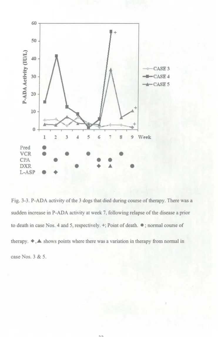
0ftbe three dogs that died, two cases
(Nos.
4 &5)
showed marked elevation in total P-ADA activityupon relapse after having relatively stabilized, however the dog with mediastinal lymphoma(case
No・3)
did not show such a pattern(Fig・
3-3)・
3.5. DISCUSSION
In this s山dy total P-ADA activities were higher in the dogs
with lympboma than the over 5 years101d controls・ ADA is found in most tissues, its activityis
greatest in the lymphoid tissues, and more specifically the T lymphocytes
(Adams
and Harkness,1976)・
These higher levels of P-ADA activitycould be a reflection of theprolifTerative activlty Of the tumors and the stage of differentiation reached by both the
nomal and the neoplastic lympboid cells.
Our results did not show any significant difference in total P-ADA between dogs with mediastinal and those with multicentric lymphoma at presentation・ According to a s山dy done in human lymph node samples by Ganeshaguru et al
(1981),
the highest levels of ADA were found in T-cell tumors of lymphoblastic anddi乱se undifferentiated types while in the B-celltype tumors, the level of ADA varied with the proportions of T-cells in the tumor. Therefore, tumor characteristics
including phenotype may be responsible for the differences seen between P-ADA
activities in the two groups.
According to Vezzoni et al
(1985),
a relationship was fわund between ADAactivityof various
histotypes
of non-Hodgkin's lymphoma in humans, and their gradeofmalignancy・ Our results did not show any association between the cancer stage and
total plasma
activityat
presentation.Total PIADA activity was seen to decrease drastically upon the
commencement of treatment but then was seen to fluctuate throughout the course and
appeared to be very variable. There was also great variabilityin the PIADA activityat presentation・ A study in humans, by Ponce et ai
(2004),
reported that significant differences seen between the lymphomasubtypes
are suggestive of inherent biologicalfeatures and clinical behavior of these tumors. These findings may account for the
variabilityseen in total P-ADA activityprior and during the chemotherapeutic regime・
The dramatic elevation of total P-ADA activity upon relapse in patients
(Case
Nos. 4 &
5)
and the strong relationship noted between lymph node size and P-ADAactivity(case
No.4)
reiterates the potential use of this enzyme in patient monitoring・ADA activl_ty Of ceiis缶.om human neoplastic lymph nodes is related to the proliferative activityof lymphomas
(Ungerer
et al,1992)・
This probably in turnaffects the plasma and a similar assumptlOn could be made in dogs・
ADA may be as useful in the diagnosis of canine lymphoma like it is in human
medicine. However the tumor
histologicaltype
may be amajor
determining factor,because ADA has been reported as a marker of human lymphoblastic lymphoma that showed unusually bigb enzyme levels in a study by Vezzoni et al
(1984)・
Since there is no information in the literature on ADA activityin canine lymphoma or itsresponse to cbemotherapeutic treatment, this preliminary s山dy provides valuable insight on the potential oftbis enzy皿e in the diagnosis and assessment of the canine
patient with lymphoma・ Our study lS, however, limited by the lack of tumor
characterization. Based on these results, further studies were carried out to investlgate the role of lymphocytes and neutrophils in P-ADA activityin canine lymphoma・
Table 3-1. Profile ofdogswith lymphoma at first presentation
CASE BREED AGE SEX LYMPHOMA CANCER Initial
NO. (yrs) STAGE ADA
(IU/L) 1 2 3 4 5 6 Corgi Golden Retriever
Mixed breed dog
Maltese Poodle Sbi tzu Yorkshire terrier 4 8 ll 12 7 9 F F M M M M Multicentric Mediastinal Mediastinal Multicentric Mu lticentric Multicentric 5 1 5 5 4 5
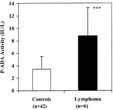
′ヽ ,A ii=ヨ こ) ■■l ㌔-′ ヨ! .t>・ 'B U i < (∋
i
B< 8 6 4 2 0 Controls Lymphoma(n-42)
(n=6)
Fig. 311. P-ADAー' activityof controls and dogs with lymphoma・ Dogs with lymphoma had higher P-ADA activity than controls
(***P<0・005)・
This result showed that冒
至】 巳.普
> :a U < <葛
i
1 2 3 4 5 Pred + VCR + + + CPA ● DXR L-AS】) ● 6 0 4 3 ′-ヽ 【;7 日 り ヽ-′ 43 月 【アノ P二 pせ ○ 貞 J∃ A 百 ∃l J 6 7 Ⅵ7eek ●Fig. 3-2. Strong relationship between P-ADA activityand lymph node size in case No・ 4withmulticentric lymphoma・ The corresponding course of chemotherapeutic
treatment undertaken isalso shown. Pred. was administered per os every other day・
冒
i) 亡.杏
T 'j: もl < <守
I ≡≡ --(.A邪S +('A5F J +CAS君5 き 6 ? $ 9 Week Pred ● VC見 ● ● ● CPA ● DX乱 しAS‡I ● ◆ ● ●Fig. 313. PIADA activityofthe 3 dogs that died dming course of therapy. There was a
sudden increase in P-ADA activityatweek 7, following relapse of the disease aprior to death in case Nos. 4and 5, respectively. +; Point of death. + ;normal course of
therapy. +
,A shows points Where there
was a variation in therapy &om normal in
CHAPTER
4INVESTIGATION
OF
ADAACTIVITY
IN LYMPHOCYTES
ANDNEUTROPHILS
INNORMAL
ANDDISEASED
ANIMALS
4.1. INTRODUCTION
In view of the fact that PIADA activityhas some potential in canine
lymphoma, we further investigated its source in peripheral blood cells・ Numerous
reports suggest that spontaneous山mors in humans are recognlZed as antlgenic by the
host. In many Instances, however, the malignant tissue fails to evoke an immune
response capable of destroying the neoplastic cells. It has been suggested in a study
by Green and °han
(1973),
that increased levels orAdo in lympbocytes may result inan inabilityof the cells to divide. The level of ADA activitymight influence the
capability of the immune system to respond
(Uberti
et al,1976).
FIV is atypical lentivirus that replicates preferentially in T cells and is
stmc山rally similar to HIV
(Bendinelli
et al,1995)・
Tberefbre in both human beingsand cats, the disease is characterized by severe impalrment Of T cell functions and
cellular immune response due to infection of CD4'ce11s
(Ackley
et al, 1990, Joshi etal,
2004).
Gradual reduction in both the percentage and the absolute number of CD4'T cells is one of the most striking lmmunOloglCal consequences of FIV infection
resulting in the reduction of the CD4/CD8 ratio
(Hoffman-Fezer
et al,1992)・
increased incidence of lymphoma. Unlike HIV infection where the pnmary receptor for HIV is CD4,
Shimojima
et al(2004)
identified that the primary receptor thatpromotes viral binding and renders CD4+ cells permissive to infection, as CD134 in
FIV. Despite the progressive deterioration of T cell function, the abilityof B cells to
recognize and respond to T-independent antigenic stimulus was not affected
(Torten
et ai,
i991).
FIV aiso replicates in macrophages and astrocytes. Primary infection ofcats by FIV is associated with a protracted asymptomatic phase of several months or
years that in some cats culminates in the development of immunosuppression・ Ishida and Tomoda
(1990)
have proposed classi丘cation of FIV stages as fわllows: Primaryinfection, AC, PGL characterized by generalized lymphadenopathy, ARC characterized by weight loss, bacterial and viral infections, and FAIDS characterized
by severe secondary and opportunistic chronic infections, tumors and wastlng・
_fiematol_og!c man_ifestations of FIV infection include anemia, 1ymphopenia, nentropenia and thrombocytopenia.
4.2. OBJECTIVE
To investigate ADA activity in blood peripheral cells in canine lymphoma and FIV. The diseases chosen were based on the results丘om the survey obtained in the previous chapter.
4.3。 MATERIALS AND METHODS 4。31. ANIMALS
Heparinized blood was collected from ll healthy dogs
(5
males and 6females)
aged 5.7j=3.0 years・ Eleven dogs with lymphoma(7
males and 4females)
aged 7・3 ±2・3 years・ Lymphoma diagnosis was made by血e-needle asplration biopsy, wbicb was then stained and examined.
Heparinized blood was also collected from 13 healthy controls and ll FIV positive cats・ The control group was made up of 6 males and 7 females and the age
ranged丘om 6・7±4・5 years・ The FIV positive cats Ⅵ′ere comprised of 5 males and 6
females and the age ranged &om 9・Oj=2・5 years・ Of the positive cats, 7 were in the
AC stage of infection while 4 were in the ARC stage・ All the cats included in this
work were seronegative for Feline leukemia virus antigen and the FIV-positive group
was seropositive fわr FIV antibody
(IDEXX
Laboratories, Portland,Maine)・
4.32. CELL SEPARATION
pBMC and PMNC were separated using the double densityHistopaque@
(sigma-Aldrich,
St. Louis, MO,USA)
separation according to the method described by Strasser et al(1998).
Hl.077 was carefully layered onto 4 ml Hl・1 19 and stored at4oC until use・ The columns were kept on ice in separate conical 12 ml centrifugal
polypropylene山bes・ 4 ml ofblood was layered on the low gradient solution uslng a
21G hypodermic needle on a synnge・ The tubes were centrifuged at 350 x g for 30
min at room temperature with a swing-Out rotor and the process terminatedwithout applying brakes.
was layered over the Hl・119・ Six milliliters of whole blood was layered over the
Hl.077 and. centrifuge at700 x g for 20 minuteswith no brake atroom temperature.
After centrifugation the top Inter-Phase layer consisting Of PBMC and the second layer of PMNC were collected and transferred to separate 50 ml centrifuge
山bes fわr washing. Washing was done with D-PBS
(Sigma-Aldrich,
St. Louis, MO,TuTSA)
and centrifuged at 200 x g for lOmirluteS. Red bl100d ceiis were iysed usingammonium chloride buffer solution and the cells washed twice. The PBMCs were
cultured for 1 hour in RPMI-1640 media containing 10% FBS
(Sigma-Aldrich,
St. Louis, MO,USA)
at 37oC and 5% CO2 in a humidified incubator to removemonocytes. Lymphocytes were then harvested for ADA analysis.
4.33. MEASUREMENT OF LYMPHOCYTE and NEUTROPHIL ADA ACTIVITY
The cells were counted uslng a hemocytometer and lysed by ultrasound. The
suspension was then centrifuged and the cell lysate-supernatant was analyzed for
ADA activitycontent. P-ADA activitywas measured as earlier described in chapter 2・
Statistical analyses were carried out uslng Student's T test and Pearson's
correlation. Results were presented as mean±SD and P<0.05 was considered to be of
statistical signi丘cance.
4.4. RESULTS
4.41. CANINE LYMPHOCYTE/NEUTROPHIL ADA ACTVITY
control groupしADA activity was 2.6± 1.6
(Ⅳ/106
cells)
and that of dogsADA activity of the dogs with lymphoma when compared with the control group
(P<0.05,
Fig.4-1).
N-ADA activity was 1.2±0.52
(IU/106 cells)
in the controlsand 0.4±0.23
(IU/106 cells),
in the dogs with lymphoma・ Healthy controls had higher N-ADAactivitythan dogs with lymphoma
(P<0・0001,
Fig.4-2).
L-ADA was significantlyhigher than N-ADA in both groups
(P<0・05).
There was, however, no significantcorrelation between L-ADA and P-ADA activities in neither group・
4.42。 FELINE LYMPHOCYTE/NEUTROPHIL ADA ACTIVITY
Tbe control cats'L-ADA activity was 0.78±0.67
(IU/106
cells)
and ofFIV-positive cats was 1,4±2.06
(Ⅳ/106 cells)
and is shown in Fig. 4-3. There was nosignificant difference between FIV positive and FIV negative L-ADA activity.The
AC groupしADA activity was 0.68±0.28
(IU/106 cells)
and ARC group was 3.55±3・096
(IU/106 cells).
There was a significant diffei・enCe betweerl the ARC group andthe AC and control groups
(P<0.05,
Fig.4-3).
N-ADA activity in the nomal cats was 0.19±0.22
(IU/106 cells)
and 0.22±o・36
(〟/106 cells)
in the FIV positive cats・ In AC and ARC groups, N-ADA was 0.09j=0・15
(IU/106
cells)
and O・98-+0・54(IU/106 cells),
respecfively・ N-ADAactivity
was slgnificantly higher in cats in the ARC stage of FIV infection than the other groups
(P<0・05,
Fig・4-4).
L-ADA activitywas significantly higher than N-ADA in all groups(P<0・05)・
There was a negative co汀elation between P-ADA activity andしADA activityin the control group
(r--0.55, P<0.05, Fig.
4-5),
but there was none found in the FIV positive cats・ Similarly there was no relationship fTound betweenP-4.5. DISCUSSION
This study reported for the first time, L-ADA activity,in both the canine and feline species・ In case of the dogs, L-ADA was slgnificantly elevated in the dogs with
lympboma compared with the healthy controls
(P<0.05).
The result concurred with that of Meier et ai(i976)
who reported high L-ADAactivityvaiues
in human patients with iymphoma・ Muller et al(1982),
on the other hand, reported reduced levels ofADA activity in Hodgkin lympboma. Carter et al
(1986)
have shown that there arestrong similarities of morphology and behavior between human non-Hodgkin's lympbomas and canine lympbomas. Uberti et al
(1976)
have suggested that variations in purine or pynmidine concentrations within the lympbocytes may result in reduced immune response directed agalnSt山mor antlgenS and neoplastic cells. In addition, previous studies in human have suggested that L-ADA activity may Offer insight into molecular aspects of the immune mechanism and hosト山mor interactions(Su丘・in
et al, 1977 and1978)・
The increase inしADA activity in the dogs with lympboma may be largely a肘ibuted to山mor cells present in the peripheral blood atpresentation.The results showed a significant difference in L-ADA activitybetween ARC
group and the other groups
(P<0.05).
Our results concur with those of Christensen etal
(1988)
who fわund that L-ADA activity was increased in HIV patients and that this increase was only slgnificant in the AIDS and ARC patients. Previous studies haverepo朽ed that mitogen-stimulated lymphocytes produce increased ADA
(Hovi
et al,1976)・
Therefore, the results seen in cats in the ARC stage of infection may be areflection of the on-golng Immune aCtivation and virus multiplication that has been reported in this phase of the disease.
IncreasedしADA activity has been reported in leprosy
(Sebgal
et al,1992),
typhoid fever
(Galanti
et al,1981),
and HIV infection(Christensen
et al,1988)I
Decreased L-ADA activityhas been fわund in diseases causing an impairment of the
immune response, such as acute lymphocytic leukemia in children
(Zimmer
et al,1975),
tumor patients(Uberti
et al,1976),
glomerulonephritis(Klinger
et al,1983),
chronic active liver diseases
(Nardieiio
et ai,1983)
and renal adenocarcinoma(Sufrirl
et al,1978).
The results show that canine and feline neutrophils also produce ADA・ However, in both cases L-ADA activity Was Significantly higher than that of
neutrophils. This finding concurs with the findings of other authors who have suggested that lymphocytes are an important component of ADA activlty・Erel et al
(1998)
made similar obseⅣations where L-ADA activity was signi丘cantly higher than N-ADA activity in both controls and patients with leisbmaniasis. In dogs Ⅵ′itb lymphoma, N-ADA activitywas seen to significantly decrease compared to thecontrols. S山dies in bumans with lymphoma, have shown some degree of neutrophil
dysfunction exists including reduced chemotaxis and adherence to nylon wool in
some patients which improved following commencing therapy
(Fliedner
et al, 1979 and McCoⅢnack et al,1978).
In addition, one s山dy showed a marked increase in cellcount, enzyme release, phagocytosis and killing, following stimulation
(Fossat
et al,1994).
These findings suggest that, like in the case in humans, neutrophils in caninelymphoma may also exist in a depressed state resulting ln a reduction in enzyme
synthesis including ADA・ On the other hand, cats in the ARC stage of FIV infection had significantly elevated N-ADA activity1evels. In HIV infection, patients had activated neutrophils that showed increased apoptosis, decreased viability and
(pitrak,
1999 and Kubes et al,2003)・
In cats therefore the increased N-ADA activity may be due activation status of the cells in FIV infection resulting ln increaseden2γme release・
Despite bigb nomal P-ADA activity in cats, L-ADA activity was relatively
low, contrary to the case in dogs. Tanabe
(1993)
also obseⅣed that tissue ADAactivlty Was much l10Wer thanP-ADA activity・In dogswith lymphoma, elevated L-ADA activity may largely be a renectiorl Of tumor cell presence in the peripheral
blood・ In this regard LIADA activity may be useful in distinguishing lymphoma from
mere lymphocytosis due to other causesI In cats L-ADA activitymay be an indicator
of the immune system status and phase of FIV infection・ Further experiments were
carried out to investlgate the role of T or B-1ympnocytes in the selected canine and ll
1S ′ヽ
芸16
V言14
▼■5
12 ■■こ
川 :E: .>_◆■ f; U < 6i4
1
2 0 Controls(I)-1I)
Lymphoma(m≡ll)
Fig・ 411 I Sig山ficant increase in L-ADA activityin dogswith lymphoma compared to
the healthy controls
(*P<0・05)A
Dogswith lymphoma have higher LIADA activity than healthy controls.′ヽ 7J 4) U ∈> ■■
5
l■ll ヽ■′倉
> :J5 くJ < < 負 < 1 2: 1.8 1.6 1.4 1.2 I 0.8 0.6 0.4 0.2 0 Controls(n=11)
Lympho ma(n=11)
Fig・ 4-2. N-ADA activities in healthy controls and dogs with lymphoma, Dogswith lymphoma had a significantly lower N-ADAthanthe control group
(****P<0.0001).
ゴ y. FF] E∃
邑
王、 ●芦 ミロ tJ < < 良 < iCoJltT'OIs FIV+ AC ARC
(n=1 3) (n-11) (n=7) (n≡ヰ)
Fig1 4-3・ L-ADA activity of controls, FⅣ+, AC and ARC stages of FIV infection・ The LIADA of the ARC group was significantly elevated compared to the other two
groups
(*P<0・05).
Increase in LIADA occurs in cats inthe ARC stage of FIVJ.t[ 富1・4 4) 1'e ).三 ;⊃
盲1
-≡慧o・8
<!
o・6 2: 0.4 Controls FTV十 AC ARC (n-;13) (b-11) (I)ヒ7) (n-4)Fig・ 4-4・ N-ADA activities of controls, FⅣ-positive, AC and ARC stages of FIV infection・ N-ADA activityinthe ARC group was significantly higherthanthe
other groups
(*P<0.05).
′■■\ ∽ l■■l lllll■■ q) U \丘 ∈〉 I■・・{ i? i) ■■ ヽ■′ iヽ .t> 1■■ U < < `∋ < l J 0 0 20 40 60 80 100
P-ADA Activity
(IU/L)
Fig・ 4-5・ Negative correlation between P-ADA activity and L-ADA activity ln nOmal cats
(rニー0.55,P<0.05).
CHAPTER
5
INVESTIGATION
OF
ADENOSINEDEAMINASE
IN T AND B
LYMPHOCYTES
5.1. INTRODUCTION
In the previous chapterしADA activity was shown to be the
major
componentof ADA activity, we further investigated the relationship between T and
B-1ymphocytes and ADA activity.
ADA is an enzyme capable of catalyzlng the cataboiism of purine bases and
whose prlnCIPal biologic activity is detected in T lymphocytes・ The role of this enzyme in cellular immune function was highlighted followlng the discovery of reduced levels in patients with SCID. In humans, ADA activityis 5120-fold higher in
T lymphocytes than B lymphocytes
(Sullivan
and Osbourne,1977)・
In humans, ADA has been considered a marker of cell-mediated immunity(Baganha
et al,1990).
Low T-ADA activity has also been reported in patients with multiple sclerosis(Vivekanandhan
et al,2005).
A negative correlation was observed between serumADA and T lymphocytes percentage in human patients wi也nephrotic syndrome
(Misra
et al,1997).
ADA activityhas been identified as a marker of T cell activation(Rose
et al,2001).
Fur也ennore, several reports have observed that ADA interacts with CD 26 on the surface of T cells surface resulting ln a COIStimulatory effect in the activation or T cells(Blazquez
et al, 1992, Morimoto and Schlossman, 1998, Cordero et al, 2001 and Pacheco et al,2005).
Most of the lymphomas described in canines onglnate from B lymphomas
(Teske, 1994).
These lymphomas react much better to chemotherapy than do T cell lymphomas, which make up 10-38% of the cases and have a much worse prognosis(Hahn
et al,1992).
It is generally accepted that FIV may be useful as a model for AIDS・ Several
authors have reported that serum ADA activity is elevated in HIV infection
(Gakis
etal, 1989, Goto et al, i992 and Inigo et al,
1992).
Furthermore, a relationship has beendescribed in which serum ADA correlated with retroviral infection in HIV infection
(Mastrioanni
et al,1987).
ADA is also reported to be a useful marker of progressionEXPERIMENT A: INVESTIGATION OF T AND B LYMPHOCYTE ADA ACTIVITY IN DOGS AND CATS
5.2. OBJECTIVE
The present study was undertaken to investigate the role of T and B lymphocyte in ADA activityin dogs and cats.
5.3。 MATERIALS AND METHODS 5.31. ANIMALS
Heparinized blood was collected from 10 healthy dogs of various breeds, and
8 dogs with lymphoma. The control group composed of 6 males, 4 fTemales,with an
average age of 3,7± 1.8 years. The group of dogs with lymphoma composed 5 males,
3 females and an average age of 7.5j=2.1 years. None of the dogs were receiving
immunosuppressive medication atthe time of sampling.
Heparinized blood was collected from 10 healthy FIV-negative and 8
FIV-infected cats. All the cats included in this work were seronegative for Feline leukemia
virus antigen and the FIV-positive group was seropositive fわr FIV antibody
(IDEXX
Laboratories, Portland,
Maine).
The cats were of mixed-breed with the FIV-negativegroup composed of 6 males, 4 females, with an average age of4・7±4・2 years・ The
FIV positive group composed 5 males, 3 females and an average age of 7・5±2・1