Stability of Orbital Floor Fracture Fixation After Endoscope-Assisted Balloon Placement
日本大学医学部形成外科学系形成外科学分野
樫村 勉
申請年 2017年
指導教員 仲沢 弘明
Copyright © 2017 Mutaz B. Habal, MD. Unauthorized reproduction of this article is prohibited.
Stability of Orbital Floor Fracture Fixation After Endoscope-
Assisted Balloon Placement
Tsutomu Kashimura, MD, Kazutaka Soejima, MD, Yuji Kikuchi, MD, and Hiroaki Nakazawa, MD
Abstract: In recent years, endoscope-assisted balloon fixation using transantral and endonasal approaches has gained popularity as a minimally invasive treatment for orbital floor fractures. How- ever, the optimal duration for balloon placement and the efficacy of the method have not been fully evaluated. The authors report their assessment of this method using postoperative and chronological measurements of the maxillary sinus volume.
Fourteen patients with blowout fracture of the orbital floor who underwent reduction using endoscopic transantral and endonasal approaches followed by 6-week fixation with a balloon were evaluated. The volume of the maxillary sinus was measured for comparison using computed tomography at the time of balloon removal and 6 months after the surgery.
The ratio of change in the maxillary sinus volume (maxillary sinus volume 6 months after surgery/maxillary sinus volume at balloon removal) for all subjects was 0.90 to 1.04 (0.960.44, meanSD). No postoperative reduction in volume was detected, indicating satisfactory fixation. Postoperative computed tomogra- phy showed bone regeneration in the orbital floor in all patients in whom the fractured bone fragments were removed. No subjects had remaining enophthalmos greater than 2 mm.
The postoperative change in the maxillary sinus volume was small, confirming the efficacy of 6-week balloon placement. This method was effective even in patients in whom fractured bone fragments were removed. Therefore, it is advisable to remove the fractured bone fragments if there is concern that the fragments will stray into the orbit due to inflation of the balloon.
Key Words:Balloon, maxillary sinus volume, orbital floor fracture, transantral and transnasal approaches
O
rbital floor fractures develop due to a blunt injury, resulting in ocular motility disorder and/or enophthalmos. Various treat- ment methods for this type of fracture have been reported, and discussions continue to this day regarding operative indications, methods, and timing.Fracture reduction using endoscopic transantral and transnasal approaches followed by fixation with the balloon method was reported recently. We employ the balloon method routinely as a minimally invasive treatment for orbital floor fractures. We
reported that shortening of the connecting tube by enlarging the natural ostium of the maxillary sinus and hiding the tube in the nasal cavity enable long-term placement of the balloon, resulting in a good treatment outcome.1However, the balloon method has been insufficiently examined, especially with regard to the optimal duration for balloon placement and the efficacy in fixing the orbital floor.
In this study, we examined the efficacy of the balloon method for fixing the orbital floor using postoperative and chronological measurements of the maxillary sinus volume.
METHODS
The subjects consisted of 14 patients with blowout fracture of the orbital floor, who underwent reduction at our institution using endoscope-assisted transantral and endonasal approaches followed by fixation with the balloon method between October 2010 and September 2016. The fracture was diagnosed using computed tomography (CT). The subjects underwent ophthalmologic inspec- tions, that is, visual acuity test, eye movement test, exophthalmo- metry, and field diplopia test. The indication for surgery with the balloon method using endoscopic transantral approaches was the presence of diplopia or enophthalmos greater than 2 mm compared with the unaffected side approximately 2 weeks after injury.
Patients who developed missing muscle syndrome due to contrac- tion of the extraocular muscle were excluded.2Pediatric patients were excluded because of the possibility of problems with post- operative tube management due to a high incidence of linear fractures in young patients.3
The subjects consisted of 10 men and 4 women, aged 15 to 68 years (45.912.9, meanSD). The causes of injury were home accidents and falling from heights (6 patients, 42.8%), sports (4 patients, 28.6%), assault (2 patients, 14.3%), and traffic accident (2 patients, 14.3%). The mean period from the time of injury to surgery was 20.99.1 (meanSD) days. Six patients (42.8%) were diag- nosed as having enophthalmos of greater than 2 mm, and 10 patients (71.4%) had persistent diplopia before the surgery. Patients were informed about the potential risks and benefits of all treatment options, and written informed consent was obtained from all patients or their parents. The fractured bone fragments were removed in 8 patients and balloon placement was maintained for 6 weeks in all patients.
Surgical Technique
The subjects underwent surgery under general anesthesia. After exposing the anterior wall of the maxillary sinus by incising the oral vestibule, an area measuring 1.51.5 cm was fenestrated. After approaching the orbital floor from the maxillary sinus side, the floor was reduced endoscopically. In patients in whom the surgeon judged that the fractured bone fragments would stray into the orbit based on preoperative CT results, the fragments were removed while preserving the periosteum. The maxillary sinus membrane- like portion was then incised from the nasal cavity to approach the interior of the maxillary sinus. A balloon (#3007, Koken Co, Tokyo, Japan) was inserted into the maxillary sinus and an injection tube was guided into the nasal cavity through the maxillary sinus membrane-like portion. Saline was injected through the tube while checking the exophthalmos as well as the traction.
One day postoperatively, the subjects underwent CT to check the degree of reduction. The volume of saline injected into the balloon was adjusted as necessary. Thereafter, the injection tube was ligated, cut apart, and hidden in the nasal cavity.
Six weeks postoperatively, the balloon was removed without anesthesia on an outpatient basis. The subjects underwent CT preoperatively, 1 day postoperatively, at the time of balloon removal, and 6 months postoperatively.
From the Department of Plastic and Reconstructive Surgery, Nihon University School of Medicine, Tokyo, Japan.
Received February 24, 2017.
Accepted for publication March 19, 2017.
Address correspondence and reprint requests to Tsutomu Kashimura, MD, Department of Plastic and Reconstructive Surgery, Nihon University School of Medicine, 30-1 Ooyaguchikami-cho, Itabashi-ku, Tokyo 173- 8610, Japan; E-mail: [email protected]
The authors report no conflicts of interest.
Copyright#2017 by Mutaz B. Habal, MD ISSN: 1049-2275
DOI: 10.1097/SCS.0000000000003826
The Journal of Craniofacial Surgery Volume 28, Number 7, October 2017 Brief Clinical Studies
#2017 Mutaz B. Habal, MD
e669
Copyright © 2017 Mutaz B. Habal, MD. Unauthorized reproduction of this article is prohibited.
Measurement of Maxillary Sinus Volume
The maxillary sinus volume was measured using data from 0.5 mm slice CT images of the frontal plane and Ziostation (Ziosoft, Tokyo, Japan), the analytical software for CT images (Fig. 1). The maxillary sinus volumes at the time of balloon removal and 6 months after the surgery were measured. The maxillary sinus volume 6 months after the surgery was divided by the volume at the time of balloon removal to calculate the volume change ratio. A smaller value indicates re-recession in the orbital floor caused by the reduction of the maxillary sinus volume after balloon removal.
Assessment of Degree of Exophthalmos
Exophthalmometry was based on CT data and was conducted according to the report by Sung et al.4
RESULTS
The maxillary sinus volume for all subjects at the time of balloon removal was 16.13 to 31.79 mL (23.144.94, meanSD), whereas the maxillary sinus volume 6 months after injury was 15.09 to 29.36 mL (22.314.79, meanSD). The difference in the maxillary sinus volume (maxillary sinus volume upon balloon removal maxillary sinus volume 6 months after injury), that is, the amount of enlargement in the orbital volume was1.16 to 2.43 mL (0.831.09, meanSD). The ratio of change in the maxillary sinus volume for all subjects was 0.90 to 1.04 (0.960.41, meanSD) (Fig. 2). No postoperative reduction in the maxillary sinus volume was observed, indicating satisfactory fixation using the method.
The ratio of change in the maxillary sinus volume for patients in whom the fractured bone fragments were preserved and those in whom the fragments were removed was 0.91 to 1.00 (0.960.04, meanSD) and 0.90 to 1.04 (0.970.05, meanSD), respect- ively (Fig. 3). Thus, satisfactory fixation occurred in both patients.
Additionally, postoperative CT showed bone regeneration in the orbital floor in all patients in whom the fractured bone fragments were removed.
Six months postoperatively, diplopia was found in 2 subjects.
None of the subjects had remnant enophthalmos of 2 mm or greater.
No correlation was found between the change in maxillary sinus volume and the degree of exophthalmos 6 months after injury.
CLINICAL REPORT
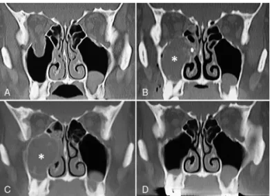
A 37-year-old female visited our department because she was hit by a baseball. The initial examination found upward gaze diplopia,
2.21 mm enophthalmos compared with the unaffected side, and sensory disturbance in the right cheek. Because CT showed a wide range of right orbital floor fractures, surgery was indicated (Fig. 4A).
Thirty days after the injury, she underwent surgery under general anesthesia. Since the orbital floor bone was blown out, there was concern that the bone would stray into the orbit due to inflation of the balloon. Therefore, the fractured bone fragments were removed.
The balloon was inserted into the maxillary sinus, followed by injection of 14 mL of saline (Fig. 4B).
Six weeks postoperatively, the balloon in the sinus was removed.
Computed tomography performed at the time of the removal showed that the orbital floor bone had regenerated (Fig. 4C). Six months after the injury, another CT scan showed that the bone had regenerated without re-recession (Fig. 4D). The ratio of change in the maxillary sinus volume 6 months after the surgery was 0.964, indicating satisfactory fixation. In addition, neither diplopia nor enophthalmos was found.
FIGURE 1. Measurement of maxillary sinus volume. (A) Tracing of the maxillary sinus on a computed tomography image of the frontal plane. (B) Measurement of the volume of the maxillary sinus using image analysis software.
FIGURE 2.Ratio of change in the maxillary sinus volume (maxillary sinus volume in balloon removal/maxillary sinus volume 6 mo after surgery) for all subjects.
FIGURE 3.Ratio of change in the maxillary sinus volume (maxillary sinus volume upon balloon removal/maxillary sinus volume 6 mo after surgery) for patients in whom fractured bone fragments were retained and patients in whom the fragments were removed.
Brief Clinical Studies The Journal of Craniofacial Surgery Volume 28, Number 7, October 2017
e670
#2017 Mutaz B. Habal, MDCopyright © 2017 Mutaz B. Habal, MD. Unauthorized reproduction of this article is prohibited.
DISCUSSION
Many surgical treatment methods for the orbital floor fracture have been reported. Thus far, methods involving a transorbital approach with subciliary incision and/or transconjunctival incision have been widely utilized. However, the incisions may cause postoperative scars and/or complications such as ectropion of the lid and gran- ulomatosis.5In addition, in the event of fractures in the posterior part of the orbit, these incisions cannot secure a sufficient operative field in many patients, increasing the risk of optic nerve injury.6In the 1980s, treatment methods for fractures by means of endoscopic paranasal sinus surgery were developed, and treatment of orbital floor fractures using an endoscopic transantral approach was reported by Saunders et al7 in 1997. The transantral approach has the advantage of avoiding eyelid complications because it requires no incision of the exposed portion of the face. In addition, the approach can secure a good operative field for fractures in the posterior part of the orbit. Jin et al8reported that the approach could better improve diplopia and enophthalmos.
Various fixation methods that can be employed after the reduction of the orbital contents using the transantral approach have also been reported. Above all, use of a urethral catheter is common.9A urethral catheter is generally placed for around 2 weeks because it has a large portion exposed off the nostril and is difficult to cut short. Fixation using a balloon is still under debate regarding the duration of balloon placement.6However, a short duration is known to cause enophthal- mos due to re-recession after balloon removal.10We used a balloon developed for the maxillary sinus (#307, Koken Co, Tokyo, Japan) for fixation. By confirming the proper balloon inflation by means of CT 1 day postoperatively, we cut the tube and hid it in the nasal cavity, enabling placement of the balloon without restricting daily activities for as long as 6 weeks after the surgery.
In this study, we examined the efficacy of the balloon method for fixing the orbital floor through comparison of the maxillary sinus volume by means of postoperative CT. We postulated that the maxillary sinus surrounded by bones would reflect changes in the orbital floor more accurately because it is more similar to a closed cavity than the orbit. The maxillary sinus volume 6 months after the surgery, long after the 6-week balloon placement, showed neither
reduction nor re-recession compared with the volume at the time of balloon removal, indicating satisfactory fixation.
With regard to the degree of exophthalmos, enophthalmos of 2 mm or greater becomes an esthetic problem and is an indication for further treatment.4,11Raskin et al12mentioned that an increase in the orbital volume by 1 mL results in 0.47 mm enophthalmos. In the present study, a 2.43 mL maximal increase in the orbital volume was observed 6 months postoperatively, whereas none of the subjects developed enophthalmos of 2 mm or greater.
In this study, an increase in the maxillary sinus volume was observed in 3 subjects. It was previously reported that atrophy of the orbital contents occurs following orbital floor fracture.13 We speculate that this atrophy was responsible for the apparent increase in the maxillary sinus volume. We also speculate that the lack of correlation between the change in the maxillary sinus volume and the degree of exophthalmos was influenced by the atrophy-induced change in the volume of the orbital contents.
During fixation using the balloon method, there is a possibility that bone fragments will stray into the orbit because of balloon inflation, leading to complications due to visual disturbance.14Far- well and Hinohira et al reported that fractured bone fragments need to be removed through endoscopic surgery for blowout orbital floor fractures.10,15We placed the balloon after removing only the frac- tured bone fragments, preserving the periosteum where possible in patients in whom there was concern that the fragments would stray into the orbit. In this study, even in the patients in whom the fragments were removed, no postoperative reduction in the maxillary sinus volume was observed, indicating satisfactory fixation. Moreover, the orbital bone was found to have regenerated in patients in whom the fragments were removed. This is likely because preservation of the periosteum when removing the fractured bone fragments allowed bone regeneration through membranous ossification.
The efficacy of the balloon placement method for fixing the orbital floor after orbital floor fractures was evaluated through chronological measurements of the maxillary sinus volume. Comparison of the volume 6 months after the surgery and at the time of balloon removal showed minimal change in the volume, indicating satisfactory effi- cacy for the 6-week balloon placement. Even in patients in whom the fractured bone fragments were removed, sufficient fixation was observed. Therefore, it is advisable to remove fractured bone frag- ments in patients in whom there is concern that the fragments will stray into the orbit due to inflation of the balloon.
REFERENCES
1. Soejima K, Shimoda K, Kashimura T, et al. Endoscopic transmaxillary repair of orbital floor fractures: a minimally invasive treatment.J Plast Surg Hand Surg2013;47:368–373
2. Wachler BS, Holds JB. The missing muscle syndrome in blowout fractures: an indication for urgent surgery.Ophthal Plast Reconstr Surg1998;14:17–18
3. Kwon JH, Moon JH, Kwon MS, et al. The differences of blowout fracture of the inferior orbital wall between children and adults.Arch Otolaryngol Head Neck Surg2005;131:723–727
4. Sung YS, Chung CM, Hong IP. The correlation between the degree of enophthalmos and the extent of fracture in medial orbital wall fracture left untreated for over six months: a retrospective analysis of 81 cases at a single institution.Arch Plast Surg2013;40:335–340
5. Gosau M, Schoneich M, Draenert FG, et al. Retrospective analysis of orbital floor fractures—complications, outcome, and review.Clin Oral Investig2011;15:305–313
6. Jeon SY, Kwon JH, Kim JP, et al. Endoscopic intranasal reduction of the orbit in isolated blowout fractures.Acta Otolaryngol Suppl
2007;558:102–109
7. Saunders CJ, Whetzel TP, Stokes RB, et al. Transantral endoscopic orbital floor exploration: a cadaver and clinical study.Plast Reconstr Surg1997;100:575–581
FIGURE 4. Clinical report. (A) A CT image at the time of injury. (B) CT image obtained 1 day postoperatively. The fractured bone fragments are missing because they were removed. The asterisk () indicates the balloon. (C) Computed tomography image obtained 6 weeks postoperatively (at the time of balloon removal). The asterisk () indicates the balloon. Regeneration of the orbital bone is visible. (D) Computed tomography image obtained 6 months postoperatively. CT, computed tomography.
The Journal of Craniofacial Surgery Volume 28, Number 7, October 2017 Brief Clinical Studies
#2017 Mutaz B. Habal, MD
e671
Copyright © 2017 Mutaz B. Habal, MD. Unauthorized reproduction of this article is prohibited.
8. Jin HR, Yeon JY, Shin SO, et al. Endoscopic versus external repair of orbital blowout fractures.Otolaryngol Head Neck Surg 2007;136:
38–44
9. Otori N, Haruna S, Moriyama H. Endoscopic endonasal or transmaxillary repair of orbital floor fracture: a study of 88 patients treated in our department.Acta Otolaryngol2003;123:718–723 10. Hinohira Y, Yumoto E, Shimamura I. Endoscopic endonasal reduction
of blowout fractures of the orbital floor.Otolaryngol Head Neck Surg 2005;133:741–747
11. Koo L, Hatton MP, Rubin PAD. When is enophthalmos ‘‘significant’’?
Ophthal Plast Reconstr Surg2006;22:274–277
12. Raskin EM, Millman AL, Lubkin V, et al. Prediction of late enophthalmos by volumetric analysis of orbital fractures.Ophthal Plast Reconstr Surg1998;14:19–26
13. Kim SM, Jeong YS, Lee IJ, et al. Prediction of the development of late enophthalmos in pure blowout fractures: delayed orbital tissue atrophy plays a major role.Eur J Ophthalmol2017;27:104–108
14. Rosbe KW, Meredith SD, Holmes DK. Complication of maxillary sinus Foley balloon placement for orbital floor support.Otolaryngol Head Neck Surg1997;117:S148–S150
15. Farwell DG, Strong EB. Endoscopic repair of orbital floor fractures.
Otolaryngol Clin North Am2007;40:319–328
Brief Clinical Studies The Journal of Craniofacial Surgery Volume 28, Number 7, October 2017
e672
#2017 Mutaz B. Habal, MDAuthors: Tsutomu Kashimura, Kazutaka Soejima, Yuji Kikuchi, Hiroaki Nakazawa
Title of article: Stability of Orbital Floor Fracture Fixation After Endoscope-Assisted Balloon Placement Title of journal: The Journal of Craniofacial Surgery
October 2017 - Volume 28 - Issue 7 - p e669–e672 Website URL to the journal page:
http://journals.lww.com/jcraniofacialsurgery/Abstract/2017/10000/Stability_of_Orbital_Floor_Fracture_Fixation_After.86.aspx
1 Abstract
Background
In recent years, endoscope-assisted balloon fixation using transantral and endonasal approaches
has gained popularity as a minimally invasive treatment for orbital floor fractures. However, the
optimal duration for balloon placement and the efficacy of the method have not been fully evaluated.
We report our assessment of this method using postoperative and chronological measurements of
the maxillary sinus volume.
Method
Fourteen patients with blowout fracture of the orbital floor who underwent reduction using
endoscopic transantral and endonasal approaches followed by 6-week fixation with a balloon were
evaluated. The volume of the maxillary sinus was measured for comparison using computed
tomography at the time of balloon removal and 6 months after the surgery.
Result
The ratio of change in the maxillary sinus volume (maxillary sinus volume 6 months after surgery
/ maxillary sinus volume at balloon removal) for all subjects was 0.90 - 1.04 (0.96 ± 0.04, mean
± SD). No postoperative reduction in volume was detected, indicating satisfactory fixation.
Postoperative CT showed bone regeneration in the orbital floor in all cases in which the fractured
bone fragments were removed. No subjects had remaining enophthalmos greater than 2 mm.
Conclusion
The postoperative change in the maxillary sinus volume was small, confirming the efficacy of 6-
2
week balloon placement. This method was effective even in cases in which fractured bone fragments
were removed. Therefore, it is advisable to remove the fractured bone fragments if there is concern
that the fragments will stray into the orbit due to inflation of the balloon.
本文 背景
眼窩底骨折は、殴打やスポーツなどの眼窩部への鈍的外力に起因する眼窩内圧の上昇によっ て生じる骨折である。眼窩底骨折は、CT により診断され、骨折部の形態により線状型骨折と打ち抜 き型骨折に分類される(Figure 1)。
(Figure 1)
眼窩底骨折の画像診断による分類 左:線状型骨折、右:打ち抜き型骨折眼窩底骨折の症状には、眼球運動障害、複視、眼球陥凹、嘔吐、三叉神経第2枝領域の知覚障 害、鼻出血などが挙げられる。これらの中で、臨床上問題となり手術適応となるのは、複視と眼球陥 凹である(Figure 2 上)。骨折部に外眼筋や眼窩脂肪がトラップされることにより、眼球運動障害を生 じる。眼球運動障害は、上下方向に出現することが多く、上下方向の注視時に複視を生じる。また、
眼窩内容が上顎洞に逸脱することにより、眼球陥凹を生じる(Figure 2下)。
3
(Figure 2)
上:右眼窩底骨折により上方視で、右の上転障害と複視を認める。下:左の眼窩底骨折により右側と比較して
3
㎜の眼球陥凹を認める。眼窩底骨折の治療は、前述の骨折の型により異なる。線状型骨折は、眼窩底の欠損がなく、眼窩 底の再建が不要であり、逸脱した組織の整復のみを行う。一方、打ち抜き型骨折では、眼窩底の欠 損が大きく、眼窩底の再建が必要となり、骨移植や人工物などが用いられる。打ち抜き型骨折の手 術は、睫毛下切開や結膜切開による経眼窩アプローチが行われる。眼窩底にアプローチし、逸脱し た眼窩内容を眼窩内に整復し、骨欠損部を露出する。骨欠損部へは、肋軟骨や腸骨などの自家骨 や人工物を移植し、眼窩底の再建を行う。しかし、このような従来法には、いくつかの問題点が報告 されている[1]。経眼窩アプローチでは、露出部の瘢痕、眼瞼の変形、肉芽腫形成などの合併症の可 能性があり、骨移植は、健常部位への侵襲が必要であり、肋軟骨では気胸、腸骨では外側大腿皮神 経の損傷などが危惧される。また、人工物を用いた再建では、感染や露出の可能性がある。
1980
年代に内視鏡補助下での副鼻腔手術による治療法の開発が盛んにおこなわれ、1997年に内視鏡補助下に経上顎洞アプローチによる眼窩底骨折の治療が
Saunders
らにより報告された[2]。4
従来のバルーン留置法では、尿道バルーンが多く用いられていたが、尿道バルーンは、術後に体 外に大きく露出し日常生活に支障を来すため、留置期間が2週間程度に制限されていた[3, 4]。そ のため、バルーン抜去後の不安定さから、再陥凹の問題が残っていた[5]。
われわれは、従来法に改良を加えた内視鏡補助下上顎洞バルーン留置法について報告してき た[6]。本法では、バルーンを短切し鼻腔内に格納することで長期間の留置を可能にした。さらに上 顎洞自然孔を拡大しバルーンを留置することにより、バルーンの抜去の困難さと上顎洞炎を予防す ることで、バルーンの安全な長期間の留置を可能とした。本法は、1、顔面皮膚切開が不要である。2、
固定材料を必要としない。3、バルーンの注入量を変化させることで術後固定位置の調整が可能で ある。4、内視鏡補助下での経上顎洞アプローチでは、眼窩後方の骨折の整復を確実に行うことが できる[7]。5、退院後、バルーンの体外への露出がない。6、外来で麻酔を使用しないでバルーンの 抜去が可能である。7、バルーンの長期間の留置が可能である。といった利点を有する。
内視鏡補助下上顎洞バルーン留置法は、低侵襲であり多くの利点を有するが、バルーン抜去後 の再陥凹を予防し得る眼窩底の固定性の検討は不十分であり、いまだ定量的な評価に関する報
告はない。われわれは、内視鏡補助下上顎洞バルーン留置法において、術後の
CT
デジタルデータより上顎洞容積を測定し、眼窩底の固定性について検討した。
対象
2010
年からの6
年間に内視鏡補助下上顎洞バルーン留置法により治療した眼窩底骨折14
例を対象とし
Retrospective
に検討を行った。男性が10
例、女性が4
例、平均年齢は、45.9歳であった。転倒や転落による受傷が
6
例(42.8%)、スポーツによる受傷が4
例(28.6%)、暴力による受傷が2
5
例(14.3%)、交通事故による受傷が
2
例(14.3%)であった。術前症状として10
例に複視を認め、6例に健側と比較して
2mm
以上の眼球陥凹を認めた。受傷から手術までの平均日数は、20.9±9.1 日であった。6例で眼窩底の骨折片を温存し、8例で骨折片を除去した。バルーンは、全例で
6
週間の留置を行った。
手術手技
全身麻酔下に手術を施行した。患側の歯肉頬移行部より
5mm
程度口唇側から、口腔前庭切開を行い、経上顎洞アプローチを行った。切開部から上顎骨へと到達し、上顎洞前壁を広く剥離した。
眼窩下神経より尾側で
1.5
㎝程度の大きさで、上顎洞を開窓した。開窓した部分から内視鏡を上顎洞内に挿入し、逸脱した眼窩内容の整復を行った。術前の
CT
で骨折片が眼窩内に迷入することが危惧された症例は、骨膜を温存するよう愛護的に骨折片を除去した。続いて鼻腔内アプローチを行 った。ここでは、主に上顎洞膜様部の開窓による上顎洞自然孔の拡大を行った。次に、バルーンの 挿入を行った。上顎洞用のバルーン(#3007, Koken Co, Japan)を上顎洞内に挿入し、注入用チュー ブを拡大した上顎洞自然孔より鼻内から体外に誘導した。眼球陥凹ならびに眼球の動きが改善さ れたことを確認しつつ、注入用チューブより生理食塩水を注入した。口腔前庭切開部分を縫合し、
手術を終了した。
術後
1
日目にCT
で生理食塩水の注入量の確認を行い、必要に応じて調整を行った。注入量が適切であれば、バルーンのチューブ゙を切断し鼻腔内に格納した。術後
6
週間、外来で麻酔を使用しないでバルーンを抜去した。
6
手術適応と禁忌1、眼窩縁や上顎洞前壁に骨折を伴わない単純な眼窩底骨折(Pure Type)を適応とした。眼窩縁や
上顎洞前壁の骨折を伴う場合には、これらの骨折部位の固定の際に、上顎洞バルーンの損傷が危 惧されるためである(Figure 3 a, b)。2、外眼筋が眼窩内より上顎洞へと逸脱し、CTで眼窩内から外 眼筋が消失する
missing muscle syndrome
は、線状型骨折に合併しやすい[8]。線状型骨折では、バルーンにより逸脱した外眼筋や組織が絞扼され病態の悪化を招く可能性があるため、適応から 除外した(Figure 3 c)。3、小児例においては、前述の線状型骨折が多く、術後のバルーン留置中の
管理の問題もあり、適応から除外した[9]。さらに、受傷後
2
週間で複視もしくは2mm
以上の眼球陥凹が残存している症例を適応とした。
(Figure 3)
本法の適応と禁忌方法
術前後の臨床症状(複視・眼球陥凹)および
CT
をRetrospective
に検討した。上顎洞容積の計測
上顎洞容積は、0.5mm スライスの
CT
のデジタルデータより、CT 画像の解析ソフトウェアである7
Zio Station(Zio Soft
社製)を用い、前額断面の画像から上顎洞辺縁をトレースし計測した。バルーン抜去時の上顎洞容積と術後
6
カ月の上顎洞容積を測定した。術後6
カ月の上顎洞容積をバルーン抜去時の上顎洞容積で割り、上顎洞の容積比を算出した。上顎洞容積の比が小さいほど、バル ーン抜去後に上顎洞容積が縮小し、眼窩底が再陥凹していることを示す。
眼球突出度の評価
眼球突出度は、CTのデジタルデータより
Sung
らの報告に準じて測定を行った[10]。結果
全症例のバルーン抜去時の上顎洞容積は、16.13 - 31.79 ml(23.14 ± 4.94, mean ± SD)、受傷
後
6
カ月の上顎洞容積は、15.55 - 29.36 ml(22.31 ± 4.79, mean ± SD)であった。術後6
カ月の上顎洞容積は、Wilcoxonの符号付順位和検定で有意に縮小していた(Figure 4)。上顎洞容積の差
(バルーン抜去時の上顎洞容積―受傷後 6
カ月の上顎洞容積)、すなわち眼窩容積の拡大量は、-1.16 – 2.43 ml (0.83 ± 1.09, mean ± SD)であった。
(Figure 4)
全症例のバルーン抜去時と術後6
カ月の上顎洞容積8
骨温存例のバルーン抜去時の上顎洞容積は、16.68 - 27.0 ml(21.28 ± 4.02, mean ± SD)、受
傷後
6
カ月の上顎洞容積は、16.75 – 24.74 ml(20.46 ± 3.61, mean ± SD)であった。術後6
カ月の上顎洞容積は、Wilcoxonの符号付順位和検定で有意差に縮小していた(Figure 5)。
(Figure 5)
骨温存例のバルーン抜去時と術後6
カ月の上顎洞容積骨除去例のバルーン抜去時の上顎洞容積は、16.13 - 31.79 ml(24.53 ± 5.10, mean ± SD)、受 傷後
6
カ月の上顎洞容積は、15.55 - 29.36 ml(23.70 ± 5.09, mean ± SD)であった。術後6
カ月の上顎洞容積は、Wilcoxonの符号付順位和検定で有意な縮小を認めなかった(Figure 6)。
9
(Figure 6)
骨除去例のバルーン抜去時と術後6
カ月の上顎洞容積全症例の上顎洞容積の比は、0.90 - 1.04 (0.96 ± 0.04, mean ± SD)であった(Figure 7)。術後 上顎洞容積の縮小を認めず、良好な固定性が得られていた。
(Figure 7)
全症例の上顎洞容積の比10
骨温存例の上顎洞容積の比は、0.91 - 1.00 (0.96 ± 0.04, mean ± SD)、骨除去例は
0.90 - 1.04
(0.97 ± 0.05, mean ± SD)であった。骨除去例でも、骨温存例と同様に Mann-Whitney
の U検定で優位差なく、良好な固定性が得られていた(Figure 8)。
(Figure 8)
骨温存例と骨除去例の上顎洞容積の比骨折片を除去した全例で、術後
CT
で眼窩底の骨再生を認めた。術後
6
カ月で、2例に複視を認めた。2mm以上の眼球陥凹が残存した症例はなかった(Table1)。Table 1)
術前術後の臨床症状(n=14)11
考察今回われわれは、内視鏡補助下上顎洞バルーン留置法におけるバルーン抜去後の再陥凹につ いて、上顎洞容積を測定し検討を行った。眼窩骨骨折などの評価方法として、CT による眼窩や副
鼻腔の容積の測定を行う報告は多く見られる[11]
[12]。しかしながら、これまでにバルーンを用いた
眼窩骨骨折治療における再陥凹について、容積を測定するなど定量的な評価を行った報告はな い。上顎洞は、周囲を骨で囲まれており、バルーン抜去後の再陥凹による形状の変化は、骨折部に 限定される。そのため、上顎洞容積の変化が再陥凹を鋭敏に反映すると考え、上顎洞容積の比較 による再陥凹の評価を行った。また、眼窩骨は、体重や筋肉など外力の影響を受けづらい骨である。
そのため、術後
6
カ月を骨折部の安定性が得られる時期と考え、本研究の経過観察の終了時期とした。6カ月を超える長期的な骨折部の安定性については、今後の検討課題である。
過去
10
年間に本法を施行した眼窩底骨折例の上顎洞容積の比の散布図を示す(Figure 9)。4週間以上の留置により、概ね安定した固定性が得られていた。また、骨折片を除去した
6
週間以上の留置例で骨の再生を認め、より強固な固定性が得られると考えられた。術後
6
週間の留置は、強固な固定性が得られる最短の留置期間と考え症例数を重ね検討を行った。
12
(Figure 9)
各留置期間と上顎洞容積の比(n=48)バルーン抜去後の不安定性による再陥凹では、主に眼球
陥凹が問題となる。眼球陥凹が健側と比較して
2mm
以上となった場合、整容的に問題となり治療適応となる[10, 13]。眼
窩底骨折では、眼窩容積の
1ml
が逸脱することで0.47
㎜の眼球陥凹を生じることが報告されている[11]。つまり
4.26ml
以上の眼窩容積の逸脱により
2mm
以上の眼球陥凹を呈することになる(Figure 10)。
全症例のバルーン抜去時と術後
6
カ月の上顎洞容積の比較において、上顎洞容積の縮小に有意差を認めた。これは、バルーン抜去後に一定の再陥凹を生じていたことを示す。しかし、眼窩内容 の逸脱量は最大で
2.43ml
にとどまり、健側と比較して2mm
以上の眼球陥凹を生じること無く、臨床(Figure 10) 眼窩容積の逸脱と眼球陥凹
13
上は良好な固定性が得られていたと考える。本研究において
3
例で上顎洞容積の拡大を認めた。眼窩底骨折では、眼窩内容の萎縮を生じることが報告されている[14]。これにより、見かけ上、上顎洞容積の増大を生じた可能性があると考 える。また、上顎洞容積の変化と眼球突出度の間に相関関係を認めなかったことも、萎縮による眼 窩内容の容積の変化が影響したと考える。
本法の合併症として、前述のバルーン抜去後の再陥凹に加えて上顎洞炎、バルーンの抜去困難 などが挙げられる。これらに対しては、上顎洞自然孔を拡大してバルーンを挿入することで予防を
行っている。また、とくに重篤な合併症として、バルーンの
inflation
による骨折片の眼窩内への迷入がある。骨の迷入は、視神経への影響も危惧され、同様の症例報告も散見されている[15]。Farwell
や
Hinohira
らは、粉砕型の骨折の場合には、骨折片の除去の必要があると報告している[5, 16]。当科でも骨折片の迷入が危惧される症例では、予防的に内視鏡下に骨膜を温存しつつ骨折片を除 去しバルーンを留置している。本研究では、骨除去例においても骨温存例と比較して有意差のな い固定性が得られていた。さらに骨除去例でも全例で骨の再生を認めた。これまでにバルーン留置 法を施行した眼窩骨骨折における骨再生に関する報告はない。骨膜を温存し骨を除去した場合、
整復とバルーンのインフレーションにより、眼窩脂肪と骨膜が適正な位置に固定される。バルーン抜 去後も、温存された骨膜からの膜性骨化により骨の
再生が得られたと考える。また、再生した骨はいずれ も健側と比較して同程度の厚みであり、眼球陥凹や 突出などに影響はないと考える(Figure 11)。
(Figure 11)
術後6
カ月の CT(○再生した骨)14
結語・眼窩底骨折に対する内視鏡補助下上顎洞バルーン留置法における眼窩底の固定性について、
上顎洞容積を経時的に測定し検討を行った。
・上顎洞容積の変化は小さく、6週間のバルーンの留置で良好な固定性が得られていた。
・骨折片を除去した症例においても、充分な固定性が得られていた。眼窩内への骨の迷入が危惧さ れる症例においては、骨折片を除去することが望ましいと考えられた。
・本法は、低侵襲かつ安定した治療成績の得られる術式として、今後眼窩底骨折の標準治療となり うると考えられた。
15
1. Gosau, M., et al.,
Retrospective analysis of orbital floor fractures--complications, outcome, and review of literature.
Clin Oral Investig, 2011. 15(3): p. 305-13.2. Saunders, C.J., et al.,
Transantral endoscopic orbital floor exploration: a cadaver and clinical study.
Plast Reconstr Surg, 1997. 100(3): p. 575-81.3. Otori, N., S. Haruna, and H. Moriyama,
Endoscopic endonasal or transmaxillary repair of orbital floor fracture: a study of 88 patients treated in our department.
Acta Otolaryngol, 2003. 123(6): p. 718-23.4. Jeon, S.Y., et al.,
Endoscopic intranasal reduction of the orbit in isolated blowout fractures.
Acta Otolaryngol Suppl, 2007(558): p. 102-9.
5. Hinohira, Y., E. Yumoto, and I. Shimamura,
Endoscopic endonasal reduction of blowout fractures of the orbital floor.
Otolaryngol Head Neck Surg, 2005. 133(5): p. 741-7.6. Soejima, K., et al.,
Endoscopic transmaxillary repair of orbital floor fractures: a minimally invasive treatment.
J Plast Surg Hand Surg, 2013. 47(5): p. 368-73.7. Jin, H.R., et al.,
Endoscopic versus external repair of orbital blowout fractures.
Otolaryngol Head Neck Surg, 2007. 136(1): p. 38-44.8. Wachler, B.S. and J.B. Holds,
The missing muscle syndrome in blowout fractures: an indication for urgent surgery.
Ophthal Plast Reconstr Surg, 1998. 14(1): p. 17-8.9. Kwon, J.H., et al.,
The differences of blowout fracture of the inferior orbital wall between children and adults.
Arch Otolaryngol Head Neck Surg, 2005. 131(8): p. 723-7.10. Sung, Y.S., C.M. Chung, and I.P. Hong,
The Correlation between the Degree of
16
Enophthalmos and the Extent of Fracture in Medial Orbital Wall Fracture Left Untreated for Over Six Months: A Retrospective Analysis of 81 Cases at a Single Institution.
Arch Plast Surg, 2013. 40(4): p. 335-40.11. Raskin, E.M., et al.,
Prediction of late enophthalmos by volumetric analysis of orbital fractures.
Ophthal Plast Reconstr Surg, 1998. 14(1): p. 19-26.12. Emirzeoglu, M., et al.,
Volumetric evaluation of the paranasal sinuses in normal subjects using computer tomography images: a stereological study.
Auris Nasus Larynx, 2007.34(2): p. 191-5.
13. Koo, L., M.P. Hatton, and P.A. Rubin,
When is enophthalmos "significant"?
Ophthal Plast Reconstr Surg, 2006. 22(4): p. 274-7.14. Kim, S.M., et al.,
Prediction of the development of late enophthalmos in pure blowout fractures: delayed orbital tissue atrophy plays a major role.
Eur J Ophthalmol, 2017. 27(1):p. 104-108.
15. Rosbe, K.W., S.D. Meredith, and D.K. Holmes,
Complication of maxillary sinus Foley balloon placement for orbital floor support.
Otolaryngol Head Neck Surg, 1997. 117(6):p. S148-50.
16. Farwell, D.G. and E.B. Strong,