Molecular Characterization of the Subgroup Differentiation in Binucleate Rhizoctonia AG-Ba, causal agents of rice sclerotial diseases.
Shingo Urushizaki
1, Kimiharu Inagaki
2, Issei Kobayashi
1and Masao Arakawa
21 Graduate School of Regional Innovation Studies, Mie University, 1577 Kurimamachiya-cho, Tsu City, Mie Prefecture, Japan 514-8507 2 Faculty of Agriculture, Meijo University, 1-501 Shiogamaguchi, Tempaku, Nagoya, Japan 468-8502
E-mail: [email protected]
Abstract— Rhizoctonia spp. consists of genetically incompatible strain groups (anastomosis group, AG) distinguished by lack of anastomosis between hyphae of strains.
In recent years, nucleotide sequences of rDNA-ITS region are widely used for the analysis of molecular diagnoses, classifications of DNA barcode and evolutions of plant pathogenic fungi. In this study, six different taxa of genus Rhizoctonia, includes three genus Thanatephorus, Waitea and Ceratobasidium on the teleomorph, of rice and weed sclerotial fungi, were classified with PCR-RFLP band patterns. Eight isolates of bincleate Rhizoctonia AG-Ba, the gray sclerotial fungus, showed three banding patterns, Type I (4 isolates), Type II (2 isolates) and a mixed-banding type (2 isolates). To determine whether the mixed-banding type arose from genome mixing between Type I and II, we cloned PCR products of ITS regions include 5.8S rDNA sequence. Twelve sequences derived from each isolates were sequenced from both directions and multiply aligned. The cluster analysis based on the multiple alignment showed that sequences derived from each isolate divided into two clades. The sequences derived from mixed-banding type isolates were divided into both two clades, revealing these isolates caused from genome crossing between Type I and II. To confirm this interpretation, microsatellite fingerprints of 54 isolates of binucleate Rhizoctonia AG-Ba were investigated and the fingerprint patterns were analyzed with cluster analysis. Unexpectedly, fingerprint patterns derived from 54 isolates were divided into three clades, unlike results of ITS sequencing. The result of cluster analysis likely to cause by unique banding patterns of the mixed type isolates, although banding patterns of few isolates showed mixed character of Type I and II. Because of superior information of microsatellite fingerprinting, the results lead us to interpretation that Rhizoctonia AG-Ba isolates should be classified into at least three subgroups.
Keywords— Rhizoctonia AG-Ba, rDNA-ITS region, RFLP analysis, Sequence analysis, DNA Fingerprinting
I.
I
NTRODUCTIONRhizoctonia disease is one of major problem of crop production, such as rice cultivation, in temperate and tropical regions. Sheath blight pathogen, R. solani Kühn AG-1 IA [telemorph: Thanatephorus cucumeris (Frank)
Donk], is a second worst pathogen after rice blast in rice cultivation in Japan. Also, pseudo sheath blight, which shows similar lesion to sheath blight during a heading stage to a ripening stage of rice growth, appears in various area of Japan. Several pathogenic Rhizoctonia fungi are known as causal agents of pseudo sheath blight; R. solani Kühn AG-2-2 IIIB (telemorph: T. cucumeris Donk; a pathogen of Brown sclerotium disease), R. circinata Ryker et Gooch var.
oryzae (telemorph: Waitea circinata Warcupe et Tarbot var.
oryzae; a pathogen of Bordered sheath spot), R. circinata Voorhees var. zeae (telemorph: Waitea circinata Warcup et Talbot var. zeae; a pathogen of Brown small sclerotial disease), R. fumigata (Nakata et Hara) Gunnell and Webster (binucleate Rhizoctonia AG-Ba, telemorph: Ceratobasidium cornigerum D. P. Rogers; a pathogen of Gray sclerotium disease) and R. oryzae-sativae Mordue (binucleate Rhizoctonia AG-Bb (telemorph: C. setariae Oniki, C.
oryzae-sativae; a pathogen of Brown sclerotium disease) are known as pathogens that causes a pseudo sheath blight (Inagaki, 2001). Optimum growth temperature of Rhizoctonia spp. is 28-32C, and disease severity is accelerated by high temperature, humidity and close planting condition, causing yield and quality loss of rice. In a traditional way, Rhizoctonia spp. has been classified according to 14 anastomosis groups (AGs; AG-1 to AG13 and AG-BI) (Carling et al., 2002) Also, binucleate Rhizoctonia fungi are divided into 19AGs from AG-A to AG-S. Identification of Rhizoctonia AGs are based on observation of characteristic hyphal anastomosis reactions on culture media or symptoms on rice plants (Ogoshi, 1976;
Inagaki, 2001). However, there are some classes (sub-crowd) in one species, because each Rhizoctonia strain shows wide variety of characters, such as optimum temperature, colony morphology, culture type and so on. By these reasons, it is important to have practical experience and technique for identification of various Rhizoctonia symptoms. Therefore, more convenience identification method is anticipated, although determination of lipid composition of mycelia and molecular biological tests has been trying.
In this study, six taxa of rice pathogenic Rhizoctonia spp.
were tried to identify by a molecular biological method,
#4
IWRIS2010, October 14-15, Mie University 13
comparing restriction fragment length polymorphism (RFLP) band patterns of ribosomal DNA internal transcribed spacer (rDNA-ITS). As a result, ITS region of Rhizoctonia AG-Ba showed different three RFLP patterns, and the result suggests that AG-Ba is divided into three subgroups. To confirm this result, genetic distances were determined by phylogenetic analysis of DNA sequences.
However, it is difficult to evaluate the intra-specific differentiation in a certain species by comparison of a specific genetic locus or phenotype. It is extremely important to examine whether an ability of genetic cross is maintained between certain isolates or not, although sequences of rDNA are useful information for identification of species, determination of relatedness and clarification of sub-species differentiation. In the present study, to confirm the correctness of subgroup classification, fingerprint patterned of microsatellite primed PCR (MP-PCR) or inter simple sequence repeat PCR (ISSR-PCR) of the entire genome DNA were observed.
II.
R
ESULTSMolecular identification of pathogenic fungi produces more clear-cut results during shorter period than conventional morphological identification. Moreover, small amounts of samples make enough results, and only basic knowledge is required for the analysis.Use of PCR is one of practical attempts of molecular identification of pathogenic Rhizoctonia. RFLP analysis and DNA sequencing of PCR-amplified DNA, using ITS1-F/4B primer set that can be used for whole Basidiomycota, are becoming a common method for identification of Rhizoctonia spp. (Chen et al., 1996; Chen et al., 2009, Gardes and Bruns, 1993). In our previous work, Matsubara (2005) reported about PCR-RFLP analysis of rDNA-ITS region of Rhizoctonia spp. which causes various sclerotium disease of rice. It was found that RFLP banding patterns of rDNA-ITS region for Hae III and Hinf I were altered among Rhizoctonia isolates previously classified to AG-Ba. In the present study, to confirm subgroup formation of Rhizoctonia AG-Ba suggested by the previous results, we analyzed further molecular characteristics of AG-Ba isolates.
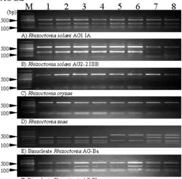
A. Size comparison of rDNA-ITS region in various Rhizoctonia spp.
When Hae III was used for digestion, different PCR-RFLP pattern was observed among isolates of a Rhizoctonia AG-Ba (Fig. 1). No differences of PCR-RFLP banding patterns were not found in other 5 taxa of isolates.
Unique RFLP banding pattern was observed in each
rDNA-ITS region of R. solani AG1 IA, R. solani AG2-2 IIIB, R. circinata var. oryzae, R. circinata var zeae and binucleate Rhizoctonia AG-Bb (550, 250, 100 bp,650, 250 bp,850 bp,850 bp and 550, 250 bp, respectively). On the other hand, three types of RFLP banding pattern, Type I (600,250 bp), Type II (450,250,150 bp) and Type Mix (600,450,250,150 bp), which seems likely to hetero type of Type I and Type II, were observed in Rhizoctonia AG-Ba.
Fig. 1. Electrophoretic profiles of PCR-RFLP products digested by Hae III.
DNA samples were extracted from major 6 anastomosis group of Rhizoctonia spp. Lane M: Molecular weight marker of 100bp ladder. 1-8:
Isolates of each taxon
B. Molecular phylogenetic analyses based on multiple alignments of rDNA-ITS region..
DNA sequences of rDNA-ITS region of 8 AG-Ba isolates (four Type I, two Type II and two Type Mix isolates) of Rhizoctonia AG-Ba were analyzed. PCR products of rDNA-ITS region derived from each isolates were TA-cloned to pCR2.1 plasmid and sequenced with ITS1-F/ITS4-B primers (Gardes and Bruns, 1993) by 3130xl Genetic analyzers (Applied Biosystems), because of expected difficulties of direct-sequencing for Type Mix (expected hetero genotype) isolates. Sizes of rDNA-ITS regions derived from Type I and Type II isolates were 848-852bp and 857-858bp, respectively, and the most of all PCR products showed almostsame size. Partially altered sequences were found in rDNA-ITS region of particular Type Mix isolates. DNA sequence homology of whole ITS
#4
IWRIS2010, October 14-15, Mie University 14
region (848-858 bp) among clones derived from unique Type Mix isolates was 95.4 to 100%. Results of BLAST search indicate that sequences of these clones had the highest homology with the ITS sequence of Rhizoctonia AG-Ba. A neighbor-joining tree was generated with software CLUSTAL W ver.1.83 (http://clustalw.ddbj.nig.ac.
jp/top-j.html) based on multiple alignment of the sequences (Fig. 2). All sequences derived from Rhizoctonia AG-Ba isolates formed one completely independent cluster from the outgroup species. Type I and II isolates belonged to each clade of Clade I (bootstrap value was 93.9%) and II (bootstrap value was 54.7%), respectively. Sequences derived from Type Mix isolate belonged to clades of Clade I or II, depending on sequence similarity to each type of sequence. However, some sequences derived from specific clones, such as Ba-Mix-03 01 and Ba-Mix-03 06, formed another clade at an intermediate position of two clades. All sequences derived from rice isolates were unevenly
distributed in the clade of Clade I.
C. DNA fingerprint analyses of the subgroups by microsatellite primed PCR (MP-PCR).
To select appropriate primer sets and annealing conditions for fingerprint analyses of Rhizoctonia AG-Ba, seven primers were tested under various conditions to DNA derived from isolates of Clade I and II. As a result, five primers [(CA)8CT, (CA)8G, (CAGC)4, (GCC)5 and (GTG)5] were decided to use for fingerprint analyses at 50, 50, 60, 65 and 50C of annealing temperature, respectively. Sizes of PCR-amplified fragments in electrophoretic gels were recorded and data matrixes were formed by whether specific size of fragments were observed or not. By the fingerprint analyses by MP-PCR with the five primers, 129 fingerprint markers were detected in total [26, 29, 31, 19 and 24 markers with primers (CA)8CT, (CA)8G, (CAGC)4, (GCC)5
AG-Ba outgroup
Ba-Mix-01 02BaI-30 05●BaI-04 01●BaI-04 04●BaI-33 06BaI-33 01
BaI-04 06●BaI-27 02●BaI-27 03●BaI-27 04●BaI-27 01●BaI-33 08BaI-33 05Ba-Mix-01 05
BaI-30 03●BaI-33 04
BaI-30 02●BaI-33 02BaI-33 03BaI-33 07
BaI-30 04●BaI-30 07●BaI-27 05●
Ba-Mix-03 02BaI-30 06●Ba-Mix-01 04BaI-30 01●BaI-04 02●
BaI-04 05●BaI-04 07●BaI-04 08●BaI-04 03●Ba-Mix-03 06
Ba-Mix-03 01BaII-07 08BaII-04 05Ba-Mix-03 03
BaII-04 01BaII-07 03
Ba-Mix-01 03Ba-Mix-03 04
BaMix-03 07BaII-07 04BaII-04 03BaII-07 01BaII-07 06
BaII-04 06BaII-04 02
BaII-07 02Ba-Mix-01 01
BaII-04 04BaII-07 07BaII-07 05Ba-Mix-03 05Bo-11 06Bo-33 07Bo-11 08Bo-33 01Bo-33 09Bo-11 04Bo-11 01Bo-33 06Bo-33 02Bo-33 05Bo-02 01Bo-02 03Bo-02 02Bo-25 09Bo-25 06Bo-25 05Bo-25 08
Bo-25 10Bo-25 04Bo-25 01Bo-25 03Bo-25 07Bo-11 09Bo-11 02
Bo-11 05Bo-33 03Bo-33 10Bo-33 11Bo-33 04Bo-11 07Bo-11 03Bo-33 08Bo-25 02Bb-01C-01 02C-01 03C-01 05C-01 01C-01 04
1-IA-020.01 63.1
54.7 93.9
65.4 88.9
60.7
57
99.5 62.1
62.4
62.6
53.9 100
87.7 5883.163.264.3
81.1 63.8
51.8 50.4
100
66.2
98.9 63.897.8
100
Clade I Clade II
BaI-02●BaI-06 ●BaI-04 ●BaI-05●BaI-01●BaI-09 ●
BaI-10 ●BaI-03BaI-11 ●BaI-15 ●BaI-16 ●BaI-13●BaI-14 ●BaI-18 ●BaI-19●BaI-17 ●BaI-08 ●BaI-20 ●BaI-23 ●BaI-21 ●BaI-25 ●BaI-26 ●BaI-22 ●BaI-31 ●BaI-32 ●BaI-30 ●BaI-29 ●BaI-27 ●BaI-28 ●BaI-24 ●BaI-07●BaI-12 ●BaI-33BaI-34BaI-35BaI-36BaI-37BaI-38BaII-03
BaII-07BaI-39BaII-06BaII-04BaII-05BaII-02BaII-01Ba-Mix-01Ba-Mix-02Ba-Mix-04Ba-Mix-03BaI-42BaI-43BaI-41BaI-40
Bo-01Bo-03Bo-07
Bo-02Bo-06Bo-04Bo-05C-011-IA-02
0.1
Subgroup I
Subgroup II
Subgr oup II I Subgroup I V
outgroup
Fig. 2. A neighbor- joining tree generated with CLUSTAL W software based on rDNA-ITS sequences. Bootstrap values (>50%) based on 1000 replications are shown at alongside of branches. The isolates marked with “●” symbols were collected from rice plants.
Fig. 3. A neighbor- joining tree generated with CLUSTAL W software based on MP-PCR fingerprint data matrixes. The isolates marked with “●” symbols were collected from rice plants and the clade shown with shade.
#4
IWRIS2010, October 14-15, Mie University 15
and (GTG)5, respectively] within a range of 250-5500 bp.
Isolaes of Rhizoctonia AG-Ba formed an unique major clade consisted by four minor clades in a phylogenetic tree generated by neighbor-joining method and the clade contained no outgroup species (Fig. 3). All of Subgroup I and IV isolates, including all rice isolates of Rhizoctonia AG-Ba were belong to Clade I of Fig. 2. Moreover, all of Subgroup II isolates were settled to Clades II in Fig. 2.
III.
D
ISCUSSIONCapability of identification by PCR-RFLP pattern with Hae III was verified at first. As a result, a characteristic band pattern was observed in all taxa, except for two taxa whose rDNA-ITS region have no Hae III restriction sites.
These two taxa were not able to be distinguished from each other by only PCR-RFLP pattern with Hae III, because possible sequence similarity of rDNA-ITS region in phylogenetically close two taxa. Therefore, to distinguish these two taxa, we used combinations of Hae III and Hinf I or Hae III and Hha I for PCR-RFLP analyses. However, for the practical method of soil diagnostics or taxa identification, use of each taxon specific primers developed by Arakawa et al. (2006) and Toda et al. (2004) may be much better in cost and easy to handle.
Subgroup differentiation in Rhizoctonia AG-Ba was analyzed by molecular phylogenetic method, based on PCR-RFLP patterns, sequences of rDNA-ITS region and microsatellite primed PCR (MP-PCR) patterns.Three RFLP patterns, Type I, Type II and Type Mix, whose RFLP pattern seems likely to be hetero type of Type I and II, were found by the PCR-RFLP analyses of Rhizoctonia AG-Ba ITS region. On the other hand, by phylogenetic analysis based on sequence similarity of rDNA-ITS region, sequences derived from Type I and Type II isolates belong to clades of Clade I and II, respectively, while sequences derived from Type Mix isolates were divided into clades of Clade I and II. Type I and II isolates classified by RFLP patterns formed independent clusters in a phylogenetic tree based on MP-PCR. However, unlike the result of phylogenetic analyses of rDNA-ITS sequences, Type Mix isolates of RFLP classification formed a unique clade. All together, present results strongly suggest the possibility of subgroup differentiation of Rhizoctonia AG-Ba, although it will be a future work to examine whether Type Mix is an independent subgroup or not.
The present results of molecular phylogenetic analyses suggest the possibility of parasitic differentiation of Rhizoctonia AG-Ba. As an evidence of the differentiation, all rice isolates of AG-Ba belong to a unique clade of Clade
I in the phylogenetic tree based on sequences of rDNA-ITS region (Fig.2). Also, all of 31 rice isolates and an isolate collected from Job’s tears (Coix lacryma-jobi) formed one unique clade in the phylogenetic tree based on MP-PCR.
Moreover, all of 11 weeds isolates formed other clades than rice isolates in the phylogenetic tree based on MP-PCR (Fig.3). Genome region closely related to parasitic differentiation of Rhizoctonia spp. is remain to be elucidated. The nomenclature and grouping of each anastomosis group or subgroup of Rhizoctonia spp. might be reviewed at some stage, because, as Kuninaga (2002) pointed out, it is expected that discovery of new anastomosis groups or subgroups accompanied with rise of new diseases will increase in the future. Understandings of diversification of taxa are expected to develop into regional innovations, because it will be lead to predict possible environmental adaptation ability of potential pathogens.
A
CKNOWLEDGMENTWe thank Dr. A. Morikami and Dr. T. Hirano in Meijo University for technical review of the manuscript.
R
EFERENCES[1] Arakawa, M., Sagara, Y. and Inagaki, K. Development of Specific PCR Primers Designed for the Detection of Rice Sclerotial Disease Fungi.
Jpn. Phytopathol. 72: 276 (2006)
[2] Carling, D. E., Baird, R. E., Gitaitis, R. D., Brainard, K. A. and Kuninaga, S. Characterization of AG-13, a newly reported anastomosis group of Rhizoctonia solani. Phytopathology 92:893-899 (2002)
[3] Chen, C.-M., de la Cerda, K. A., Kaminski, J. E., Douhan, G. W. and Wong, F. P. Geographic distribution and rDNA-ITS region sequence diversity of Waitea circinata var. circinata isolated from annual bluegrass in the United States. Plant Dis. 93: 906-911 (2009) [4] Chen, W., Gray L. E. and Grau, C. R. Molecular differentiation of fungi
associated with brown stem rot and detection of Phialophora gregata in resistant and susceptible soybean cultivars. Phytopathology 86:
1140-1148 (1996)
[5] Gardes, M. and Bruns, T. D. ITS Primers with enhanced specificity for basidiomycetes-application to the identification of mycorrhizae and rusts. Mol. Ecol. 2: 113-118 (1993)
[6] Inagaki, K. Outbreaks of rice sclerotium diseases in paddy fields and physiological and ecological characteristics of these causal fungi. Sci.
Rept., Fac. Agr., Meijo Univ. 37, 47-56 (2001)
[7] Kuninaga, S. Current situation of the taxonomy of the genus Rhizoctonia and the R. solani species complex. Jpn. Phytopathol. 68:
3-20 (2002)
[8] Ogoshi, A. Studies on the Grouping of Rhizoctonia solani Kühn with Hyphal Anastomosis and on the Perfect Stages of Groups. Bul. Nat.
Ins. Agr. Sci. Series C. 30: 1-63 (1976)
[9] Toda, T., Mghalu, J., Hayakawa, T. and Hyakumachi, M. Development of specific primers for each variety of Rhizoctonia circinata.
Inoculum 55: 36-37 (2004)