Acta Med. Nagasaki. 23 : 1-13
Epidemiological Study of Hepatitis B Virus in Nagasaki Area of Japan
Yoshito TANAKA*
Department of Bacteriology, Nagasaki University School of` Medicine,
Nagasaki, 852 JAPAN
Received for publication, August 25, 1977
The epidemiological studies of Hepatitis B Virus (HBV) in Nagasaki Prefecture well- known for the high incidence of liver cirrhosis, primary hepatoma and other liver diseases have been carried out by means of detecting several viral and related markers, such as the surface antigen of HBV (HBs Ag), the antibody to HBs Ag (anti-HBs), the antibody
to the core of HBV(anti‑HBc), e antigen(e Ag)and the antibody to e Ag(anti‑e), in
the sera among healthy blood donors, healthy inhabitants and hospitalized patients.
HBs Ag was detected in 3.8% of 15,149 healthy persons (blood donors and inhabitants) and in 4.8% of 1,192 patients by means of immunoelectrosyneresis (IES). The positive rate (3.8%) in healthy persons was apparently higher than that in those of other areas in Japan. 1.1% of healthy persons and 5.9% of hospitalized patients were found to be anti- HBs-positive by IES. The HBs Ag-positive frequency was significantly higher in male than in female, but the anti-HBs-positive frequency was lower in male than in female.
Dividing Nagasaki Prefecture into five areas, i. e. , Nagasaki City, Gotoh islands, the North, the East and the West areas, the prevalence of HBs Ag tended to be higher in the West and lower in the East. Especially, the positive occurence of HBs Ag in Gotoh area showed the highest value.
Among subtypes of 359 HBs Ag‑positive donor sera, HBs‑antigenic determinants d
(d,99%;y,1%)and γ(γ,93%;ω,7%)were dominant.
The positive frequencies of e Ag and anti‑e in 297 HBs Ag‑positive donor sera were 53(17.8%)and 141(47.5%)respectively. Both e Ag and anti‑e(e markers)were never detected in HBs Ag‑negative sera. No relationship was found between the presence of e
markers and HBs Ag subtypes.
Although no HBc Ag was detected in any sera tested, anti-HBc was detected in all three classified groups of sera, i. e., negative group for both HBs Ag and anti-HBs, positive group for only anti-HBs, and positive group for only HBs Ag. In the first group, 17.7% of samples was anti-HBc-positive, and 81.3% and 100% of samples in the second
*田 中 義 人
and the third groups respectively were also positive.
Therefore, about 39% of healthy persons was immersed with HBV in past by calculating from the data described above.
INTRODUCTION
The presence of hepatitis virus has long been believed from the epidemiological observations of hepatitis, and many investigators have made efforts to discover it in vain. 12)43)46)55)
In 1965, an antigen, which showed a precipitin line in agar against the serum of multiply transfused American haemophiliacs, was found in the serum of an Australian aborigine. This antigen was designated as `Australia' antigen (Au-antigen, at present called as Hepatitis B surface antigen : HBs Ag) by BLUMBERG.6) Subsequent studies on the distribution of the HBs Ag in normal population revealed that this HBs Ag was rarely found in the sera of North American and European people, but was frequently found in the sera of people living in the tropics and Asia. It was also frequently detected in patients with Down's syndrome, leukemia and hepatitis .2)1)11) Afterward, PRINCE42) and OKOCHI40) pointed out independently the intimate relationship between HBs Ag and hepatitis, particularly serum hepatitis. KRUGMANN,23) BARKER4) and others") directly established that HBs Ag-positive serum contained an infectious agent in infection experiments.
Three different morphological particles, i. e. , small spherical particle with appro- ximately 20 nm in diameter, tubular form with 20 x 50-230 nm in lengths) and large spherical particle with approximately 40 nm in diameter first shown by DANE (so-called
"Dane particle")9) were found in the HBs Ag -positive serum . Dane particle with inner core became to be regarded as a human type B hepatitis virus candidate by several investigators") 19)20)44)45) and the inner core (HBc Ag) was antigenically distinct from the surface of Dane particle. 1)8)10) 14)
On the other hand, in 1972, MAGNIUS et al .21) demonstrated a new antigen-antibody system distinct from HBs Ag and HBc Ag in HBs Ag-positive serum and designated the new antigenic determinant as"e". NIELSEN et al. '14) MAGNIUS et al .21) and other workers39)47) hypothetically stressed that e antigen was associated with the infectivity of HBs Ag-positive sera, because the e antigen might be efficiently produced in parallel with Dane particle synthesis. 36)
It is well known that in Nagasaki Prefecture located in western Japan, patients with primary hepatoma and liver cirrhosis have occurred most frequently in Japan. 16) At present, Dane particle, i. e., hepatitis B virus (HBV), is registered as the pathogenic agent of type B acute and chronic hepatitis, liver cirrhosis as well as primary hepatoma.
Although the pathogenesis of HBV among cirrhosis and hepatoma was not yet directly
established, vertical transmission from HBs Ag-positive mother followed by healthy carrier
state of HBV would play a significant role.") Therefore, it is important to study the
dissemination status of HBV among healthy persons, patients with liver diseases and various
patients without liver diseases, and finally to elucidate the relationship between HBV infection and many kinds of liver diseases including hepatoma.
In this report, the epidemiological studies of HBV among healthy persons and various patients in Nagasaki area are presented. The evidences of HBV infection among healthy blood donors, inhabitants and various patients admitted at the Nagasaki University Hospital were obtained by using three antigen-antibody systems, i . e . , HBs Ag : anti- HBs, HBc Ag : anti-HBc and e Ag : anti-e as markers of HBV-infection.
MATERIALS AND METHODS
(1) Serum samples
Blood donors' sera : 13,555 donor sera collected at the Nagasaki Red Cross Blood Center from August 1972 to July 1973 were studied.
Inhabitants' sera : 1, 594 sera were obtained from the local health centers appropriately located in Nagasaki Prefecture.
Patients' sera : From March to May 1974 and from July to August 1976, 1,192 inpatients' sera which were submitted to the Biochemical Section of the Central Laboratory in the Nagasaki University Hospital for testing liver functions, were randomly collected and used in this study.
(2) Methods used for detecting HBs Ag and anti-HBs
Immunoelectrosyneresis (IES) method was mainly used for detecting HBs Ag and anti-HBs. Passive hemagglutination (PHA) method was also used for detecting anti-HBs.
PHA cells were kindly supplied from Dr. Mayumi of Jichi Medical University and Eisai Company Ltd. IES was performed on glass slides (25 x 75 mm) overlayed with 1 % agar gel in M/100 barbitalsodium-Barbital buffer (pH 8.6, ionic strength 0.05) under a constant current of 2mA/cm for 45 minutes at room temperature. The final decision was made after the reacted agar gel slide was incubated in a moisture chamber for 24 hours at 4-60C.
Several appropriate high-titered HBs Ag-positive blood donor sera were pooled and used as reference antigen. They were stored at -20°C and were diluted into 5 or 10 times for use. Three reference antibodies were used for detecting HBs Ag by IES, i. e. , one from pooled anti-HBs-positive blood donor sera, another from Dr. Okochi of Kyushu University (designated as Yamakawa) and the other from hyperimmune anti- HBs rabbit serum prepared in this laboratory. PHA was performed by using microtiter hemagglutination technique and antibody-positive sera agglutinated the PHA cells at a dilution of 1 : 16 or more within 1 hour.
(3) Methods for detecting e Ag or anti-e, and for subtyping HBs Ag
Immunodiffusion (Micro-Ouchterlony : M. 0.) method was performed in 0.9%
agarose gel in 0.01M Tris buffer supplemented with O.IM NaCl and 2% Dextran A, at
pH 7.8 by incubation in the moisture chamber for 24 hours at 37°C. Standard e Ag and anti-e reagents were at first kindly supplied from Dr. Okochi and thereafter, originally prepared from HBs Ag-positive donor plasma. For subtyping HBs Ag, anti-ad and anti- ay guinea pig sera were obtained from the United States National Institutes of Health (V801-502-058, V802-501-558) and anti-ar rabbit serum was obtained from immunized animals with partially purified HBs Ag (subtype : adr) prepared from HBs Ag-positive blood donor sera by using CsCI cushion, buoyant density gradient and sucrose stepwise gradient ultracentrifugation in this laboratory.
(4) Methods.for detecting HBc Ag and anti-HBc
HBc Ag was assayed by a reversed passive hemagglutination (r-PHA) technique and anti-HBc was assayed by the reversed passive hemagglutination inhibition (r-PHAI) technique developed in this laboratory as described elsewhere (Igarashi, et al., to be published). Essentially, the tannic acid-treated human type 0 red blood cells were coated with anti-HBc IgG prepared from HBs Ag-positive blood donor sera by sedimentation with saturated ammonium sulfate and by purification using DEAE-cellulose column chromato- graphy, and those were used to detect HBc Ag by hemagglutination. When anti-HBc is added to this system, hemagglutination should be inhibited.
RESULTS
Incidence of HBs Ag and anti-HBs
The results of testing sera of healthy persons (blood donors and inhabitants) for HBs Ag and anti-HBs by IES and PHA are shown in Tables 1 and 2. From these results, the HBs Ag-positive frequency is significantly higher in male than in female. To the contrary, the anti-HBs-positive frequency by IES is significantly lower in male than in female.
In Table 2, the higher anti-HBs-positive rate in blood donors than in inhabitants by both IES and PHA would be responsible for the different composition of sex and age
Table 1. Positive frequencies of HBs Ag anti-HBs in healthy persons (blood donors and inhabitants) tested by IES
No. positive sera / No. tested (%, a=0.05)
569/15,149 (3.77±0.3)
HBs Ag male 451/10
,944 (4.14±0.37)
(female 117/ 4,113 (2.89±0.51)
166/15,149 (1.11±0.17)
Anti-HBs male 97/10
,944 (0.9 ±0.18)
(female 62/ 4,113 (1.55±0.37)
Table 2. Positive frequencies of HBs Ag and anti-HBs in blood donors and inhabitants tested by IES and PHA
Blood donors I Inhabitants I (Healthy persons) Total
HBs Ag 526/13
,555a) ( 3.9%)b) 43/1,594 (2.7%)b) 569/15;149 ( 3.8%) (by IES)
Anti-HBs 132/13
,555 ( 1.0%)-) 34/1,594 (2.1%)" 166/15,149 ( 1.1%) (by IES)
Anti-HBs 667/ 3
,787 (17.6%a) 200/ 825 (24.2%)(1) 867/ 4,612 (18.8%) (by PHA)
a) No. positive sera/No. tested b) Xo=5.531 p<0.02
c) Xo=17.13 p-<0.001 d) X02== 19.5 p<0.001
between the two groups. Tables 3 and 4, and Fig. 1 show age distributions of HBs Ag and of anti-HBs in healthy persons. In these results, prevalence of HBs Ag tends to
Table 3. Age distribution of HBs Ag-positive frequencies in
blood donors and inhabitants tested by IES
\roup Blood donor Inhabitant
Age "~~ ~ male female male female Total
<15 - - 0/ 3a1 0/ 2 0/ 5
16-21 129/ 2,720(4.74)b) 47/1,521(3.09) 5/106(4.72) 1/ 95(1.05) 182/ 4,442(4.10) 22-30 150/ 3,413(4.39) 26/ 867(3.00) 4/152(2.63) 3/159(1.89) 183/ 4,591(3.99) 31-40 92/ 2,364(3.89) 11/ 466(2.36) 10/142(7.04) 3/114(2.63) 116/ 3,086(3.76) 41-50 41/ 1,290(3.18) 11/ 388(2.84) 4/166(2.41) 2/118(1.69) 58/ 1,962(2.96) 51-60 10/ 319(3.13) 9/ 165(5.45) 4/118(3.39) 1/ 94(1.06) 24/ 696(3.45) 61< 0/ 34 0/ 8 1/ 83(1.20) 1/100(1.00) 2/ 225(0.89) total 422/10,140(4.16) 104/3,415(3.05) 28/770(3.64) 11/682(1.61) 565/15,007(3.76)
a) No. positive sera/No. tested
b) Number in parentheses :
Table 4. Age distribution of anti-HBs-positive frequencies in
blood donors and inhabitants tested by PHA
Group
male Blood donor female male Inhabitant female Total Age
<15 - - 0/ 2a) 0/ 1 0/ 3
16-21 111/ 680(16.32)b) 58/349(16.62) 14/ 75(18.67) 6/ 40(15.00) 189/1,144(16.52) 22-30 179/ 966(18.53) 51/272(18.98) 26/137(18.98) 9/ 55(16.36) 265/1,430(18.53) 31-40 117/ 727(16.09) 23/140(16.43) 24/ 89(26.97) 14/ 40(35.00) 178/ 966(17.87) 41-50 52/ 317(16.40) 29/114(25.44) 11/ 52(21.15) 17/ 58(29.31) 109/ 541(20.15) 51-60 17/ 84(20.24) 9/ 33(27.27) 10/ 26(38.46) 17/ 45(37.78) 53/ 188(28.19) 61< 0/ 7 2/ 2(100) 5/ 24(20.83) 13/ 62(20.97) 20/ 95(21.05) total 476/2,781(17.12) 172/910(18.90) 90/405(22.22) 76/301(25.25) 814/4,397(18.51)
a) No. positive sera/No. tested
b) Number in parentheses :
Fig. 1. Age distributions of positive frequencies of HBs Ag tested by IES and of Anti-HBs tested by
PHA in healthy persons (a=0.05)
(0 HBs Ag )
0 Anti-HBs
decrease and that of anti-HBs to increase as the age advances.
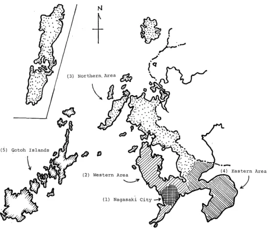
In Table 5, HBs Ag- and anti-HBs-positive frequencies in healthy persons in five divided areas of Nagasaki Prefecture, i. e. , (1) Nagasaki City, (2) the Western area, (3) the Nothern area, (4) the Eastern area and (5) Gotoh islands in Fig. 2 are presented.
HBs Ag-positive rates in Nagasaki City and Gotoh islands are higher than those in other areas, especially significantly higher than those in the Nothern and the Eastern areas, Table 5. Regional positive frequencies of HBs Ag tested by IES
and of anti-HBs tested by PHA in healthy persons
Areas (1) Nagasaki City (2) Western area (3) Northern area (4) Eastern ar ea (5) Gotoh Islands Total
HBs Ag 357/8,523 48/1,376a~ 22/890 87/3,262 42/754 556/14,805
(by IES) (4.2±0.4) (3.6±1.0)b~ (2.7±1.0) (2.7±0.6) (5.8±1.7) (3.8±0.3)
Anti-HBs 455/2,529 91/ 478 62/286 190/1,025 69/294 867/ 4,612
(by PHA) (18.0±1.5) (19.3±3.5) (22.1±4.8) (18.7±2.4) (23.8±4.8) (18.8±1.1) a) No. positive sera/No. tested
b) Number in parentheses : % (a=0.05)
Fig. 2. Map of Nagasaki Prefecture divided into five areas
but in anti-HBs-positive rates, no significant differences are observed as shown in Table 5. The relationships among prevalences of HBs Ag and anti-HBs, occurence rate of hepatitis and death rate of liver cirrhosis and hepatoma in each area are illustrated in Fig.
3 in which death rate of liver disease (liver cirrhosis plus hepatoma), HBs Ag-positive frequency and anti-HBs-positive one in Gotoh islands are markedly high.
In Table 6, positive frequencies of HBs Ag and anti-HBs in hospitalized patients are presented. Comparing the result in Table 6 with that in Table 1, positive frequency of anti-HBs in various patients is significantly higher than in healthy persons as shown in Table 7. The age distribution of HBs Ag-and anti-HBs-positive patients is not shown here, because the number of samples was too small for statistical distribution analyses.
HBs Ag subtype
Subtype analyses of HBs Ag obtained from healthy persons and hospitalized patients in Nagasaki Prefecture are summarized in Table 8. The HBs Ag-subtypes commonly detected are adr and adze, and ayr is present in small size, and ayw subtype is absent.
There is no significant difference in distribution of HBs Ag-subtypes between the two
groups.
Fig. 3. Correlations among prevalences of HBs Ag and Anti-HBs, hepatitis-occurence rate and death rate of hepatoma and
liver cirrhosis in each area of Nagasaki Prefecture
Table 6. Positive frequencies of HBs Ag and anti-HBs in hospitalized patients tested by IES
No. positive sera / No. tested (%, a=0.05) 50/1,192 (4.34± 1.15)
HBs Ag male 35/611 (6 .0±1.86)
(female 15/581 (2.9±1.32)
70/1,192 (6.10±1.34)
Anti-HBs male 35/611 (6 .0±1.86)
(female 35/581 (6.3±1.95)
Table 7. Comparisons between HBs Ag-and anti-HBs-positive frequencies in healthy persons and in hospitalized patients tested by IES
HBs Ag Anti-HBs
Patients 50/1,192a) (4.2%) I 70/1,192 (5.9%) Healthy Persons I 569/15,149 (3.8%) I 166/15,149 (1.1%)
Xo=0.58 p<O.5 X2 0=177.14 p<0.001
a) No. positive sear / No. tested
Table 8. Subtypes of HBs Ag in healthy persons and hospitalized patients Healthy Persons
d y total
r 331(92%) 4 (1%) 335 (93%)
w 24 (7%) 0 24 (7%)
total - '- 355 (99%) - - 4 - - (1/)- --I - 359 (100%)
Patients