Phenolic acids as potential protective agents against reactive oxygen species and
acetaldehyde
September, 2018
WENSI XU
Graduate School of
Environmental and Life Science (Doctor’s Course)
OKAYAMA UNIVERSITY
PREFACE
The experiments described in this dissertation were carried out at the Graduate school of Environment and Life Science (Doctor Course), Okayama University, Japan, from October 2015 to September 2018, under the supervision of Professor Yoshimasa Nakamura. These studies are original work by the author and any other assistance and collaboration from others are specially acknowledged.
This dissertation has not been submitted previously whole or in part to the council, a university or any other professional institution for a degree, diploma or other professional qualification.
WENSI XU September, 2018
CONTENTS
PREFACE ... i
CONTENTS ... ii
LIST OF FIGURES ... vi
ABBREVIATIONS ... ix
ABSTRACT...x
CHAPTER 1 ...1
General Introduction ...1
1.1 Food phytochemicals ...1
1.1.1 Lycii Fructus ...1
1.1.2 Chlorogenic acid ...2
1.1.3 Quercetin ...2
1.2 Oxidative stress ...3
1.2.1 Reactive oxygen species ...4
1.2.2 Ultraviolet B ...4
1.2.3 Antioxidants ...5
1.3 Ethanol metabolism ...5
1.3.1 Aldehyde dehydrogenase ...5
1.3.2 ALDH2 mutation...6
1.4 Nrf2/ARE and AhR/XRE pathways ...6
1.4.1 Keap1/Nrf2/ARE pathway ...7
1.4.2 AhR/ARNT/XRE pathway ...7
1.5 Study outlines ...8
CHAPTER 2 ... 10
Lycii fructus extract ameliorates hydrogen peroxide-induced cytotoxicity through indirect antioxidant action ... 10
2.1 Introduction ... 10
2.2 Materials and methods ... 12
2.2.2 Cell culture ... 12
2.2.3 Reverse transcription-polymerase chain reaction (RT-PCR) ... 12
2.2.4 Glutathione titration... 13
2.2.5 Western blot analysis ... 13
2.2.6 Cell viability determination ... 14
2.2.7 Trypan blue dye exclusion assay ... 14
2.2.8 Statistical analysis ... 14
2.3 Results ... 15
2.3.1 Lycii fructus extract induces the gene expression of several drug-metabolizing enzymes ... 15
2.3.2 Lycii fructus induces nuclear translocation of Nrf2 and AhR ... 17
2.3.3 Lycii fructus shows an antioxidant effect after a 24-h incubation ... 18
2.3.4 Chlorogenic acid is one of the predominant antioxidants in LFE ... 19
2.4 Discussion ... 22
2.5 Conclusion ... 24
CHAPTER 3 ... 25
Lycii Fructus extract and its phytochemicals ameliorate UVB-induced cytotoxicity in Human keratinocytes HaCaT cells ... 25
3.1 Introduction ... 25
3.2 Materials and methods ... 27
3.2.1 Materials ... 27
3.2.2 Cell culture ... 27
3.2.3 Ultraviolet B irradiation ... 27
3.2.4 Reverse transcription-polymerase chain reaction (RT-PCR) ... 27
3.2.5 Western blot analysis ... 28
3.2.6 Statistical analysis ... 28
3.3 Results ... 29 3.3.1 LFE, CGA, quercetin and DOPAC dose-dependently inhibited the hydrogen
3.3.2 LFE, CGA, DOPAC and quercetin significantly inhibited the UVB-induced
cytotoxicity. ... 30
3.3.3 LFE, CGA, DOPAC and quercetin stimulated the gene expression of phase 2 drug-metabolizing enzymes, including HO-1, NQO1 and GCLC. ... 31
3.3.4 LFE, CGA, DOPAC and quercetin induced the nuclear translocation of AhR and Nrf2. ... 34
3.4 Discussion ... 37
3.5 Conclusion ... 38
CHAPTER 4 ... 39
Chlorogenic acid as a potential protective agent against acetaldehyde-induced cytotoxicity in murine hepatoma cells ... 39
4.1 Introduction ... 39
4.2 Materials and methods ... 41
4.2.1 Reagents and antibodies ... 41
4.2.2 Cell culture ... 41
4.2.3 Cell viability determination ... 41
4.2.4 Determination of ALDH activity ... 42
4.2.5 Western blot analysis ... 42
4.2.6 Reverse transcription-polymerase chain reaction (RT-PCR) ... 42
4.2.7 Small interfering RNA transfection ... 43
4.2.8 Statistical analysis ... 43
4.3 Results ... 44
4.3.1 CGA and its metabolites impaired the cytotoxicity induced by acetaldehyde through the enhancement of the ALDH enzymatic activity ... 44
4.3.2 CGA enhanced the ALDH enzymatic activity through a transcriptional regulation ... 47
4.3.3 CGA activated the Nrf2- and AhR-dependent pathways ... 47 4.3.4 Silencing Nrf2 affected the effects of CGA on the acetaldehyde-induced
4.3.5 Silencing AhR affected the effects of CGA on the acetaldehyde-induced
cytotoxicity and the ALDH activity ... 53
4.4 Discussion ... 55
4.5 Conclusion ... 58
CONCLUSION ... 59
ACKNOWLEDGMENTS ... 62
REFERENCES ... 63
LIST OF FIGURES
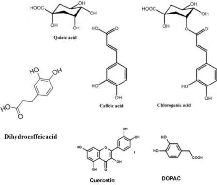
Fig. 1.1. Structures of some polyphenols. ...3 Fig. 1.2. Diagram of the direct and indirect antioxidative action. ...8 Fig. 2.1. Modulating effects of Lycii fructus extract (LFE) on the gene expression of
the drug metabolizing enzymes. ... 16 Fig. 2.2. Modulating effects of Lycii fructus extract (LFE) on the intracellular GSH
level. ... 17 Fig. 2.3. Modulating effects of Lycii fructus extract (LFE) on the Nrf2 and AhR levels
in Hepa1c1c7 cells... 18 Fig. 2.4. Effect of Lycii fructus extract (LFE) on the hydrogen peroxide-induced
cytotoxicity in Hepa1c1c7 cells. ... 19 Fig. 2.5. Effect of chlorogenic acid (CGA) on the hydrogen peroxide-induced
cytotoxicity and the gene expression of HO-1 and NQO1. ... 20 Fig. 2.6. Effect of chlorogenic acid (CGA) on the Nrf2 and AhR levels in Hepa1c1c7
cells. ... 21 Fig. 3.1. Impairing effects of Lycii fructus extract (LFE), chlorogenic acid (CGA),
quercetin and 3,4-dihydroxyphenylacetic acid (DOPAC) on the cytotoxicity induced by hydrogen peroxide. ... 29 Fig. 3.2. Impairing effects of Lycii fructus extract (LFE), chlorogenic acid (CGA),
quercetin and 3,4-dihydroxyphenylacetic acid (DOPAC) on the cytotoxicity induced by UVB. ... 30 Fig. 3.3. Modulating effects of Lycii fructus extract (LFE) on the gene expression of
representative phase 2 drug-metabolizing enzymes. ... 32 Fig. 3.4. Modulating effects of chlorogenic acid (CGA) on the gene expression of
representative phase 2 drug-metabolizing enzymes. ... 33 Fig. 3.5. Modulating effects of quercetin on the gene expression of representative phase
2 drug-metabolizing enzymes. ... 33 Fig. 3.6. Modulating effects of 3,4-dihydroxyphenylacetic acid (DOPAC) on the gene
Fig. 3.7. Modulating effects of Lycii fructus extract (LFE) on the aryl hydrocarbon receptor (AhR) and nuclear factor (erythroid-derived 2)-like 2 (Nrf2) level. ... 35 Fig. 3.8. Modulating effects of chlorogenic acid (CGA) on the aryl hydrocarbon
receptor (AhR) and nuclear factor (erythroid-derived 2)-like 2 (Nrf2) level. ... 35 Fig. 3.9. Modulating effects of quercetin on the aryl hydrocarbon receptor (AhR) and
nuclear factor (erythroid-derived 2)-like 2 (Nrf2) level. ... 36 Fig. 3.10. Modulating effects of 3,4-dihydroxyphenylacetic acid (DOPAC) on the aryl
hydrocarbon receptor (AhR) and nuclear factor (erythroid-derived 2)-like 2 (Nrf2) level. ... 36 Fig. 4.1. Impairing effects of Lycii fructus extract (LFE) and chlorogenic acid (CGA)
on the cytotoxicity induced acetaldehyde or ethanol through the enhancement of the total ALDH enzyme activity. ... 45 Fig. 4.2. Impairing effects of catabolites of chlorogenic acid (CGA) on the cytotoxicity
induced acetaldehyde through the enhancement of the total ALDH enzyme activity. ... 46 Fig. 4.3. Modulating effects of chlorogenic acid (CGA) on the protein levels of
representative ALDH enzymes. ... 48 Fig. 4.4. Modulating effects of chlorogenic acid (CGA) on the gene expression of
representative ALDH enzymes. ... 49 Fig. 4.5. Modulating effects of chlorogenic acid (CGA) on the gene expression of
representative phase 2 drug-metabolizing enzymes. ... 50 Fig. 4.6. Modulating effects of chlorogenic acid (CGA) on the aryl hydrocarbon
receptor (AhR) and nuclear factor (erythroid-derived 2)-like 2 (Nrf2) level. ... 51 Fig. 4.7. Modulating effects of Nrf2 knockdown on the chlorogenic acid (CGA)- mitigated acetaldehyde-induced cytotoxicity and ALDH activity. ... 52 Fig. 4.8. Modulating effects of Nrf2 knockdown on the gene expression of ALDHs.
Hepa1c1c7 cells were seeded at 6-well plates and transfected with the Nrf2 siRNA or control scrambled siRNA. ... 53
Fig. 4.9. Modulating effects of AhR knockdown on the chlorogenic acid (CGA)- mitigated acetaldehyde-induced cytotoxicity and ALDH activity. ... 54 Fig. 4.10. Modulating effects of AhR knockdown on the gene expression of ALDHs.
... 55
ABBREVIATIONS
AA, Acetaldehyde;
AhR, Aryl hydrocarbon receptor;
ALDH, Aldehyde dehydrogenase;
ARE, Antioxidant response element;
CA, Caffeic acid;
CGA, Chlorogenic acid;
CYP1A1, Cytochrome P450 1A1; GSH, glutathione;
DHCA, Dihydrocaffeic acid;
DOPAC, 3,4-dihydroxyphenylacetic acid;
EtOH, Ethanol;
GCLC, Glutamate-cysteine ligase catalytic subunit;
HO-1, Heme oxygenase-1;
Keap1, Kelch-like ECH-associated protein 1;
LFE, Lycii fructus extract;
NQO1, NAD(P)H: quinone oxidoreductase 1;
Nrf2, Nuclear factor (erythroid-derived 2)-like 2;
QA, Quinic acid;
ROS, Reactive oxygen species;
xCT, Cystine/glutamate antiporter (cystine/glutamate exchanger);
XRE, Xenobiotic response element.
ABSTRACT
Lycii Fructus, also known as wolfberry, Goji berries and Gou Qi Zi, possesses various characteristics including radical scavenging, anti-aging, anti-tumor, cytoprotective, neuroprotective, antiglaucoma and immunomodulatory activities, and improves control of glucose and other diabetic symptoms. Polysaccharides, carotenoids, ascorbic acid and its glycosylated precursors, phenolic acids, and flavonoids have been reported to represent biologically-active components isolated from Lycii Fructus.
Chlorogenic acid (CGA), a predominant phenolic acid in the Lycii Fructus, an ester of caffeic acid and (−)-quinic acid. In addition to CGA, quercetin glycosides are also the main polyphenol compound in wolfberry. Quercetin glycosides are converted into phenolic acids, including 3,4-dihydroxyphenylacetic acid (DOPAC), by colonic microflora. In addition to their radical scavenging potential, phenolic acids might have a protective effect against oxidative stress via up-regulation of the cytoprotective enzymes. However, to the best of my knowledge, it has not been fully elucidated whether phenolic acids can potentiate the gene expression of the drug-metabolizing enzymes. In this study, I have focused on the induction of phase 2 drug-metabolizing enzymes and aldehyde dehydrogenase by Lycii Fructus extract (LFE), CGA, and DOPAC to prevent the hydrogen peroxide- and acetaldehyde-induced cytotoxicity.
In the Chapter 2, I investigated the role of the indirect antioxidative action in the cytoprotective effect of LFE in murine hepatoma Hepa1c1c7 cells. LFE significantly enhanced the expression of the drug-metabolizing enzyme genes, such as heme oxygenase-1 (HO-1), NAD(P)H: quinone oxidoreductase 1 (NQO1), glutamate- cysteine ligase catalytic subunit (GCLC), cystine/glutamate exchanger (xCT) and cytochrome P450 1A1 (CYP1A1), and intracellular glutathione level in Hepa1c1c7 cells. Western blot analysis revealed that LFE stimulated the nuclear translocation of nuclear factor (erythroid-derived 2)-like 2 (Nrf2) as well as aryl hydrocarbon receptor (AhR), suggesting that the activation of both the Nrf2- and AhR-dependent pathways
Cell viability experiments revealed that the pretreatment of LFE for 24 h, but not for 30 min, completely inhibited the cytotoxic effect of hydrogen peroxide. Furthermore, CGA also exhibited cytoprotective effects against hydrogen peroxide-induced cytotoxicity with up-regulation of the phase 2 drug-metabolizing enzyme genes. The present study provided biological evidence that LFE and CGA induce cytoprotective responses against oxidative stress through the inducible expression of the antioxidant enzyme genes.
In the Chapter 3, I investigated the effects of LFE, CGA, DOPAC and quercetin against ultraviolet B (UVB)-induced cytotoxicity in human keratinocyte HaCaT cells.
Preliminary experiments showed that not only LFE and CGA, but also quercetin and DOPAC dose-dependently inhibited the hydrogen peroxide-induced cytotoxicity.
LFE and the three phenolic compounds stimulated the expression of antioxidant enzyme genes, including HO-1, NQO1 and GCLC in human keratinocytes. They also mitigated cytotoxicity induced by UVB irradiation (50 mJ/cm2) in HaCaT cells.
These results suggested that the inducible expression of the antioxidant enzyme genes might be effective on the protection against UVB-induced oxidative stress.
In the Chapter 4, I examined the modulating effects of LFE as well as CGA and its catabolites on the acetaldehyde-induced cytotoxicity in Hepa1c1c7 cells. Cell viability assay showed that LFE, CGA and its catabolites ameliorated the acetaldehyde- cytotoxicity. In addition, CGA significantly protected the ethanol-induced cytotoxicity. LFE, CGA and its catabolites enhanced the total ALDH activity, suggesting that not only LFE, but also CGA and its metabolites, have an ability to impair the acetaldehyde-induced cytotoxicity through the enhancement of the ALDH enzymatic activity. RT-PCR and western blot experiments revealed that CGA significantly increased the gene and protein expression of ALDH1A1, ALDH2 and ALDH3A1. CGA significantly stimulated the nuclear translocation of Nrf2 and AhR.
Moreover, silencing Nrf2 counteracted the protective effect of CGA on the acetaldehyde-induced cytotoxicity. The enhanced ALDH enzymatic activity and
strongly suggested that CGA ameliorates the acetaldehyde-induced cytotoxicity, possibly through the enhancement of the ALDH expression with activation of the Nrf2- dependent pathway.
The present study provides biological evidence that (1) LFE and its major constituent, CGA, exhibits cytoprotective effects on hydrogen peroxide-cytotoxicity through an indirect antioxidant action in murine hepatoma Hepa1c1c7 cells; (2) LFE and CGA mitigates the cytotoxicity induced by not only hydrogen peroxide, but also UVB with the transcriptional regulation of Nrf2 and AhR in human keratinocyte HaCaT cells; (3) CGA shows the cytoprotection against acetaldehyde with the enhanced ALDH enzymatic activity. In conclusion, the present study represents a potentially efficient strategy to prevent the oxidative stress- and alcohol-induced cytotoxicity by dietary phenolic acids.
CHAPTER 1 General Introduction 1.1 Food phytochemicals
Food phytochemicals are compounds that are produced by plants ("phyto" means
"plant"). They are found in fruits, vegetables, grains, beans, and other edible plants.
Some of these phytochemicals such as polyphenols, flavanols, glucosinolates are believed to protect cells from damage that could lead to chronic diseases of cancer (Scalbert et al., 2011). Phytochemicals has a reputation of “the cancer fighters in your foods” Some researchers think that you could reduce your cancer risk by as much as 40% by eating more vegetables, fruits, and other plant foods that have certain phytochemicals in them. Because these phytochemicals are in the fruits, vegetables, beans, and grains you eat, it's fairly easy to include them in your diet. Phytochemicals under research can be classified into major categories, such as carotenoids and polyphenols, which include phenolic acids, flavonoids, and stilbenes/lignans (Heneman et al., 2008).
1.1.1 Lycii Fructus
Lycii Fructus, a kind of edible plant whichalso known as wolfberry, Goji berries and Gou Qi Zi, is the dried ripe fruits of Lycium chinense and L. barbarum, which has long been used as a traditional food material with various health-promoting characteristics in East Asia (Potterat, 2010). Lycii Fructus has long been used as a traditional food and folk medicine in East Asia, it possesses various health-promoting characteristics including antioxidant, antiaging, anti-tumor, cytoprotective, neuroprotective, antiglaucoma and immunomodulatory activities, and promotes endurance and improves control of glucose and other diabetic symptoms (Zhang et al.
2010; Li et al. 2007, 2011; Tang et al. 2011). Polysaccharides, carotenoids, ascorbic acid and glycosylated precursors, phenolic acids, and flavonoids have been reported to represent biologically-active components isolated from Lycii Fructus (Le et al., 2007).
1.1.2 Chlorogenic acid
Chlorogenic acid (CGA), the predominant phenolic acid in the lycii fructus, an ester of caffeic acid and (−)-quinic acid. It is an important antioxidant compound found in foods, such as lycii fructus, coffee, apples, pears, etc. CGA has been reported to be a potent polyphenol antioxidant, because it has a catechol group, which can scavenge hydroxyl radicals and superoxide anion radicals to protect cells from oxidative injury.
CGA also possesses anti-bacterial and anti-carcinogenic properties (Kono et al., 1997;
Santos et al., 2006; Ozyürek et al, .2008). CGA has shown its photo-protection against UV-induced skin damage in animal model and displayed the suppression on UVB- related ROS mediated cellular processes in vivo (Feng et al., 2005; Kitagawa et al., 2011). CGA prevents acetaminophen-induced liver injury through inhibiting its metabolism and involved with CYP450 metabolic enzymes and some antioxidant signals (Pang et al., 2015). CGA protects MC3T3-E1 cells against H2O2-induced oxidative damage by enhancing Nrf2 nuclear translocation and upregulating HO-1 induction via PI3K/Akt signaling pathway (Han et al., 2017). CGA protects against acetaminophen-induced hepatotoxicity and alcohol-induced liver injuries through antioxidative pathway in liver cells (Wei et al., 2018; Kim et al., 2018).
Around 65% of ingested CGA is metabolized in the colon and converted into caffeic acid (CA) and quinic acid (QA) (Pari et al., 2007). CA protects H2O2-induced cell damage in human lung fibroblast cells (Kang et al., 2006) as well as has a beneficial effect in reducing the adverse effect of alcohol (Pari et al., 2007). Furthermore, Dihydrocaffeic acid (DHCA) is a metabolite of CA with potent antioxidant properties.
It is a potential candidate for photo-protection by interfering with the events initiated after UV exposure in keratinocytes (Poquet et al., 2008). QA, another main metabolite of CGA, is a normal constituent of our diet, capable of conversion to tryptophan and nicotinamide via the gastrointestinal (GI) tract microflora (Pero et al., 2009).
1.1.3 Quercetin
Quercetin is the aglycone form of flavonoid glycosides, which is one of the
study showed that quercetin 4′-glucoside were converted into smaller phenolic acids by colonic microflora, 3,4-dihydroxyphenylacetic acid (DOPAC) is one of the main metabolites (Mullen et al., 2008). It has been reported that DOPAC was identified as a predominant bioactive catabolite of quercetin glycosides (Tang et al., 2016). DOPAC also has been reported to ameliorate acetaldehyde-induced cytotoxicity by enhancing aldehyde dehydrogenase activity in murine hepatoma cells (Liu et al., 2017).
Fig. 1.1. Structures of some polyphenols.
1.2 Oxidative stress
Oxidative stress has been defined as a disturbance in the balance between the production of reactive oxygen species (free radicals) and antioxidant defenses, which may lead to tissue injury (Betteridge, 2000). The shift in the balance between oxidants and antioxidants in favor of oxidants is termed “oxidative stress”.
Disturbances in the normal redox state of cells can cause toxic effects through the production of peroxides and free radicals that damage all components of the cell, including proteins, lipids, and DNA. Oxidative stress from oxidative metabolism
causes base damage, as well as strand breaks in DNA. Further, some reactive oxidative species act as cellular messengers in redox signaling. Thus, oxidative stress can cause disruptions in normal mechanisms of cellular signaling.
1.2.1 Reactive oxygen species
Reactive oxygen species (ROS) are produced by living organisms as because of normal cellular metabolism and environmental factors, such as air pollutants or cigarette smoke. ROS are highly reactive molecules and can damage cell structures such as carbohydrates, nucleic acids, lipids, and proteins and alter their functions (Esra Birben et al., 2012). ROS are produced by cellular metabolic activities and environmental factors, such as air pollutants or cigarette smoke. ROS are highly reactive molecules because of unpaired electrons in their structure and react with several biological macromolecules in cell, such as carbohydrates, nucleic acids, lipids, and proteins, and alter their functions. ROS also affects the expression of several genes by upregulation of redox-sensitive transcription factors and chromatin remodeling via alteration in histone acetylation/deacetylation. Regulation of redox state is critical for cell viability, activation, proliferation, and organ function (Esra Birben et al., 2012).
1.2.2 Ultraviolet B
Ultraviolet B (UVB), one of the three types of invisible light rays (together with ultraviolet A and ultraviolet C) given off by the sun. Although UVC is the most dangerous type of ultraviolet light in terms of its potential to harm life on earth, it cannot penetrate earth's protective ozone layer because of the shortest wavelength. Therefore, it poses no threat to human, animal or plant life on earth. UVA and UVB, on the other hand, do penetrate the ozone layer in attenuated form and reach the surface of the planet.
UVA has the longest wavelength and reaches deep into the layers of skin (dermis) causing aging or wrinkling. Whereas, UVB has largest effect on the top layer of skin (epidermis), causing redness, burning and skin cancer. Thus, scientists long blamed UVB as the sole culprit in deep into skin, generating ROS and causing skin cancer in
CGA has shown its photo-protection against UV-induced skin damage in animal model and displayed the suppression on UVB-related ROS mediated cellular processes in vivo (Feng et al., 2005; Kitagawa et al., 2011).
1.2.3 Antioxidants
The human body is equipped with a variety of antioxidants that serve to counterbalance the effect of oxidants (Esra Birben et al., 2012). Antioxidants protect your body's cells from free radicals — unstable molecules created during normal cell functions. Pollution, radiation, cigarette smoke, and herbicides also can create free radicals in your body. Free radicals can damage a cell's genetic parts and may trigger the cell to grow out of control. These changes may contribute to the development of cancer and other diseases. As a rule, dark-colored fruits and vegetables have more antioxidants than other fruits and vegetables.
1.3 Ethanol metabolism
Ethanol, also called alcohol or drinking alcohol, is mainly metabolized in the liver.
Ethanol metabolism occurs in two steps: Ethanol is metabolized quickly by alcohol dehydrogenase (ADH) to generate acetaldehyde, then acetaldehyde is metabolized by the mitochondrial aldehyde dehydrogenase (ALDH) to acetate acid. Acetaldehyde is very diffusible and crosses biological membranes, can be circulated in the blood. Its toxicity increases the risk of tissue injury, which leads to steatohepatitis, cirrhosis, liver failure or hepatocellular carcinoma (Ishii et al., 1997; Lee et al., 2013; Chen et al., 2014).
Simultaneously, excessive consumption of alcohol promotes oxidative stress, which enhanced the generation of reactive oxygen species (ROS) or depletion of the antioxidant defense system, causing an imbalance between pro-oxidants and antioxidants (Reinke et al., 1987; Zeng et al., 2013).
1.3.1 Aldehyde dehydrogenase
The aldehyde dehydrogenase (ALDH) superfamily is composed of NAD(P)➕- dependent enzymes that catalyze aldehyde oxidation. To date, 19 ALDH genes have
conversion from toxic acetaldehyde to acetic acid. ALDH1A1 is an effective enzyme in the metabolism of lipid aldehydes (Makia NL et al., 2012). ALDH2 expresses at a high level in the liver tissue of humans and play a major role in the acetaldehyde metabolism (Agarwal et al., 1981). ALDH3A1 belongs to the phase 2 drug- metabolizing enzymes and assists ALDH2 in the metabolism of acetaldehyde and ethanol in vivo (Muzio G et al., 2011; Chen et al., 2015). ALDH3A1 also protects airway epithelial cells from cigarette smoke-induced DNA damage and cytotoxicity through AhR-mediated pathway (Jang J H et al., 2014). Benzyl isothiocyanate ameliorates acetaldehyde-induced cytotoxicity by enhancing aldehyde dehydrogenase activity in murine hepatoma Hepa1c1c7 cells (Liu et al., 2017).
In addition, some ALDHs may play a critical role in cellular homeostasis by maintaining redox balance, a number of ALDH genes are upregulated as a part of the oxidative stress response. However, ALDH genes mutation or ALDH polymorphism certainly contributes to the positive association with abnormal reaction or liver injuries in Asian populations.
1.3.2 ALDH2 mutation
Deficiency in mitochondrial aldehyde dehydrogenase (ALDH2), a tetrameric enzyme, results from inheriting one or two ALDH2*2 alleles. In Asian countries, about 40% of the population have the mutation of ALDH2 (ALDH2*2), which causes significant damage to the liver and facial flushing, vasodilation and tachycardia after alcohol consumption (Xiao et al., 1995; Chen et al., 2014; Gross et al., 2015).
Rutin attenuates neurotoxicity induced by ethanol and plays a pivotal role as a key activator of ALDH2, resulting in conversion of the toxic ethanol metabolite acetaldehyde to nontoxic acetic acid (Song et al., 2014).
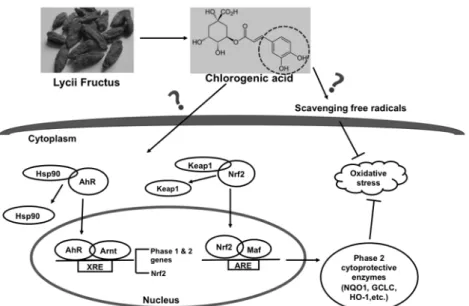
1.4 Nrf2/ARE and AhR/XRE pathways
It well known that the Nrf2/ARE and AhR/XRE pathways represent the important antioxidative defense mechanisms. Nuclear factor (erythroid-derived 2)-like 2 (Nrf2)
of the drug-metabolizing enzyme gene expressions (Nakamura et al., 2010). A ubiquitous flavonoid, quercetin, has been shown to activate the transcription factor Nrf2 (Surh et al., 2008) and to act as an exogenous ligand of AhR (Satsu et al., 2015). Cross talk between the AhR and Nrf2 pathways is plausible because the gene promoter of Nrf2 has a functional XRE (Ashida et al., 2008). Thus, the regulation of AhR by non- toxic food phytochemicals has been of considerable interest because it is likely that detoxification of toxic xenobiotics could be enhanced by concerted induction of phase 1 and 2 enzymes by AhR-dependent mechanisms (Nakashima et al., 2016).
1.4.1 Keap1/Nrf2/ARE pathway
Nrf2 binds to antioxidant response element (ARE) and induces a number of Nrf2- dependent phase 2 drug-metabolizing genes, including NQO1, HO-1, GCLC, etc.
(Nakamura et al., 2010). Han found that CGA protects MC3T3-E1 cells against H2O2- induced oxidative damage by enhancing Nrf2 nuclear translocation and upregulating HO-1 induction via PI3K/Akt signaling pathway (Han et al., 2017). Caffeic acid phenethyl ester (CAPE) was reported to exert indirect antioxidant capacity by up- regulating the expression of phase 2 antioxidant and detoxifying enzyme HO-1 via the ERK-Nrf2 signaling pathway (Kim et al., 2014). In addition, Ellagic acid protects human keratinocyte cells against UVA-induced oxidative stress and apoptosis through the upregulation of the HO-1 and Nrf2 antioxidant genes (Hseu et al., 2012).
Chitooligosaccharides can protect against ethanol-induced oxidative damage in human L02 normal liver cells through inhibition of ROS generation, activation of Nrf2 and reduction of MAPK phosphorylation (Luo et al., 2014).
1.4.2 AhR/ARNT/XRE pathway
AhR, a ligand-dependent transcription factor activated by a variety of synthetic and natural molecules, binding to xenobiotic responsive element (XRE), mainly contributes not only to the inducible expression of phase 1 enzyme genes including CYP1A1 (Nakamura et al., 2018), but also to the Nrf2-mediated gene expression of the phase 2 enzyme genes (Ciolino et al., 1999; Miao et al., 2005). It has reported that CGA
prevents acetaminophen-induced liver injury through inhibiting its metabolism and involved with CYP450 metabolic enzymes and some antioxidant signals (Pang et al., 2015).Jang J H demonstrated that CSE robustly upregulates ALDH enzymatic activity in human bronchial epithelial cells through a mechanism dependent on AhR-mediated ALDH3A1 expression (Jang J H et al., 2014).
Fig. 1.2. Diagram of the direct and indirect antioxidative action.
1.5 Study outlines
In this study, I have focused on the induction of phase 2 drug-metabolizing enzymes and aldehyde dehydrogenase by Lycii Fructus extract (LFE), chlorogenic acid (CGA) and 3,4-dihydroxyphenylacetic acid (DOPAC) to prevent the hydrogen peroxide- and acetaldehyde-induced cytotoxicity.
I determined the role of the indirect antioxidative action in the cytoprotective effect of LFE in murine hepatoma Hepa1c1c7 cells. Cell viability experiments revealed that the pretreatment of LFE and CGA for 24 h completely inhibited the cytotoxic effect of hydrogen peroxide with up-regulation of the phase 2 drug- metabolizing enzyme genes.
I investigated the effects of LFE, CGA, DOPAC and quercetin against ultraviolet B (UVB)-induced cytotoxicity in human keratinocyte HaCaT cells. LFE and the three
phenolic compounds stimulated the expression of antioxidant enzyme genes, including HO-1, NQO1 and GCLC in human keratinocyte HaCaT cells. They also mitigated cytotoxicity induced by UVB irradiation. These results suggested that the inducible expression of the antioxidant enzyme genes might be effective on the protection against UVB-induced oxidative stress.
I examined the modulating effects of LFE, CGA and its catabolites on the acetaldehyde-induced cytotoxicity in Hepa1c1c7 cells. LFE, CGA and its catabolites ameliorated the acetaldehyde-cytotoxicity with the enhanced ALDH activity by transcriptional regulation.
In conclusion, the present study represents a potentially efficient strategy to prevent the oxidative stress- and alcohol-induced cytotoxicity by dietary phenolic acids.
CHAPTER 2
Lycii fructus extract ameliorates hydrogen peroxide-induced cytotoxicity through indirect antioxidant action
2.1 Introduction
Lycii Fructus, also known as Gou Qi Zi, is the dried ripe fruits of Lycium chinense and L. barbarum, both of which are called wolfberry or Goji berries and belong to the Solanaceae family and are closely related species with similar uses (Potterat et al., 2010). Lycii Fructus has long been used as a traditional food and folk medicine in East Asia. Recent studies have found that Lycii Fructus possesses various health- promoting characteristics including antioxidant, neuroprotective, and immunomodulatory activities in the context of age-related diseases including atherosclerosis, cancer, neurodegeneration and diabetes (Potterat et al., 2010; Amagase et al., 2011). Polysaccharides, carotenoids, ascorbic acid and glycosylated precursors, flavonoids, and phenolic acids have been reported to represent biologically-active components isolated from the fresh wolfberry (Amagase et al., 2011; Le et al., 2007).
Among the biological activities, the antioxidant properties have been well investigated by various in vitro and in vivo assays. The mechanisms of the antioxidant action are suggested to involve reducing capacity, metal ion chelation and radical scavenging activity (Le et al., 2007). The fresh L. chinense fruit extract also recovers the cellular activities of several antioxidant enzymes decreased by hydrogen peroxide (Zhang et al., 2010). In a mouse skin model, the oral administration of the fresh wolfberry juice significantly inhibited lipid peroxidation induced by UVA irradiation, possibly through production of endogenous skin antioxidants (Reeve et al., 2010).
These results led us to hypothesize that, in addition to its radical scavenging properties, the dried Lycii Fructus extract (LFE) might have a protective effect against oxidative stress via induction of the antioxidant enzyme activities. However, to the best of our knowledge, it has not been elucidated whether LFE alone can potentiate the gene expression of the drug-metabolizing enzymes.
In the present study, we investigated the significant role of the indirect antioxidative action in the LFE-induced cytoprotective effect in vitro. We demonstrated for the first time that LFE enhanced the expression of the drug- metabolizing enzyme genes, such as NAD(P)H: quinone oxidoreductase 1 (NQO1), heme oxygenase-1 (HO-1), glutamate-cysteine ligase catalytic subunit (GCLC), a component of cysteine/ glutamate antiporter (cystine/glutamate exchanger; xCT) and cytochrome P450 1A1 (CYP1A1), as well as the intracellular glutathione (GSH) level in mouse hepatoma Hepa1c1c7 cells with the nuclear translocation of aryl hydrocarbon receptor (AhR) as well as nuclear factor (erythroid-derived 2)-like 2 (Nrf2). Cell viability experiments revealed that the pretreatment of LFE for 24 h completely inhibited the cytotoxic effect of hydrogen peroxide. Furthermore, the effect of a main constituent of LFE, chlorogenic acid (CGA), on the hydrogen peroxide-induced cytotoxicity and the drug-metabolizing enzyme gene expression was examined. This study provides evidence showing that LFE represents a potential antioxidant material with indirect enhancement of the cytoprotective gene expression.
2.2 Materials and methods
2.2.1 Materials
The 50%-ethanol extract of Lycii Fructus (edible quality grade, Kukoshiekisu A), the dried fruits of L. barbarum and L. chinense originated from Ningxia Hui Autonomous Region, China, was obtained from Matsuura Yakugyo (Nagoya, Japan).
The extract was frozen dried before experiment. Chlorogenic acid was obtained from MP Biomedicals, LLC (Santa Ana, CA, USA). α-Minimum essential medium (α- MEM), and Trizol reagent were purchased from Life Technologies (Carlsbad, CA, USA). Fatal bovine serum (FBS) was purchased from Nichirei Corporation (Tokyo, Japan). Primary antibodies against AhR (Cat No. sc-133088) and Lamin B1 (Cat No.
sc-377000) were purchased from Santa Cruz Biotechnology (Santa Cruz, CA, USA).
Antibodies against Nrf2 (Cat #12721) was purchased from Cell Signaling Technology (Beverly, MA, USA). Pierce™ BCA Protein Assay Kit was purchased from Thermo Scientific (Meridian Rd., Rockford. USA). All other chemicals were purchased from obtained from Wako Pure Chemicals Industries (Osaka, Japan) or Nacalai Tesque (Kyoto, Japan).
2.2.2 Cell culture
The mouse hepatoma cell line Hepa1c1c7, obtained from the American Type Culture Collection, was grown and maintained at 37oC in a 5% CO2 atmosphere in α- MEM containing 10% FBS, 4 mM L-glutamine, 100 U/ml penicillin, and 100 µg/ml streptomycin. For experiments, cells were seeded in complete medium and treated with reagent or dimethyl sulfoxide (DMSO) vehicle (final 0.1%, v/v).
2.2.3 Reverse transcription-polymerase chain reaction (RT-PCR)
Total cellular RNA was isolated using Trizol reagent according to the manufacturer’s recommendations. RNA was quantified by measuring absorbance at 260 nm with a spectrophotometer (V630 Bio, JASCO Corporation, Tokyo, Japan).
Total RNA (8 µg) was reverse transcribed to cDNA using M-MLV reverse transcriptase.
PCR amplification was then performed with Taq polymerase and specific primers.
5ʹ- TgAAgAgAgTACA TggAgCC – 3ʹ (20 cycles, product size 290 bp); mGCLC, (F)
5ʹ – ggCgATgTTCTTgAgACTCTgC – 3ʹ and (R) 5ʹ –
TTCCTTCgATCATgTAACTCCCATA – 3ʹ (26 cycles, product size 99 bp); mHO-1, (F) 5ʹ-gTgATggAgCgTCCACAgC – 3ʹ and (R) 5ʹ- TggTggCCTCCTTCAAgg – 3ʹ (26 cycles, product size 66 bp); mxCT, (F) 5ʹ – CCTggCATTTggACgCTACAT – 3ʹ and (R) 5ʹ- TgAgAATTgCTgTgAgCTTgCA −3ʹ (25 cycles, product size 182 bp);
mCYP1A1, (F) 5ʹ- CCTCTTTggAgCTgggTTTg – 3ʹ and (R) 5ʹ- TgCTgTgggggATggTgAAg −3ʹ (24 cycles, product size 229 bp). The PCR products were separated on an agarose gel (3% or 4%), stained with ethidium bromide, and visualized under UV light (Luminescent Image Analyzer LAS-3000, FUJIFILM Corporation, Tokyo, Japan). The relative density analysis of the bands was carried out using the Image J Software Program.
2.2.4 Glutathione titration
GSH contents were determined using 5,5ʹ-dithiobis (2-nitrobenzoic acid) and GSH reductase according to the method of Baker et al (Baker et al., 1990).
2.2.5 Western blot analysis
Cells were washed with ice-cold phosphate buffered saline (PBS) (-). For separation of nuclear fraction, the cells were first suspended with buffer-1 (10 mM HEPES, pH 7.9, 10 mM KCl, 0.1 mM EDTA, 0.1 mM EGTA, 1 mM DTT, 10 mM NaF, 1 mM Na3VO4 and PMSF) containing PIC. After 0.4% NP-40 were added, cell lysates were centrifuged at 500 g for 4 min. The supernatant was prepared as cytoplasmic extractions. After washing with buffer-1 twice again, precipitates were suspended in buffer-2 (20 mM HEPES (pH 7.9), 400 mM NaCl, 1mM EDTA, 1mM EGTA, 1 mM DTT, 10 mM NaF, 1 mM Na3VO4 and 1 mM PMSF) containing PIC, then centrifuged 18,000 g for 20 min. The supernatant containing 10 µg protein were prepared for SDS-PAGE. Protein concentration in the nuclear fraction was quantified using a BCA protein assay. After SDS-PAGE purification, proteins were transferred to nitrocellulose membranes (Bio-Rad Laboratories, Cat No. #1620094, Hercules, CA,
primary antibody overnight at 4°C followed by an appropriate secondary antibody.
Secondary antibody binding was visualized using a Chemi-Lumi One Super under a chemiluminescence system (Luminescent Image Analyzer LAS-3000, FUJIFILM Corporation, Tokyo, Japan). The relative density analysis of the bands was carried out using the Image J Software Program (National Institutes of Health, Bethesda, MD, USA).
2.2.6 Cell viability determination
Hepa1c1c7 cells (5 × 10⁴) were suspended in a 96-well plate. After overnight preculture, the cells were incubated with LFE or CGA for 30 min or 24 h. After washing with PBS, the cells were treated with hydrogen peroxide (100 µM) for 6 h. After stimulation, 10 µL of an MTT solution was added to each well, and the absorbance was measured with a microplate reader (Benchmarkplus, Bio-Rad laboratories, Hercules, CA, USA) at 530 nm after incubation at 37°C for 2–3 h in a humidified CO2 incubator.
The obtained values were compared with each of the controls incubated with vehicle only.
2.2.7 Trypan blue dye exclusion assay
Trypan blue dye exclusion assay was carried out for quantitative analysis of cell viability. Cell suspensions were mixed with 0.4% Trypan blue stain. Viable cells (cells that excluded blue dye) were counted using a hemocytometer (Bürker-Türk) under a light microscope.
2.2.8 Statistical analysis
All values were expressed as means ± SD. Statistical significance was analyzed by a one-way ANOVA followed by Tukey’s HSD using XLSTAT software.
2.3 Results
2.3.1 Lycii fructus extract induces the gene expression of several drug- metabolizing enzymes
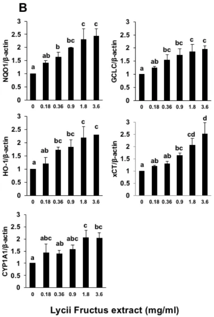
We investigated whether LFE (commercially available and edible quality grade) has the ability to induce the gene expression of the drug-metabolizing enzymes, including NQO1, GCLC, xCT, HO-1, and CYP1A1. The incubation of Hepa1c1c7 cells with LFE for 24 h led to a significant increase in the mRNA levels of not only phase 2 drug-metabolizing enzyme genes, such as NQO1, GCLC, HO-1 and xCT, but also a phase 1 gene, CYP1A1 (Figure 2.1). We also confirmed the dose-dependent effect of LFE on the expression of these drug-metabolizing enzymes (Figure 2.1). These results suggested that LFE might induce the expression of the drug-metabolizing enzymes through the common pathway(s) for both phase 1 and 2 gene induction.
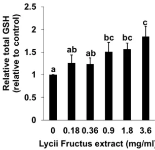
Because LFE induced the gene expression of GCLC and xCT, both of which play an important role in the GSH biosynthesis, the effects of LFE on the intracellular total GSH level were determined. As shown in Figure 2.2, the treatment of Hepa1c1c7 cells with LFE for 24 h exhibited a significant and dose-dependent enhancement in the intracellular GSH level. These results suggested that LFE might enhance the cellular GSH level through a transcriptional regulation of the GSH biosynthesis related genes.
Fig. 2.1. Modulating effects of Lycii fructus extract (LFE) on the gene expression of the drug metabolizing enzymes. The total RNA was extracted from Hepa1c1c7 cells treated with LFE at the indicated concentrations for 24 h, then a RT-PCR analysis for NQO1, GCLC, HO-1, xCT and CYP1A1 was carried out. All values were expressed as means ± SD of three separate experiments. Different letters above the bars indicate significant differences among the treatments for each condition (p < 0.05).
Fig. 2.2. Modulating effects of Lycii fructus extract (LFE) on the intracellular GSH level. Hepa1c1c7 cells were treated with LFE for 24 h and the total GSH level was measured by a DTNB assay. All values were expressed as means ± SD of three separate experiments. Different letters above the bars indicate significant differences among the treatments for each condition (p < 0.05).
2.3.2 Lycii fructus induces nuclear translocation of Nrf2 and AhR
Nrf2 and AhR play a pivotal role in the transcriptional regulation of the drug- metabolizing enzyme gene expressions (Nakamura et al., 2010; 2018). Nrf2 binds to antioxidant response element (ARE) and induces a number of Nrf2-dependent phase 2 drug-metabolizing genes, including NQO1, HO-1, GCLC, etc (Nakamura et al., 2010;
2018). AhR, the transcription factor binding to xenobiotic responsive element (XRE), mainly contributes not only to the inducible expression of phase 1 enzyme genes including CYP1A1, but also the Nrf2-mediated gene expression of the phase 2 enzyme genes (Ciolino et al., 1999; Miao et al., 2005). Thus, we examined the nuclear translocation of Nrf2 and AhR by Western blotting. As shown in Figure 2.3, the treatment of LFE significantly increased the nuclear level of AhR as well as Nrf2. These results suggested that LFE actually activated both the Nrf2/ARE and AhR/XRE pathways, possibly through enhancement of their nuclear translocation.
Fig. 2.3. Modulating effects of Lycii fructus extract (LFE) on the Nrf2 and AhR levels in Hepa1c1c7 cells. Nuclear cell lysates (10 µg/lane) were analyzed by immunoblotting using antibodies that specifically recognize AhR, Nrf2, and Lamin B1.
(A) Representative blots and quantitative data for (B) AhR and (C) Nrf2. All the values were expressed as means ± SD of three separate experiments. Different letters above the bars indicate significant differences among the treatments for each condition (p <
0.05).
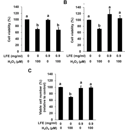
2.3.3 Lycii fructus shows an antioxidant effect after a 24-h incubation
LFE has been reported to show a significant scavenging effect against free radicals (Zhang et al., 2010). LFE also exhibited the inducing effect on some enzymes (HO-1 and GCLC) producing endogenous antioxidants such as biliverdin and GSH (Figures 2.1 and 2.2). These results prompted us to examine whether LFE protects the cells from oxidative stress through its direct radical scavenging action or indirect antioxidant action regulated by the transcriptional level. The inhibitory effect of LFE on the hydrogen peroxide-induced cytotoxicity was evaluated by an MTT assay. Although the incubation of LFE (0.9 mg/ml), 30 min before the hydrogen peroxide treatment, showed no effect on the cytotoxicity (Figure 2.4 (A)), pretreatment with LFE for 24 h inhibited
it (Figure 2.4 (B)). As shown in Figure 2.4 (C), the data of a trypan blue dye exclusion assay showed the similar result to the MTT assay data. Thus, the treatment with LFE at the concentration required for the inducible expression of the drug-metabolizing enzymes actually exhibited an antioxidant effect in Hepa1c1c7 cells.
Fig. 2.4. Effect of Lycii fructus extract (LFE) on the hydrogen peroxide-induced cytotoxicity in Hepa1c1c7 cells. Hepa1c1c7 cells were pretreated with LFE (0.9 mg/mL) for 30 min (A) or 24 h (B and C), then treated with hydrogen peroxide (100 µM) for 6 h. Cell viability was measured using an MTT assay (A and B) or a Trypan blue dye exclusion assay (C). All values were expressed as means ± SD of three separate experiments. Different letters above the bars indicate significant differences among the treatments for each condition (p < 0.05).
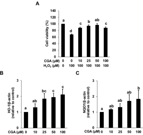
2.3.4 Chlorogenic acid is one of the predominant antioxidants in LFE
Since CGA is the most abundant phenolic acid in the fresh wolfberry (Zhang et al., 2016), the effect of CGA on the hydrogen peroxide-induced cytotoxicity was
examined. As shown in Figure 2.5 (A), the pretreatment of CGA for 24 h significantly impaired the hydrogen peroxide-induced cytotoxicity. Also, the treatment of CGA for 24 h significantly induced the gene expression of HO-1 and NQO1 in a dose-dependent manner (Figure 2.5 (B) and (C)). Furthermore, we examined whether CGA induces the nuclear translocation of Nrf2 and AhR. As shown in Figure 2.3, the treatment of LFE significantly increased the nuclear level of AhR as well as Nrf2. These results suggested that CGA actually activated both the Nrf2/ARE and AhR/XRE pathways, possibly through enhancement of their nuclear translocation, similar to the action of LFE. Rutin, one of the major flavonoids in LFE (Zhang et al., 2016), showed little effect on the HO- 1 and NQO1 gene expression (data not shown). Thus, CGA was identified as one of the possible predominant antioxidants in LFE.
Fig. 2.5. Effect of chlorogenic acid (CGA) on the hydrogen peroxide-induced cytotoxicity and the gene expression of HO-1 and NQO1. (A) Hepa1c1c7 cells were pretreated with CGA at the indicated concentration for 24 h, then treated with hydrogen peroxide (100 µM) for 6 h. Cell viability was measured using an MTT assay. (B) and
(C) The total RNA was extracted from Hepa1c1c7 cells treated with CGA at the indicated concentrations for 24 h, then a RT-PCR analysis for HO-1 and NQO1 was carried out. All values were expressed as means ± SD of three separate experiments.
Different letters above the bars indicate significant differences among the treatments for each condition (p < 0.05).
Fig. 2.6. Effect of chlorogenic acid (CGA) on the Nrf2 and AhR levels in Hepa1c1c7 cells. Nuclear cell lysates (10 µg/lane) were analyzed by immunoblotting using antibodies that specifically recognize AhR, Nrf2, and Lamin B1. (A) Representative blots and quantitative data for (B) AhR and (C) Nrf2. All values were expressed as means ± SD of three separate experiments. Different letters above the bars indicate significant differences among the treatments for each condition (p < 0.05).
2.4 Discussion
In the present study, we identified LFE as one of the promising materials from medicinal foods, in terms of the inducible potentials of the antioxidant enzyme gene expression. Previous study demonstrated that, among the 45 common vegetable samples, only the 8 extracts at a concentration of 200 µg/mL significantly induced NQO1 activity in vitro (Hashimoto et al., 2002). The effective concentration of the tested LFE required for the phase 2 enzyme induction (>180 µg/mL, Figure 2.1) is quite similar with those of the vegetables such as broccoli, celery, parsley, etc. (Zhang et al., 2010), supporting an idea that LFE is an equivalent source of the phase 2 enzyme inducers to such vegetables.
LFE significantly and dose-dependently enhanced not only the mRNA levels of all the tested genes (Figure 2.1), but also the intracellular GSH level (Figure 2.2). The concentration dependency of LFE for the intracellular GSH level was closely correlated with that for the gene expression of xCT and GCLC. Furthermore, the antioxidant effect in Hepa1c1c7 cells was actually exhibited by the 24-h treatment of LFE at the concentration required for the induction of the GSH-related genes, such as xCT and GCLC, as well as the GSH up-regulation, suggesting the significant role of GSH in the cellular antioxidant action.
Various molecular targets for the drug-metabolizing effects of food phytochemicals have been identified. For example, a ubiquitous flavonoid, quercetin, activates the transcription factor Nrf2 and acts as an exogenous ligand of AhR (Surh et al., 2008; Ciolino et al., 1999). Nrf2 induces a number of phase 2 drug-metabolizing genes, including NQO1, GSTs, HO-1, and GCLC. AhR plays an important role in regulating the expression of not only CYPs including CYP1A1, but also the phase 2 genes, such as NQO1 and GSTA1 (Ciolino et al., 1999). Cross talk between the AhR and Nrf2 pathways is plausible because the gene promoter of Nrf2 has a functional XRE (Miao et al., 2005). Thus, the regulation of AhR by non-toxic food phytochemicals has been of considerable interest because it is likely that detoxification
enzymes by AhR-dependent mechanisms (Satsu et al., 2015). Thus, LFE might be able to activate both the Nrf2- and AhR-dependent pathways (Figure 2.3) and thus coordinately enhance the phase 2 drug-metabolizing enzyme expression.
Among the food constituents, aromatic compounds with the coplanar or non-bulky conformation, including flavonoids, indole compounds, curcumin, and phenolic acids, have been reported to act as AhR ligands as well as Nrf2 activators (Nakamura et al., 2010; Ashida et al., 2008; Nakashima et al., 2016; Liu et al., 2017). We observed that CGA, the major phenolic acid in the fresh wolfberry and also in LFE (Zhang Q et al., 2016; Zhang XF et al., 2018), showed the significant cytoprotective effect on the hydrogen peroxide-induced cytotoxicity with the enhanced gene expression of HO-1 and NQO1 (Figure 2.5). CGA showed the similar effects on the nuclear translocation of AhR and Nrf2 as well as the HO-1 and NQO1 gene expression (Figures 2.5 and 2.6) to those of LFE (Figures 2.1 and 2.4). These results suggested that CGA might be one of the predominant antioxidants in LFE.
The fresh wolfberry represents an excellent source of CGA, because the content of CGA in the extract of fresh wolfberry is 113–526 µg/g (Zhang Q et al., 2016) and comparable to the polyphenol-rich foods, such as instant coffee, apple juice, Maté as well as blueberry (Clifford et al., 2017; Gavrilova et al., 2011). However, other LFE constituents might contribute to the total antioxidant activity of LFE, because the CGA content in the fresh wolfberry is not enough to explain it. In addition to CGA, rutin is also the main polyphenol compound in the fresh wolfberry, accompanied by small amounts of hyperoside, quercetin, and morin (Potterat et al., 2010; Amagase et al., 2011). Considerable amounts of flavonoid glycosides including rutin can pass through the small intestine, and the conjugated glycosides are removed from the flavonoid molecules by bacterial enzymes in the colon (Murota et al., 2018). We previously reported that their aglycone quercetin and catabolites produced by gut microbiota showed an enhancing effect on the gene expression of the phase 2 drug-metabolizing enzymes and protected from hydrogen peroxide-induced cytotoxicity in mouse
hepatoma hepa1c1c7 cells (Tang et al., 2016), suggesting that rutin could also be a potential in vivo antioxidant of LFE.
2.5 Conclusion
The present study provided biological evidence that LFE and CGA induces cytoprotective responses against oxidative stress through the inducible expression of the antioxidant enzyme genes. Since LFE and CGA have some advantages for application as a food material without severe toxicity, future efforts will be concerned with further understanding the signaling pathway of the antioxidant enzyme induction as well as identification of the other compounds contributing to the indirect antioxidative action.
CHAPTER 3
Lycii Fructus extract and its phytochemicals ameliorate UVB- induced cytotoxicity in Human keratinocytes HaCaT cells 3.1 Introduction
Reactive oxygen species (ROS) are produced by living organisms because of normal cellular metabolism and environmental factors, such as air pollutants, cigarette smoke or Ultraviolet (UV) irradiation. ROS are highly reactive molecules and can damage cell structures such as carbohydrates, nucleic acids, lipids, and proteins and alter their functions (Esra Birben et al., 2012). UV irradiation is the most common environmental factor that damages the human skin. Excessive exposure to UV irradiation generates ROS in skin and leads to the activation of transcription factors that induces the expression of pro-inflammatory cytokines and metalloproteinase (MMPs) resulting cellular senescence. Collagen-degrading MMP-1 is upregulated and serves as the primary MMP in UV-exposed skin. Therefore, excessive degradation of collagen and matrix by UV-induced MMPs is a characteristic feature of photo-damaged skin, and MMP is used as a major marker of UVB-induced photoaging as well as skin inflammation (Pillai et al., 2005; Podhaisky et al., 2002).
Ultraviolet B (UVB), one of the three types of invisible light rays (together with ultraviolet A and ultraviolet C) given off by the sun. Although UVC is the most dangerous type of ultraviolet light in terms of its potential to harm life on earth, it cannot penetrate earth's protective ozone layer because of the shortest wavelength. Therefore, it poses no threat to human, animal or plant life on earth. UVA and UVB, on the other hand, do penetrate the ozone layer in attenuated form and reach the surface of the planet.
UVA has the longest wavelength and reaches deep into the layers of skin (dermis) causing aging or wrinkling. Whereas, UVB has largest effect on the top layer of skin (epidermis), causing redness, burning and skin cancer. Thus, scientists long blamed UVB as the sole culprit in deep into skin, generating ROS and causing skin cancer in persons with a history of sunburn and repeated overexposure to ultraviolet radiation.
Lycii Fructus has long been used as a traditional food and folk medicine in East Asia, it possesses various health-promoting characteristics including antioxidant, antiaging, anti-tumor, cytoprotective, neuroprotective, antiglaucoma and immunomodulatory activities, and promotes endurance and improves control of glucose and other diabetic symptoms (Zhang et al. 2010; Li et al. 2007, 2011; Tang et al. 2011). Chlorogenic acid (CGA) has shown its photo-protection against UV- induced skin damage in animal model and displayed the suppression on UVB-related ROS mediated cellular processes in vivo (Feng et al., 2005; Kitagawa et al., 2011).
Quercetin is the aglycone form of flavonoid glycosides, which is one of the ubiquitous flavonoids in fruits and vegetables,especially in red onions. Previous study showed that quercetin 4′-glucoside were converted into smaller phenolic acids by colonic microflora, 3,4-dihydroxyphenylacetic acid (DOPAC) is one of the main metabolites (Mullen et al., 2008). It has been reported that DOPAC was actually identified as a predominant bioactive catabolite of quercetin glycosides (Tang et al., 2016). DOPAC also has been reported to ameliorate acetaldehyde-induced cytotoxicity by enhancing aldehyde dehydrogenase activity in murine hepatoma cells (Liu et al., 2017).
However, the biological evidence of Lycii Fructus extract (LFE), CGA and DOPAC, quercetin induce cytoprotective responses against UVB irradiation have not yet been elucidated. The present study examined the effects of LFE, CGA, DOPAC and quercetin against UVB-induced cytotoxicity in human keratinocyte HaCaT cells.
3.2 Materials and methods
3.2.1 Materials
Quercetin and DOPAC were obtained from Sigma Aldrich (St. Louis, MO, USA).
3.2.2 Cell culture
The human keratinocyte cell line HaCaT cells, obtained from the American Type Culture Coll ection, was grown and maintained at 37oC in a 5% CO2 atmosphere in DMEM containing 10% FBS, 4 mM L-glutamine, 100 U/ml penicillin, and 100 µg/ml streptomycin. For experiments, cells were seeded in complete medium and treated with reagent or dimethyl sulfoxide (DMSO) vehicle (final 0.1%, v/v).
3.2.3 Ultraviolet B irradiation
HaCaT cells were suspended at a density of 9 × 10⁴ cells per well in a 96-well plate. After 24 h pre-culture,the cells were treated with LFE (50, 100, 250, 500 and 1000 µg/ml), CGA (5, 10, 25, 50 and 100 µM), quercetin (0.5, 2.5, 5, 10 and 25 µM) or DOPAC (5, 10, 25, 50 and 100 µM) for 24 h before UVB irradiation (50 mJ/cm2) while covered with 50 µl HBSS in each well using UVB lamp. The UVB exposure was measured with a radiometer (PMA2100, Solar Light Company, Inc., Glenside, PA, USA). After UVB irradiation, the cells were washed with warm PBS, and incubated with medium for 24 h. After incubating, 10 µl of an MTT solution was added to each well at 37°C for 2 h in a humidified CO2 incubator. The MTT containing formazan crystals in living cells were solubilized in DMSO, then the absorbance was measured with a microplate spectrophotometer (Benchmark plus, Bio-Rad laboratories, Hercules, CA, USA) at 570 nm according to the manufacturer's instructions. The obtained values were expressed as percentages compared with each of the controls.
3.2.4 Reverse transcription-polymerase chain reaction (RT-PCR)
Total cellular RNA was isolated using Trizol reagent according to the manufacturer’s recommendations. RNA was quantified by measuring absorbance at 260 nm with a spectrophotometer (V630 Bio, JASCO Corporation, Tokyo, Japan).
Total RNA (8 µg) was reverse transcribed to cDNA using M-MLV reverse transcriptase.
Primers are as follows: hNQO1, (F) 5ʹ –AgAAgAgCACTgATCgTACTgg– 3ʹ and (R) 5ʹ-CgTAATTgTAAgCAAACTCTCCTATg– 3ʹ (27 cycles); hGCLC, (F) 5ʹ – ATCATCAATgggAAggAAggT– 3ʹ and (R) 5ʹ –gTCATTAGTTCTCCAgATgCTC–
3ʹ (25 cycles); hHO-1, (F) 5ʹ-gATTgCCCAgAAAgCCCTggAC – 3ʹ and (R) 5ʹ- CTgTCgCCACCAgAAAgCTgAg – 3ʹ (27 cycles); The PCR products were separated on an agarose gel (3%), stained with ethidium bromide, and visualized under UV light (Luminescent Image Analyzer LAS-3000, FUJIFILM Corporation, Tokyo, Japan). The relative density analysis of the bands was carried out using the Image J Software Program.
3.2.5 Western blot analysis See Chapter 2
3.2.6 Statistical analysis See Chapter 2
3.3 Results
3.3.1 LFE, CGA, quercetin and DOPAC dose-dependently inhibited the hydrogen peroxide-induced cytotoxicity.
Preliminary experiments (Figure.3.1) showed that not only Lycii fructus extract (LFE) and chlorogenic acid (CGA), but also quercetin and 3,4- dihydroxyphenylacetic acid (DOPAC) significantly and dose-dependently inhibited the hydrogen peroxide- induced cytotoxicity in HaCaT cells
Fig. 3.1. Impairing effects of Lycii fructus extract (LFE), chlorogenic acid (CGA), quercetin and 3,4-dihydroxyphenylacetic acid (DOPAC) on the cytotoxicity induced by hydrogen peroxide. HaCaT cells were treated with DMSO or LFE (A), CGA (B), quercetin (C) and DOPAC (D) respectively at the indicated concentrations for 24 h, then changed the medium and treated with H2O2 (100 µM) for 6 h. Cell viability was measured using MTT assay. All values were expressed as means ± SD of three separate experiments. Different letters above the bars indicate significant differences among the treatments for each condition (p < 0.05).
3.3.2 LFE, CGA, DOPAC and quercetin significantly inhibited the UVB-induced cytotoxicity.
The preliminary experiment of UVB irradiation only on the HaCaT cells (data not shown) showed that UVB irradiation led to a tendency suppression of HaCaT cell viability. Cell viability: 90%, 10 mJ/cm2; 83%, 25mJ/cm2; 69%, 50mJ/cm2; 44%, 100mJ/cm2; 28%, 150mJ/cm2. We chose 50mJ/cm2 as the intensity of UV toxicity. As shown in figure.3.2, LFE (Figure.3.2 (A)), CGA (Figure.3.2 (B)) and DOPAC (Figure.3.2 (D)) dose-dependently inhibited the UVB-induced cytotoxicity. whereas, lower concentration of quercetin significantly exhibited the ability to against the cytotoxicity induced by UVB (Figure.3.2 (C)).
Fig. 3.2. Impairing effects of Lycii fructus extract (LFE), chlorogenic acid (CGA), quercetin and 3,4-dihydroxyphenylacetic acid (DOPAC) on the cytotoxicity induced by UVB. HaCaT cells were treated with DMSO or LFE (A), CGA (B), quercetin (C) and DOPAC (D) respectively at the indicated concentrations for 24 h, then removed the medium, irradiated (50 mJ/cm2) with 50 µl HBSS using UVB lamp. After UVB irradiation, washed with warm PBS, and incubated with new medium for 24 h. Cell viability was measured using MTT assay. All values were expressed as means ± SD of
three separate experiments. Different letters above the bars indicate significant differences among the treatments for each condition (p < 0.05).
3.3.3 LFE, CGA, DOPAC and quercetin stimulated the gene expression of phase 2 drug-metabolizing enzymes, including HO-1, NQO1 and GCLC.
We then continued to investigate whether LFE, CGA, DOPAC and quercetin have the ability to induce the gene expression of the phase 2 drug-metabolizing enzymes, such as NQO1, GCLC and HO-1. The incubation of HaCaT cells with LFE (Figure 3.3) for 24 h led to a significant increase in the mRNA levels of HO-1 and GCLC (Figure 3.3 (B) and (C)). Lower concentration of LFE can increase the gene expression of NQO1, whereas, its higher concentration decreased the gene expression of NQO1 (Figure 3.3 (D)). We also confirmed the effects of CGA, quercetin and DOPAC on the expression of these representative phase 2 drug-metabolizing enzymes.
As shown in Figure 3.4, CGA increased the gene expression of HO-1 and NQO1 (Figure 3.4 (B) and (D)). Lower concentration of CGA can increase gene expression of GCLC, whereas, higher concentration had no effect on it (Figure 3.4 (C)). As Figure 3.5 shown, quercetin dose-dependently enhanced the gene expression of HO-1, NQO1 and GCLC. Although, DOPAC dose-dependently increased the gene expression of HO- 1 and GCLC (Figure 3.6 (B) and (C)), the similar action with CGA, it had no effect on the gene expression of NQO1 (Figure 3.6 (D)). These results suggested that LFE, CGA, DOPAC and quercetin might induce the expression of the drug-metabolizing enzymes through the transcriptional regulation.
Fig. 3.3. Modulating effects of Lycii fructus extract (LFE) on the gene expression of representative phase 2 drug-metabolizing enzymes. Total RNA was extracted from HaCaT cells treated with LFE for 24 h, then a RT-PCR analysis for each gene was carried out. (A) representative blots and quantitative data for (B) heme oxygenase-1 (HO-1), (C) glutamate-cysteine ligase catalytic subunit (GCLC) and (D) NAD(P)H:
quinone oxidoreductase 1 (NQO1). All values are expressed as means ± SD and analyzed by Student's t-test (*p<0.05, **p<0.01 compared to control).
Fig. 3.4. Modulating effects of chlorogenic acid (CGA) on the gene expression of representative phase 2 drug-metabolizing enzymes. Total RNA was extracted from HaCaT cells treated with CGA for 24 h, then a RT-PCR analysis for each gene was carried out. (A) representative blots and quantitative data for (B) heme oxygenase-1 (HO-1), (C) glutamate-cysteine ligase catalytic subunit (GCLC) and (D) NAD(P)H:
quinone oxidoreductase 1 (NQO1). All values are expressed as means ± SD and analyzed by Student's t-test (*p<0.05, **p<0.01 compared to control).
Fig. 3.5. Modulating effects of quercetin on the gene expression of representative phase 2 drug-metabolizing enzymes. Total RNA was extracted from HaCaT cells treated with quercetin for 24 h, then a RT-PCR analysis for each gene was carried out. (A) representative blots and quantitative data for (B) heme oxygenase-1 (HO-1), (C) glutamate-cysteine ligase catalytic subunit (GCLC) and (D) NAD(P)H: quinone oxidoreductase 1 (NQO1). All values are expressed as means ± SD and analyzed by Student's t-test (*p<0.05, **p<0.01 compared to control).
Fig. 3.6. Modulating effects of 3,4-dihydroxyphenylacetic acid (DOPAC) on the gene expression of representative phase 2 drug-metabolizing enzymes. Total RNA was extracted from HaCaT cells treated with DOPAC for 24 h, then a RT-PCR analysis for each gene was carried out. (A) representative blots and quantitative data for (B) heme oxygenase-1 (HO-1), (C) glutamate-cysteine ligase catalytic subunit (GCLC) and (D) NAD(P)H: quinone oxidoreductase 1 (NQO1). All values are expressed as means ± SD and analyzed by Student's t-test (*p<0.05, **p<0.01 compared to control).
3.3.4 LFE, CGA, DOPAC and quercetin induced the nuclear translocation of AhR and Nrf2.
We next examined the effect of LFE, CGA, DOPAC and quercetin on the nuclear translocation of AhR and Nrf2. Western blot analysis showed that the nuclear level of the AhR (Figure 3.7 (B)) and Nrf2 (Figure 3.7 (C)) protein were significantly increased by a 24 h LFE treatment in a time-dependent manner. LFE actually enhanced the nuclear translocation of AhR and Nrf2. These results suggested that LFE activated
the Nrf2- and AhR-dependent pathways possibly through the enhancement of the nuclear translocation of AhR and Nrf2. Not only LFE, but also CGA (Figure 3.8), quercetin (Figure 3.9) and its catabolite, DOPAC (Figure 3.10) exhibited the similar action on the nuclear translocation of AhR and Nrf2. These results suggested that LFE and its phytochemicals ameliorate UVB-induced cytotoxicity possibly through the enhanced expression of the phase 2 drug-metabolizing enzyme genes with the nuclear translocation of Nrf2 and AhR.
Fig. 3.7. Modulating effects of Lycii fructus extract (LFE) on the aryl hydrocarbon receptor (AhR) and nuclear factor (erythroid-derived 2)-like 2 (Nrf2) level. Nuclear fraction was separated from HaCaT cells treated with LFE for 24 h, then a Western blot analysis for each protein was carried out. (A) representative blots and quantitative data for (B) AhR and (C) Nrf2. All values are expressed as means ± SD and analyzed by Student's t-test (* p<0.05, ** p<0.01 compared to control).
Fig. 3.8. Modulating effects of chlorogenic acid (CGA) on the aryl hydrocarbon receptor (AhR) and nuclear factor (erythroid-derived 2)-like 2 (Nrf2) level. Nuclear
fraction was separated from HaCaT cells treated with CGA for 24 h, then a Western blot analysis for each protein was carried out. (A) representative blots and quantitative data for (B) AhR and (C) Nrf2. All values are expressed as means ± SD and analyzed by Student's t-test (* p<0.05, ** p<0.01 compared to control).
Fig. 3.9. Modulating effects of quercetin on the aryl hydrocarbon receptor (AhR) and nuclear factor (erythroid-derived 2)-like 2 (Nrf2) level. Nuclear fraction was separated from HaCaT cells treated with quercetin for 24 h, then a Western blot analysis for each protein was carried out. (A) representative blots and quantitative data for (B) AhR and (C) Nrf2. All values are expressed as means ± SD and analyzed by Student's t-test (* p<0.05, ** p<0.01 compared to control).
Fig. 3.10. Modulating effects of 3,4-dihydroxyphenylacetic acid (DOPAC) on the aryl hydrocarbon receptor (AhR) and nuclear factor (erythroid-derived 2)-like 2 (Nrf2) level.
Nuclear fraction was separated from HaCaT cells treated with DOPAC for 24 h, then a Western blot analysis for each protein was carried out. (A) representative blots and quantitative data for (B) AhR and (C) Nrf2. All values are expressed as means ± SD and analyzed by Student's t-test (* p<0.05, ** p<0.01 compared to control).