Case Report
Improvement of native pulmonary alveolar proteinosis after contralateral single
living-donor lobar lung transplantation: A case report
Authors
Kazuma Kobayashi1, Shinya Ohkouchi2, Yoji Sasahara3, Masahito Ebina4, Koh
Nakata5, Ryoko Saito6, Miki Akiba7, Tetsu Sado1, Hisashi Oishi1, Tatsuaki
Watanabe1, Hajime Kurosawa2, Yoshinori Okada1.
Institution
1Department of Thoracic Surgery, Institute of Development, Aging and Cancer,
Tohoku University, Department of 2Occupational Health, 3Pediatrics, and
6Pathology, Tohoku University, Graduate School of Medicine, 4Division of
Respiratory Medicine, Tohoku Medical and Pharmaceutical University,
5Bioscience Medical Research Center, Niigata University Medical and Dental
Hospital, 4Division of Organ Transplantation, Tohoku University Hospital, Miyagi,
Address
*Corresponding author: Shinya Ohkouchi, M.D., Ph.D.,
Department of Occupational Health, Tohoku University Graduate School of
Medicine, 1-1 Seiryoumachi, Aoba-ku, Sendai 980-8574, Japan.
Contributions
None of the authors have conflicts of interest with commercial companies
concerning this work. Conception and design: K.K., S.O.; data collection and
manuscript drafting for important intellectual content: K.K., S.O., Y.S., M.E., K.N.,
R.S., M.A., T.S., H.O., T.W., H.K., Y.O
Authors’ e-mail addresses
Kazuma Kobayashi: [email protected]
Youji Sasahara: [email protected]
Masahito Ebina: [email protected]
Koh Nakata: [email protected]
Miki Akiba: [email protected]
Tetsu Sado: [email protected]
Hisashi Oishi: [email protected]
Tatsuaki Watanabe: [email protected]
Hajime Kurosawa: [email protected]
Yoshinori Okada: [email protected]
Key Words
secondary pulmonary alveolar proteinosis, bone marrow transplantation, lung
transplantation, macrophages, one antigen mismatch,
Diamond-Blackfan-Anemia
Abbreviations
PAP; Pulmonary alveolar proteinosis, sPAP; secondary PAP, AMs; alveolar
macrophages, GM-CSF; granulocyte macrophage colony stimulating factor,
STAT5; signal transducer and activator of transcription 5, BMT; bone marrow
transplantation, DBA; Diamond-Blackfan-Anemia, GVHD; graft versus host
obliterans, GGO; ground glass opacification, KL-6; Krebs von den Lungen-6,
HLA; human leukocyte antigen, RPLS; reversible posterior leukoencephalopathy,
MMF; mycophenolate mofetil, HOT; home oxygen therapy, PFTs; pulmonary
function tests, CT; computed tomography, SP-A; surfactant protein A, PAS;
periodic acid Schiff, HSDT; hematopoietic stem cell transplantation.
Acknowledgments
We thank Dr. Haruyuki Ishii and Dr. Takuji Suzuki for their helpful comments on
sPAP and Ms. Miki Akiba for her work as a recipient coordinator and her
involvement in the data collection. This work is supported by grants from the
Abstract
Pulmonary alveolar proteinosis (PAP) is a rare disease characterized by the
accumulation of surfactant materials in the alveolar spaces due to the imbalance
of surfactant homeostasis (production and clearance). We herein report a case
of an eight-year-old girl who developed PAP after bone marrow transplantation
(BMT) from her mother for the treatment of Diamond-Blackfan-Anemia (DBA).
The anemia was improved by BMT; however, respiratory dysfunction due to
graft-versus-host disease gradually progressed. She eventually underwent right single
living-donor lobar lung transplantation (LDLLT) from her mother when she was
14 years old. A pathological examination of the excised lung confirmed the finding
of diffuse bronchiolitis obliterans and unexpectedly revealed widespread alveolar
proteinosis. Interestingly, the ground glass opacification (GGO) of her native left
lung on chest X-ray was improved after LDLLT. We present the very unique
clinical course of this patient and discuss the mechanisms underlying the
development of PAP after BMT and its improvement after LDLLT from the same
Introduction
Pulmonary alveolar proteinosis (PAP) is a rare disease characterized by the
accumulation of surfactant materials in the alveolar spaces. Regarding the
pathophysiology, alveolar macrophages (AMs) lose the ability to remove
excessive sediments from the alveolar spaces. PAP is commonly classified into
autoimmune (aPAP), secondary (sPAP), hereditary (hPAP), congenital (cPAP)
and unclassified PAP (uPAP). aPAP accounts for 90% of cases and is associated
with the existence of anti-GM-CSF (granulocyte-macrophage colony stimulating
factor) auto-antibody and an increased number of alveolar foamy macrophages.
GM-CSF is a monomeric glycoprotein, an activator of STAT5 phosphorylation,
and an inducer of macrophage maturation. sPAP, accounting for 10% of cases,
is accompanied by small and immature macrophages and mainly caused by
hematological diseases, such as myelodysplastic syndrome [1-4].
We herein report a case of sPAP that developed in an eight-year-old girl after
allogeneic bone marrow transplantation (BMT) from her mother for the treatment
of Diamond-Blackfan-Anemia (DBA), a disease of congenital erythroblast
deficiency. Although her anemia and hemosiderosis were improved by BMT,
progressed. She eventually received right single lung lobar lung transplantation
(LDLLT) from her mother when she was 14 years old. Unexpectedly, a
pathological examination revealed widespread sPAP coexisting with diffuse
bronchiolitis obliterans (BO), a typical pathological form of GVHD. Interestingly,
the ground glass opacification (GGO) of her native left lung on chest X-ray
consistent with PAP was improved after LDLLT. The levels of KL-6, which reflects
the severity of PAP, were also decreased dramatically. We describe the clinical
course and discuss the pathophysiological mechanisms underlying the
Case Presentation
A four-month-old girl was admitted to our hospital for the treatment of advanced
anemia. As she met the diagnostic criteria of <1 year old, macrocytic anemia with
no other significant cytopenia and normal marrow cellularity with a paucity of
erythroid precursors, the anemia was diagnosed as DBA, a congenital
erythroblast deficiency. Her familial history did not suggest any congenital
diseases, including hematological disorders. Her anemia was steroid-resistant
and rapidly progressive, so she was treated with frequent red blood cell
transfusion. As a result, she developed hemosiderosis in her organs. Therefore,
the patient received BMT at eight years old from her mother, whose HLA showed
five matched haplotypes and one mismatched haplotype from the recipient.
After BMT, her anemia and hemosiderosis were improved. However, three
months after the BMT, she showed symptoms of cough and dyspnea, and steroid
pulse therapy and augmentation of immunosuppressive therapy were
administered under the diagnosis of progressive GVHD. Cyclosporine and
tacrolimus could not be used because of the occurrence of convulsion induced
by reversible posterior leukoencephalopathy (RPLS) [5]. She was finally treated
these therapies, the severity of her dyspnea gradually progressed, and home
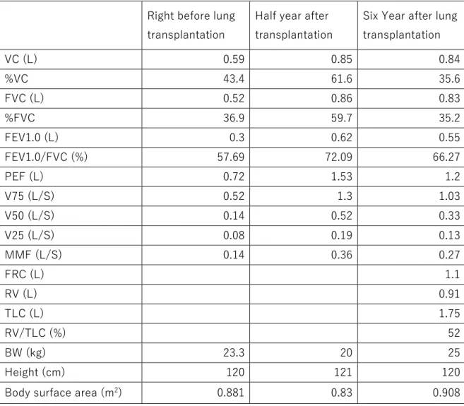
oxygen therapy (HOT) was ultimately needed at 10 years old. Her pulmonary
function tests (PFTs) immediately before lung transplantation showed severe
mixed restrictive and obstructive impairments (Table 1). She was registered to
the lung transplant waiting list at 12 years old. At that time, her height and weight
were 120 cm and 23.3 kg, respectively.
Chest computed tomography (CT) showed diffuse cystic dilation of peripheral
bronchi and widespread patchy GGO in the bilateral lung fields. While on the
waiting list, her respiratory dysfunction rapidly worsened, and she eventually
underwent right single LDLLT from her mother at 14 years old. The mother's right
lower lobe was transplanted into the patient’s right thoracic cavity.
The pathological analysis of the excised right lung showed epithelial injury and
obstruction of bronchioles with subepithelial fibrotic lesions, which was consistent
with BO (Figure 1A, B). Diffuse BO was observed in the excised right lung. In
addition, the retention of eosinophilic materials was observed in most alveolar
spaces in all fields of the excised right lung. The alveolar eosinophilic material
was positive for surfactant protein A (SP-A) and periodic acid Schiff (PAS)
negative for serum GM-CSF antibody.
She was discharged from the intensive-care unit and the hospital on days 49 and
65, respectively. She was treated with an unusual post-lung transplant
immunosuppressive regimen of only 2 mg of prednisolone and 250 mg of MMF
because the BMT and lung transplant donors were the same person, and the
HLA of her bone marrow and the right transplanted lower lobe was identical. The
courses of her postoperative imaging findings and laboratory data are shown in
Figure 2. Interestingly, the GGOs that were observed in all CT sections of the left
native lung gradually improved and eventually almost completely disappeared
(Figure 2A). The value of KL-6, a marker reflecting the severity of the PAP [2],
normalized (Figure 2B). Her hypoxia and the combined impairments with
restrictive and obstructive disturbances in the PFTs were also slightly improved
after lung transplantation (Figure 2B and Table 1). She remains alive and works
Discussion
The important problem in the present case is whether we could diagnose sPAP
before lung transplantation. Generally, typical PAP is associated with restrictive
impairments, while BO is associated with obstructive impairments in PFTs.
However, both impairments were mixed in this case. In addition, the radiographic
findings were atypical of PAP or BO. Surgical lung biopsy before lung
transplantation was difficult due to the invasiveness of the procedure. Thus, it
might have been difficult to suspect sPAP before lung transplantation in this case.
Whether or not the choice of lung transplantation using the one antigen
mismatched maternal lung could be acceptable for rescuing the patient in the
present case might be difficult to determine because of the lack of effective
treatment for the devastating situation.
Ishii reported that sPAP patients accounted for 10% of the 404 total PAP patients
(n=40) registered from 1999 to 2009 in the database of Niigata University in
Japan. Blood diseases are found in 88% of sPAP patients as causal diseases,
with the remaining 12% related to inflammatory and autoimmune diseases. No
case associated with BMT has been reported in the database [1]. However, a
stem cell transplantation (HSCT) and BMT [6-8]. These cases were rapidly
progressive with a fatal outcome, so some cases were diagnosed by an autopsy.
The authors of those reports considered that immunosuppression before and
after HSCT or BMT likely suppressed the function of AMs and played a role in the
development of PAP. Indeed, a retrospective study demonstrated that the use of
corticosteroids induced macrophage dysfunction, which caused aPAP [9].
In the present case, PAP developed after BMT, and the patient was treated with
prednisolone and MMF in an attempt to manage her GVHD. After LDLLT, the
immunosuppression was weakened because the HLA of the bone marrow and
the lung graft was identical (mother’s marrow and lung). Therefore, the
development of PAP and its remission may be attributed to the context of changes
in the strength of immunosuppression, as has been described in previous reports.
A pathological examination of the excised right lung showed the existence of
widespread PAP and diffuse BO. These findings suggest that the improvement of
GGO on CT mainly depended on the improvement of PAP, due to the diminished
extent of immunosuppressive therapy after lung transplantation.
Reconstruction of the lymphatic vessels was not performed in the present case;
vessels by mechanical ventilation might be not the principal reason of
improvement of GGO.
Another potential explanation for the observations made in the present study is
slightly complicated. Recently, the existence of two types of AMs in the lung has
been reported. One type is derived from progenitors in the fetal lung, while the
other is derived from circulating monocytes differentiated from hematopoietic
stem cells in bone marrow [10, 11]. Some clinicians have observed the
coexistence and slow replacement of donor AMs with recipient AMs in the lung
over several years in leukemia patients after BMT [12, 13]. Following BMT, the
immunocompetent cells derived from the donor bone marrow may attack recipient
resident AMs, thereby inducing the loss of the function of recipient AMs, which
causes sPAP. The improvement in PAP after LDLLT in the present case may be
explained by the hypothesis that the donor AMs from the right transplanted lung
may hematogenously or lymphogenously or trans-tracheal infiltrate the left native
lung, thereby weakening the attack of donor immunocompetent cells against
recipient AMs or inducing chimerism of AMs with dominant donor cells. However,
no conclusive studies or case reports support this hypothesis.
report describes the unique clinical course of a patient with PAP who underwent
Figure legends
Figure 1. Pathology of the excised left lung. Epithelial injury and obstruction of
bronchioles with subepithelial fibrotic lesions, which was consistent with
bronchioliits obliterans (A: hematoxylin-eosin at 100x magnification, B:
Elastica-Masson at 100x magnification). Massive eosinophilic material deposition in the
alveolar space (C, hematoxylin-eosin at 100x magnification). The material in the
alveolar space was positive on periodic acid-Schiff (PAS) stain (D).
Figure 2. The courses of her postoperative imaging findings and laboratory data.
The ground glass opacification of the left native lung on chest X-ray and CT, which
is consistent with pulmonary alveolar proteinosis, improved and eventually
disappeared after right single lung lobar lung transplantation (LDLLT) (A). The
value of KL-6, a marker reflecting the disease severity, normalized after LDLLT
References
1. Ishii H, Tazawa R, Kaneko C, Saraya T, Inoue Y, Hamano E, Kogure Y,
Tomii K, Terada M, Takada T et al: Clinical features of secondary pulmonary
alveolar proteinosis: pre-mortem cases in Japan. The European respiratory
journal 2011, 37(2):465-468.
2. Inoue Y, Trapnell BC, Tazawa R, Arai T, Takada T, Hizawa N, Kasahara Y,
Tatsumi K, Hojo M, Ichiwata T et al: Characteristics of a large cohort of patients
with autoimmune pulmonary alveolar proteinosis in Japan. American journal of
respiratory and critical care medicine 2008, 177(7):752-762.
3. Suzuki T, Trapnell BC: Pulmonary Alveolar Proteinosis Syndrome. Clinics
in chest medicine 2016, 37(3):431-440.
4. Kumar A, Abdelmalak B, Inoue Y, Culver DA: Pulmonary alveolar
proteinosis in adults: pathophysiology and clinical approach. The Lancet
Respiratory medicine 2018, 6(7):554-565.
5. Fischer M, Schmutzhard E: Posterior reversible encephalopathy
syndrome. Journal of neurology 2017, 264(8):1608-1616.
6. Pidala J, Khalil F, Fernandez H: Pulmonary alveolar proteinosis following
46(11):1480-1483.
7. Tomonari A, Shirafuji N, Iseki T, Ooi J, Nagayama H, Masunaga A, Tojo
A, Tani K, Asano S: Acquired pulmonary alveolar proteinosis after umbilical cord
blood transplantation for acute myeloid leukemia. American journal of
hematology 2002, 70(2):154-157.
8. Butnor KJ, Sporn TA: Human parainfluenza virus giant cell pneumonia
following cord blood transplant associated with pulmonary alveolar proteinosis.
Archives of pathology & laboratory medicine 2003, 127(2):235-238.
9. Akasaka K, Tanaka T, Kitamura N, Ohkouchi S, Tazawa R, Takada T,
Ichiwata T, Yamaguchi E, Hirose M, Arai T et al: Outcome of corticosteroid
administration in autoimmune pulmonary alveolar proteinosis: a retrospective
cohort study. BMC pulmonary medicine 2015, 15:88.
10. Misharin AV, Morales-Nebreda L, Reyfman PA, Cuda CM, Walter JM,
McQuattie-Pimentel AC, Chen CI, Anekalla KR, Joshi N, Williams KJN et al:
Monocyte-derived alveolar macrophages drive lung fibrosis and persist in the
lung over the life span. J Exp Med 2017, 214(8):2387-2404.
11. van de Laar L, Saelens W, De Prijck S, Martens L, Scott CL, Van Isterdael
Fetal Liver, and Adult Monocytes Can Colonize an Empty Niche and Develop into
Functional Tissue-Resident Macrophages. Immunity 2016, 44(4):755-768.
12. Nakata K, Gotoh H, Watanabe J, Uetake T, Komuro I, Yuasa K, Watanabe
S, Ieki R, Sakamaki H, Akiyama H et al: Augmented proliferation of human
alveolar macrophages after allogeneic bone marrow transplantation. Blood 1999,
93(2):667-673.
13. Nayak DK, Zhou F, Xu M, Huang J, Tsuji M, Hachem R, Mohanakumar T:
Long-Term Persistence of Donor Alveolar Macrophages in Human Lung
Transplant Recipients That Influences Donor-Specific Immune Responses.
American journal of transplantation : official journal of the American Society of Transplantation and the American Society of Transplant Surgeons 2016,
Table 1. Pulmonary function test
Right before lung
transplantation
Half year after transplantation
Six Year after lung transplantation VC (L) 0.59 0.85 0.84 %VC 43.4 61.6 35.6 FVC (L) 0.52 0.86 0.83 %FVC 36.9 59.7 35.2 FEV1.0 (L) 0.3 0.62 0.55 FEV1.0/FVC (%) 57.69 72.09 66.27 PEF (L) 0.72 1.53 1.2 V75 (L/S) 0.52 1.3 1.03 V50 (L/S) 0.14 0.52 0.33 V25 (L/S) 0.08 0.19 0.13 MMF (L/S) 0.14 0.36 0.27 FRC (L) 1.1 RV (L) 0.91 TLC (L) 1.75 RV/TLC (%) 52 BW (kg) 23.3 20 25 Height (cm) 120 121 120