• 検索結果がありません。

Recurrence of Atrial Fibrillation within Three Months after Pulmonary Vein Isolation in Patients with Paroxysmal Atrial Fibrillation : Analysis Using an External Loop Recorder with Auto-trigger Function

N/A
N/A
Protected

Academic year: 2021

シェア "Recurrence of Atrial Fibrillation within Three Months after Pulmonary Vein Isolation in Patients with Paroxysmal Atrial Fibrillation : Analysis Using an External Loop Recorder with Auto-trigger Function"

Copied!
11
0
0

読み込み中.... (全文を見る)

全文

(1)

Recurrence of Atrial Fibrillation within Three Months after Pulmonary Vein Isolation in Patients with Paroxysmal Atrial Fibrillation : Analysis Using an External Loop Recorder with Auto-trigger Function

Shiro K AWASAKI

, Kaoru T ANNO , Akinori O CHI , Koichiro I NOKUCHI , Yuta C HIBA , Yoshimi O NISHI , Yoshimasa O NUMA , Yumi M UNETSUGU , Miwa K IKUCHI ,

Hiroyuki I TO , Tatsuya O NUKI , Fumito M IYOSHI , Yoshino M INOURA , Norikazu W ATANABE , Taro A DACHI ,

Taku A SANO and Youichi K OBAYASHI

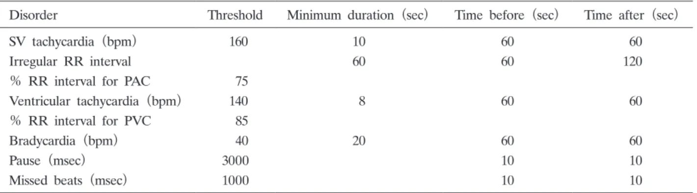
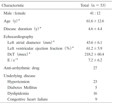
Abstract : Pulmonary vein isolation (PVI) via catheter ablation has been shown to be a highly effective option for patients with symptomatic paroxysmal atrial fibril- lation (AF) . The recurrence of AF within 3 months after PVI is not considered a failure of the ablation procedure because early recurrence of AF is not always associated with late recurrence. We examined the usefulness of an external loop recorder with auto-trigger function (ELR-AUTO) to detect AF following PVI to characterize early recurrence and determine the implication of AF within 3 months after PVI. The study included 53 consecutive patients with symptomatic paroxys- mal AF (age, 61.6 12.6 years ; 77% male) who underwent PVI, and were fitted with an ELR-AUTO for 7 2 days within 3 months after PVI. Of the 33 patients

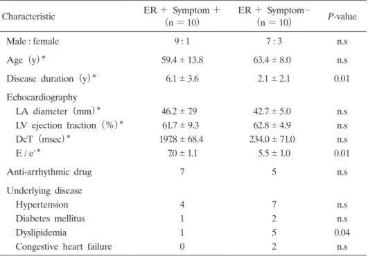
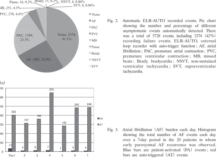
(62.2%) who did not have AF within the 3-month period, only 1 patient had AF recurrence at 12 months. Seven of 20 patients (35%) who experienced AF within 3 months had symptomatic AF recurrence at 12 months. The sensitivity, specific- ity, positive predictive value, and negative predictive value of early AF recurrence for late recurrence was 87.5%, 71.1%, 35.0%, and 96.9%, respectively. Thus, AF recurrence detected by ELR-AUTO within 3 months after PVI can predict late AF recurrence. Freedom from AF in the first 3 months following ablation significantly predicts long-term freedom from AF. An ELR-AUTO is useful for detecting symptomatic and asymptomatic AF.

Key words : atrial fibrillation, pulmonary vein isolation, external loop recorder

Introduction

 Pulmonary vein isolation (PVI) via catheter ablation has been shown to be a highly effective option for patients with symptomatic paroxysmal atrial fibrillation (AF) . The recurrence of AF in the 3-month period after PVI is common and is not considered a procedure failure of catheter ablation because the early recurrence of AF in this period is not always associated with Original

Division of Cardiology, Department of Medicine, Showa University School of Medicine, 1—5-8 Hatanodai, Shinagawa-ku, Tokyo 142-8666, Japan.

To whom corresponding should be addressed.

(2)

late AF recurrence. Therefore, this period has been called “the blanking period”

1, 2)

. However, the clinical significance of early recurrence is controversial because most studies determine AF recurrence based on patientsʼ symptoms

3)

. The incidence of symptomatic and asymptomatic AF recurrence within 3 months after PVI is not well described.

 SpiderFlash-t

TM

A-Fib (Sorin Group, Clamart, France) is an external loop recorder with an auto-trigger function (ELR-AUTO) , which detects and records any arrhythmia automatically.

We examined the usefulness of the ELR-AUTO to detect AF within 3 months after PVI to characterize early recurrence and to determine the implication of AF within 3 months after PVI.

Methods Study subjects

 Fifty-three consecutive patients who underwent PVI at our hospital, and were monitored with an ELR-AUTO within 3 months after catheter ablation were enrolled in this study from April 2012 to December 2012. The patients all had highly symptomatic AF which was refractory to drugs. Medical histories were obtained from patient interviews and medical records, such as electrocardiograms (ECGs) and Holter monitors depicting episodes of AF. Written informed consent was obtained from all subjects before catheter ablation.

Catheter ablation

 Any new oral anticoagulant drug was discontinued on the day of the procedure. Warfarin was discontinued 3 days before the procedure. The PVI catheter ablation procedure was performed with electrode catheters placed in the high right atrium, coronary sinus, and His bundle region. The left atrium (LA) was approached via an atrial septal puncture under the guidance of intravascular ultrasound. After performing an atrial trans-septal puncture, heparin was administered, and an activated clotting time of 350 seconds was maintained. Three-dimensional mapping was performed using either a CARTO (Biosense Webster, Diamond Bar, CA) or NavX

(St. Jude Medical, St. Paul, MN) system, and then a LASSO (Biosense Webster) catheter was placed in the PV, and an ablation application was delivered via an irrigation catheter, with a power setting of 30-35 W on the anterior wall and 20-25 W on the posterior wall. The end- point of the ablation was a bidirectional block at the PV-LA antrum. The procedure was considered complete when no arrhythmia induction occurred during programmed stimulation

(cycle length, 200 msec) .

Post-ablation evaluation

 For the clinical follow-up, patients were fitted with an ELR-AUTO for 1-2 weeks within 3 months of undergoing ablation. AF burden within 3 months after ablation was defined as early AF recurrence if AF lasted for 60 seconds with or without symptoms. Symptomatic AF was defined as AF-related symptoms with palpitations and chest discomfort.

 The patients visited the hospital at 1-month intervals, and underwent an ECG recording.

Late recurrence of AF was evaluated by the 12-lead ECG, Holter ECG, or ELR-AUTO. Late

(3)

recurrence was defined as AF which was detected by any method 12 months after ablation.

External loop recorder

 The ELR-AUTO was fitted by a medical technician on the first day only, and the patients were then instructed how to attach the device themselves on subsequent days. The patients put one cathode (white) and two anode (red and brown) patches on their body surface. The frequency characteristics of the ECG recordings were from 0.05-80 Hz, the dynamic range of the input was 16 mV / 300 mV, the carrier frequency was 200 Hz, and the resolution was 10 µV.

 The arrhythmia detection function of the loop recorder was set to record the ECG for 2 minutes after an event and 4 minutes before an event when the patient noticed a symptom and pressed the button of the device. For asymptomatic events, the automatic detection function of the ELR-AUTO recorded the ECG according to the settings shown in Table 1. All patient- activated events were symptomatic, but all auto-triggered were not only asymptomatic events.

The device registered an auto-triggered event at the same time as it was recording for a patient- activated event.

Statistical analysis

 The continuous variables were expressed as the mean standard deviation. A P-value of < 0.05 was used to determine significant differences. For statistical analyses, JMP software (version 10.0 ; SAS Institute, Cary, NC) was used.

Results

Patient characteristics

 Patient characteristics are shown in Table 2. The 53 patients included 41 men and 12 women, with a mean age of 61.6 12.6 years, and an average disease duration of 4.6 4.4 years. The echocardiography findings revealed that the average LA diameter was 43.6 6.1 mm and the average ejection fraction was 61.2% 5.9%.

Table 1. Automatic detection function settings for ELR-AUTO

Disorder Threshold Minimum duration (sec) Time before (sec) Time after (sec)

SV tachycardia (bpm)  160 10 60  60

Irregular RR interval 60 60 120

% RR interval for PAC  75

Ventricular tachycardia (bpm)  140  8 60  60

% RR interval for PVC  85

Bradycardia (bpm)  40 20 60  60

Pause (msec) 3000 10  10

Missed beats (msec) 1000 10  10

ELR-AUTO, external loop recorder with auto-trigger function ; SV, supraventricular ; PAC, premature atrial contrac-

tion ; PVC, premature ventricular contraction.

(4)

Catheter ablation

 There were 51 patients with complete isolation and 2 patients with incomplete isolation.

In these 2 patients, we were able to confirm only a unidirectional block and we ended the procedure due to the long procedure time. A LA roof and mitral isthmus linear ablation was performed in 6 patients because AF was induced by programmed atrial stimulation after PVI.

When common atrial flutter was induced by programmed atrial stimulation, cavo-tricuspid isthmus linear ablation was performed (n =18) .

AF within three months after ablation

 AF was recorded by the ELR-AUTO in 20 of 53 patients (37.7%) ; there was no significant difference in gender, age, echocardiographic findings, oral medications, or underlying disease between patients with and without early AF recurrence. Ten of 20 patients (50%) with early AF recurrence had both symptomatic and asymptomatic AF burden, whereas 10 (50%) had only asymptomatic AF burden. Table 3 shows the symptomatic and asymptomatic AF recurrence group (n =10) and the asymptomatic AF recurrence only group (n = 10) from among the patients in which AF was detected by ELR-AUTO monitoring. There was no significant difference in gender, age, echocardiographic findings, oral medications, or underlying disease between these two groups. However, patients with symptomatic AF burden had a longer history of paroxysmal AF than patients with asymptomatic AF (6.1 3.6 vs. 2.1 2.1 years, respectively ; P<= 0.01) . In addition, E / e' (the ratio between the velocity of the E and A waves on

Table 2. Patient Characteristics

Characteristic Total(n = 53)

Male : female 41 : 12

Age (y)

61.6 12.6

Disease duration (y)

4.6 4.4

Echocardiography

 Left atrial diameter (mm)

43.6 6.1  Left ventricular ejection fraction (%)

61.2 5.9  DcT (msec)

218.2 60.4  E / e'

7.2 6.2

Anti-arrhythmic drug 27

Underlying disease

 Hypertension 23

 Diabetes Mellitus  5

 Dyslipidemia 16

 Congestive heart failure  9 DcT, deceleration time ; E / e', ratio between velocity of E and A waves on Doppler transmitral flow.

Values are n unless otherwise indicated ;

mean standard

deviation.

(5)

Doppler transmitral flow) was higher in patients with symptomatic AF burden than in patients with asymptomatic AF (7.0 1.1 vs. 5.5 1.0, respectively ; P < 0.01) .

Patient-activated ELR-AUTO recorded events

 Patients activated the ELR-AUTO for a total of 391 events. Fig. 1 demonstrates the ECG recordings for the different events. Out of the 391 events, 177 events (45.2%) were normal

Table 3. Symptomatic and asymptomatic AF

Characteristic ER + Symptom +

(n = 10) ER + Symptom­

(n = 10) P-value

Male : female 9 : 1 7 : 3 n.s

Age (y)

59.4 13.8 63.4 8.0 n.s

Disease duration (y)

6.1 3.6 2.1 2.1 0.01

Echocardiography

 LA diameter (mm)

46.2 7.9 42.7 5.0 n.s  LV ejection fraction (%)

61.7 9.3 62.8 4.9 n.s  DcT (msec)

197.8 68.4 234.0 71.0 n.s  E / e'

7.0 1.1 5.5 1.0 0.01

Anti-arrhythmic drug 7 5 n.s

Underlying disease

 Hypertension 4 7 n.s

 Diabetes mellitus 1 2 n.s

 Dyslipidemia 1 5 0.04

 Congestive heart failure 0 2 n.s AF, atrial fibrillation ; ER, early recurrence ; ER+ Symptom+, symptomatic and asymptomatic early AF recurrence ; ER+ Symptom­, asymptomatic early AF recur- rence ; LA, left atria ; LV, left ventricle ; DcT, deceleration time ; E / e', ratio between velocity of E and A waves on Doppler transmitral flow ; n.s, not significant.

Values are n unless otherwise indicated ; *mean standard deviation.

Fig. 1.  Patient-activated ELR-AUTO recorded events.

Pie chart showing the number and percentage of

different events recorded when the patients felt

symptoms. A total of 393 events were detected,

with 43 events (11%) being recording failure

events. ELR-AUTO, external loop recorder with

auto-trigger function ; NSR, normal sinus rhythm ;

PAC, premature atrial contraction ; AF, atrial

fibrillation ; PVC, premature ventricular contraction ;

MB, missed beats.

(6)

sinus rhythm (NSR) , 88 events (22.5%) were premature atrial contractions (PACs) , 45 events

(12.5%) were AF, 37 events (9.4%) were premature ventricular contractions (PVCs) , and 43 events (10.9%) could not be identified because of high background noise.

Automatic ELR-AUTO recorded events

 Fig. 2 shows a total of 5775 recordings which were obtained by the ELR-AUTO automatically.

Each recording was confirmed and categorized by a specialist physician. Out of the 5775 recordings, 2374 recordings (41.1%) were noise, 1492 recordings (25.8%) were AF, 1349 recordings (23.3%) were PACs, 270 recordings (4.6%) were PVCs, 251 recordings (4.3%) were missed beats, 16 recordings (0.27%) were pauses, 15 recordings (0.26%) were bradycardia, 4 recordings (0.06%) were supraventricular tachycardia, and 4 recordings (0.06%) were ventricular tachycardia.

Number of AF events each day

 A total of 1537 symptomatic and asymptomatic AF events were recorded by the ELR-AUTO in 20 patients. Fig. 3 shows the incidence of AF each day during the ELR-AUTO monitoring.

Symptomatic AF was recorded 45 of 1537 times (2.9%) that AF was recorded.

Fig. 2.  Automatic ELR-AUTO recorded events. Pie chart showing the number and percentage of different asymptomatic events automatically detected. There was a total of 5728 events, including 2374 (42%)

recording failure events. ELR-AUTO, external loop recorder with auto-trigger function ; AF, atrial fibrillation ; PAC, premature atrial contraction ; PVC, premature ventricular contraction ; MB, missed beats ; Brady, bradycardia ; NSVT, non-sustained ventricular tachycardia ; SVT, supraventricular tachycardia.

Fig. 3.  Atrial fibrillation (AF) burden each day. Histogram showing the total number of AF events each day over a 7-day period in the 20 patients in whom early paroxysmal AF recurrence was observed.

Blue bars are patient-activated (PA) events ; red

bars are auto-triggered (AT) events.

(7)

Early AF and late symptomatic AF

 Fig. 4 shows the results of the long-term follow-up of the patients with early recurrence

(n =20) and those without early recurrence (n= 33) of AF. There was no significant difference between the two groups for age, disease duration, echocardiographic findings, underlying disease, or late recurrence, however, there was a significantly lower incidence of late AF recurrence at 12 months in patients without any early AF recurrence.

 Table 4 shows the characteristics of patients with and without late AF recurrence at 12 months. There was no significant difference between the two groups for age, disease duration,

Fig. 4.   Long-term follow-up results. Schema showing number and percentage of patients with early and late atrial fibrillation (AF)

recurrence.

ER+, early AF recurrence within 3 months after pulmonary vein isolation (PVI) ; ER­, early AF recurrence absent ; LR + , late AF recurrence at 12 months after PVI ; LR­, late AF recurrence absent.

Table 4. Late AF recurrence

Characteristic LR + (n = 8) LR­ (n = 45) P-value

Male : female 8 : 0 33 : 12 0.003

Age (y)

61.6 6.9 61.6 13.5 n.s

Disease duration (y)

6.1 4.2 4.7 4.6 n.s

Echocardiography

 LA diameter (mm)

47.6 6.3 42.9 5.9 0.04  LV ejection fraction (%)

59.2 6.8 61.5 5.8 n.s  DcT (msec)

204.0 49.6 220.8 62.3 n.s  E / e'

5.7 1.2 7.6 6.5 n.s

Anti-arrhythmic drug 5 22 n.s

Underlying disease

 Hypertension 5 18 n.s

 Diabetes mellitus 1  4 n.s

 Dyslipidemia 2 14 n.s

 Congestive heart failure 1  8 n.s AF, atrial fibrillation ; LR, late recurrence ; LA, left atria ; LV, left ventricle ; DcT, deceleration time ; E / e', ratio between velocity of E and A waves on Doppler transmitral flow ; n.s, not significant.

Values are n unless otherwise indicated ;

mean standard deviation.

(8)

or underlying disease, however, there was a significantly smaller LA diameter and lower male to female ratio in patients without any late AF recurrence.

 Late recurrence of AF occurred in 8 of 53 patients (15%) . Seven of the 20 patients (35%)

who experienced AF within 3 months after PVI had symptomatic AF recurrence at 12 months.

The sensitivity, specificity, positive predictive value, and negative predictive value of early AF recurrence for late AF recurrence was 87.5%, 71.1%, 35.0%, and 96.9% respectively.

Discussion Main Findings

 Most AF events were asymptomatic and freedom from AF in the first 3 months following PVI predicts long-term AF freedom.

AF recurrence within three months after PVI

 To our knowledge, there are no previous reports evaluating the recurrence of AF within 3 months after PVI using the ELR-AUTO. Previous trials reported that the recurrence rate of AF within 3 months after PVI was 35%〜65%

1, 4-7)

. In these trials, several different recording systems, such as Holter ECG monitors, event recorders, trans-telephonic monitoring, external loop recorders or implantable loop recorders (ILR) , were used to identify AF recurrence. We could not compare these trials because the type of patient, ablation strategy, definition of recurrence, or ECG monitoring was different among these trials. However, the longer the monitoring time, the higher the detection rate of AF recurrence. In addition, continuous monitoring is necessary to detect AF recurrence because most AF after PVI is asymptomatic.

 A predictor of recurrence was not identified, as in the previous trials

2)

. A few papers have reported that incomplete PVI is a predictor of recurrence

2, 8)

. Only 2 patients had incomplete isolation in this study, but they did not have early AF recurrence or late recurrence. It is possible that unidirectional block contributes to the suppression of the trigger and perpetuation of AF, while PVI has the effect of local denervation of the ganglionic plexus around the superior PV-LA junction during the ablation procedure

9)

.

Symptomatic and asymptomatic AF

 In the present study, the presence or absence of symptoms during AF recurrence was examined. The symptomatic AF recurrence group had significantly longer disease duration and higher E / e' compared to the asymptomatic AF recurrence group. E / e' reflects diastolic dysfunction. Kosiuk et al

10)

reported diastolic dysfunction is correlated with symptom severity in AF. A possible explanation of that observation would be the strong dependence of the hemodynamic performance of the whole system on intact atrial function, since the functional reserve of the impaired left ventricle is highly reduced.

 Neumann et al

5)

also examined the detection of symptomatic AF using a portable ECG event

recorder in patients that underwent PVI, and reported the detection of AF by symptoms was

inaccurate. Verma et al

11)

reported that according to the evaluation of ILRs, the incidence of

(9)

asymptomatic AF was 52.0% before PVI, but increased to 79.0% after PVI. After PVI, the number of AF events, duration of AF, and heart rate variability were decreased. In the present study, the incidence of asymptomatic AF was 97% ; therefore, we concluded that AF recurrence after PVI could not be evaluated by the patientʼs symptoms.

Early and late AF recurrence

 In the present study, we hypothesized that patients having early AF recurrences would have a significant trend toward also having late AF recurrences. Oral et al

3)

and Joshi et al

1)

reported that AF recurrence 2 weeks after ablation was the predictor of late phase recurrences. In addition, Joshi et al

1)

reported that AF recurrence from 2 weeks to less than 3 months was not related to late recurrence. On the other hand, Choi et al

12)

reported that early recurrence within 3 months after PVI was associated with late AF recurrences. In those studies, early recurrence was correlated with late recurrence. In this study, our results were similar. But the positive predictive value was very low, while the negative predictive value was high. However this finding is promising. Freedom from atrial fibrillation in the first 3 months following PVI predicts long-term AF freedom. In the previous studies, ILRs, telephonic monitoring, external loop recorders or Holter monitors detected AF over a long term, but as shown in our study, using the ELR-AUTO for only 1 week might predict late recurrence. Because most of the AF burden was asymptomatic, the auto-trigger function was also useful

1, 3-7)

.

Usefulness of ELR-AUTO to identify AF recurrence

 It is currently believed that Holter and trans-telephonic monitoring are the main modalities to identify arrhythmia recurrence. However, Ziegler et al

13)

reported that intermittent and symptom-based monitoring had lower sensitivity and lower negative predictive value for the identification of patients with AF, and underestimated AF burden compared to continuous monitoring. Verma et al

11)

evaluated AF recurrence after PVI using ILRs, and reported that the proportion of asymptomatic events increased after PVI. Kapa et al

7)

also used ILRs after catheter ablation to investigate early and late recurrence of AF. They reported that in 1-6 months, the rate of AF recurrence was 47% and in 6-12 months, the rate of AF recurrence was 18%. Reiffel et al

14)

investigated the Holter ECG monitors, ELRs, and automatic detection function ELRs and reported that the auto-detection ELRs were useful for the detection of asymptomatic arrhythmias. Continuous monitoring is necessary to identify AF recurrence.

Martinez and Sztajzel

15)

also reported the usefulness of the ELR for arrhythmia diagnoses, and both groups were able to reach a diagnosis in an average of 5 days. As asymptomatic events are reported to increase after PVI, an ELR-AUTO with an automatic detection function would be useful for the evaluation of arrhythmias after catheter ablation.

Limitations

 This was a retrospective study and the number of cases was small. The recordings may be

illegible because of high noise. The physicians and laboratory technicians confirmed all of the

(10)

waveforms. In addition, the algorithm for the detection of an irregular RR interval using the ELR-AUTO has not been published. Therefore some instances of AF might be missed.

 In conclusion, AF recurrence in the period within 3 months after PVI can predict the response to catheter ablation at 12 months. Freedom from AF in the first 3 months following ablation significantly predicts long-term AF freedom. The ELR-AUTO is useful for detecting symptomatic and asymptomatic AF.

Conflict of interest

 The authors have declared no conflict of interest.

References

1) Joshi S, Choi AD, Kamath GS, et al. Prevalence, predictors, and prognosis of atrial fibrillation early after pulmo- nary vein isolation: findings from 3 months of continuous automatic ECG loop recordings. J Cardiovasc Electro- physiol. 2009;20:1089-1094.

2) Calkins H, Kuck KH, Cappato R, et al. 2012 HRS / EHRA / ECAS expert consensus statement on catheter and surgical ablation of atrial fibrillation: recommendations for patient selection, procedural techniques, patient manage- ment and follow-up, definitions, endpoints, and research trial design: a report of the Heart Rhythm Society (HRS)

Task Force on Catheter and Surgical Ablation of Atrial Fibrillation. Developed in partnership with the European Heart Rhythm Association (EHRA) , a registered branch of the European Society of Cardiology (ESC) and the European Cardiac Arrhythmia Society (ECAS) ; and in collaboration with the American College of Cardiology

(ACC) , American Heart Association (AHA) , the Asia Pacific Heart Rhythm Society (APHRS) , and the Society of Thoracic Surgeons (STS) . Endorsed by the governing bodies of the American College of Cardiology Founda- tion, the American Heart Association, the European Cardiac Arrhythmia Society, the European Heart Rhythm Association, the Society of Thoracic Surgeons, the Asia Pacific Heart Rhythm Society, and the Heart Rhythm Society. Heart Rhythm. 2012;9:632-696.e21.

3) Oral H, Knight BP, Ozaydin M, et al. Clinical significance of early recurrences of atrial fibrillation after pulmonary vein isolation. J Am Coll Cardiol. 2002;40:100-104.

4) Lellouche N, Jais P, Nault I, et al. Early recurrences after atrial fibrillation ablation: prognostic value and effect of early reablation. J Cardiovasc Electrophysiol. 2008;19:599-605.

5) Neumann T, Erdogan A, Dill T, et al. Asymptomatic recurrences of atrial fibrillation after pulmonary vein isola- tion. Europace. 2006;8:495-498.

6) Berkowitsch A, Greiss H, Vukajlovic D, et al. Usefulness of atrial fibrillation burden as a predictor for success of pulmonary vein isolation. Pacing Clin Electrophysiol. 2005;28:1292-1301.

7) Kapa S, Epstein AE, Callans DJ, et al. Assessing arrhythmia burden after catheter ablation of atrial fibrillation using an implantable loop recorder: the ABACUS study. J Cardiovasc Electrophysiol. 2013;24:875-881.

8) Bertaglia E, Stabile G, Senatore G, et al. Predictive value of early atrial tachyarrhythmias recurrence after circum- ferential anatomical pulmonary vein ablation. Pacing Clin Electrophysiol. 2005;28:366-371.

9) Katritsis DG, Ellenbogen KA. Is isolation of all four pulmonary veins necessary in patients with paroxysmal atrial fibrillation? Pacing Clin Electrophysiol. 2004;27:938-940.

10) Kosiuk J, Van Belle Y, Bode K, et al. Left ventricular diastolic dysfunction in atrial fibrillation: predictors and rela- tion with symptom severity. J Cardiovasc Electrophysiol. 2012;23:1073-1077.

11) Verma A, Champagne J, Sapp J, et al. Discerning the incidence of symptomatic and asymptomatic episodes of

atrial fibrillation before and after catheter ablation (DISCERN AF) : a prospective, multicenter study. JAMA

Intern Med. 2013;173:149-156.

(11)

12) Choi JI, Pak HN, Park JS, et al. Clinical significance of early recurrences of atrial tachycardia after atrial fibrilla- tion ablation. J Cardiovasc Electrophysiol. 2010;21:1331-1337.

13) Ziegler PD, Koehler JL, Mehra R. Comparison of continuous versus intermittent monitoring of atrial arrhythmias.

Heart Rhythm. 2006;3:1445-1452.

14) Reiffel JA, Schwarzberg R, Murry M. Comparison of autotriggered memory loop recorders versus standard loop recorders versus 24-hour Holter monitors for arrhythmia detection. Am J Cardiol. 2005;95:1055-1059.

15) Martinez T, Sztajzel J. Utility of event loop recorders for the management of arrhythmias in young ambulatory patients. Int J Cardiol. 2004;97:495-498.

[Received January 27, 2014 : Accepted February 6, 2014]

Table  1. Automatic  detection  function  settings  for  ELR-AUTO
Table  2. Patient  Characteristics
Table  3. Symptomatic  and  asymptomatic  AF
Fig. 2.  Automatic  ELR-AUTO  recorded  events.  Pie  chart  showing  the  number  and  percentage  of  different  asymptomatic  events  automatically  detected
+2

参照

関連したドキュメント

When S satisfies the Type II condition, N is closed under both ordinary matrix product and Hadamard (entry-wise) product, and N becomes a commutative algebra (with unity element)

Most papers on economic growth considering the Solow-Swan or neoclassical model used the Cobb-Douglas specification of the production function, which describes a process with a

Zaslavski, Generic existence of solutions of minimization problems with an increas- ing cost function, to appear in Nonlinear

In [10, 12], it was established the generic existence of solutions of problem (1.2) for certain classes of increasing lower semicontinuous functions f.. Note that the

We present sufficient conditions for the existence of solutions to Neu- mann and periodic boundary-value problems for some class of quasilinear ordinary differential equations.. We

Next we integrate out all original domain wall indices α, β, γ, · · · and we get the effective weight function of M at each coarse grained (renormalized) dual link, where M is

We give a new proof of a theorem of Kleiner–Leeb: that any quasi-isometrically embedded Euclidean space in a product of symmetric spaces and Euclidean buildings is contained in a

A variety of methods have been introduced for the synchronization of chaotic systems which include complete synchronization, generalized synchronization, phase synchronization,