10
原著論文
Inhibitory effects of GlcNAc polymers on histamine release from mast cells by Datura stramonium agglutinin
Tamiko Suzuki-Nishimura *, Tomonari Hosokawa, Maiko Seno, Yuuki Kato Department of Pharmacology II, School of Pharmacy, Shujitsu University
(Received 24 October 2016; accepted 2 December 2016)
___________________________________________________________________________
Abstract: Mice and human mast cells express the G protein-coupled receptors Mrgprb2 and MRGPRX2, respectively, which are concerned with systemic pseudo-allergic or anaphylactic reactions. These G- proteins are activated by compound 48/80 and IgE-independently induce mast cell activation. The mast cell activation pathway by Datura stramonium agglutinin (DSA) is similar to that by compound 48/80. Examining the sugar residues recognized by DSA may give us information about the sugar- specificity of Mrgprb2 and MRGPRX2 or the components included in this pathway. N-acetyl glucosamine(GlcNAc) oligomers inhibited the histamine release induced by DSA, with the IC50 of di- N-acetyl chitobiose and penta-N-acetyl chitopentaose being 18.0 and 5.3M, respectively. On the other hand, cellobiose (10 mM) and chitosan dimer (10 mM) were ineffective. This suggests that N- acetyl residues and the1-4 bond are important in inhibiting the interaction between DSA and mast cells. While N-acetyl lactosamine (10 mM) inhibited the histamine release induced by DSA, Gal1-4- GlcNAc-specific Erythrina crista-galli agglutinin did not induce a histamine release in the absence of DSA, nor did it inhibit the histamine release induced by DSA, suggesting DSA recognizes GlcNAc1- 4-GlcNAc rather than Gal1-4-GlcNAc. Control of mast cells using specific sugars may prevent Mrgprb2 and MRGPRX2-dependent anaphylactic reactions.
Keywords: mast cell; Datura stramonium agglutinin; N-acetyl glucosamine; N-acetyl lactosamine;
histamine release
__________________________________________________________________________________
Introduction
Mast cells play important roles in allergic reactions by secreting various inflammatory and immunomodulatory substances. There are two mast cell activating pathways, IgE-dependent1) and IgE- independent2). Recently, it has been shown that mice and human mast cells express the G protein-coupled receptors Mrgprb2 and MRGPRX2, respectively. The
activation of Mrgprb2 or MRGPRX2 by the basic polymer compound 48/80 and inflammatory peptides substance P, as well as by many small-molecule drugs, was associated with systemic pseudo-allergic or anaphylactic reactions3). In addition to mice and humans, rats also have a G-protein in the MRG family4). Our group reported that rat mast cell activation induced by compound 48/80, bradykinin,
11 substance P, and basic polymer PEI6 was sugar- specifically inhibited by the N-acetyl glucosamine (GlcNAc)-specific lectin wheat germ agglutinin (WGA)5-7). Furthermore, the GlcNAc-oligomer specific lectin Datura stramonium agglutinin (DSA) 8) sugar-specifically induces calcium mobilization, histamine release, and PGD2 production by rat mast cells via the Gi protein pathway9-11).
The Gi-protein pathway of rat mast cells activated by DSA is similar to the G-protein pathway activated by compound 48/80, but the involvement of sugar residue in Mrgprb2-mediated mast cells activation is not clear. It is possible that DSA binds to either the sugar residues of Mrgprb2 or other components of the Mrgprb2-mediated mast cells activation pathway. In this study, we examined the sugar residues recognized by DSA in order to activate the G-protein pathway of mast cells.
Materials and Methods
Datura stramonium agglutinin (DSA), Erythrina crista-galli agglutinin (ECA), di-N-acetyl chitobiose, tri-N-acetyl chitotriose, tetra-N-acetyl chitotetraose, penta-N-acetyl chitopentaose, hexa-N-acetyl chitohexaose, cellobiose (Glc-Glc), and chitosan dimer (GlcNH2-GlcNH2) were purchased from Seikagaku Biobusiness Co. (Tokyo, Japan). GlcNAc, N-acetyl lactosamine(LacNAc)(Gal-1, 4-GlcNAc), and o-phthalaldehyde for the histamine fluorometric assay were from Wako Pure Chemical Industries Ltd.
(Osaka, Japan).
Mast cell activation was examined by the histamine release. The histamine release from the mast cells was measured as described previously6). Mast cells from the peritoneal cavity of male Sprague Dawley rats were purified using Percoll12) and were suspended in HEPES-buffered Tyrode's solution (137 mM NaCl, 2.7 mM KCl, 12 mM HEPES, 1 mM MgC12, 0.3 mM CaC12, 5.6 mM dextrose, and 0.03% bovine serum
albumin, pH 7.4). The mast cells were incubated with DSA (30g/mL) for 10 min in HEPES-buffered Tyrode's solution after 5 min incubation in the presence or absence of various sugars at 37C, and the released histamine was analyzed using a fluorometric assay13). Also, the cells were incubated with Erythrina crista-galli agglutinin (ECA) 14) for 5 min before incubation with DSA.
Animal Studies: All animal studies were previously approved by the Animal Care Committee of Shujitsu University and were conducted in accordance with the Principles of Laboratory Animal Care (NIH Publication #85 – 23). Sprague-Dawley male rats were used in the animal studies.
Statistical analysis: Values are the mean ± S.E.M. for replicated experiments. Statistical analysis was evaluated using ANOVA with Dunnett test with P=0.05 taken as the upper limit of significance.
Results
1. Inhibitory effects of GlcNAc oligomers on the histamine release induced by DSA.
Histamine release by DSA was significantly inhibited by 10 mM di-N-acetyl chitobiose, 10 mM tri- N-acetyl chitotriose, 10 mM tetra-N-acetyl chitotetraose, 5 mM penta-N-acetyl chitopentaose, and 5 mM hexa-N-acetyl chitohexaose (Fig. 1)
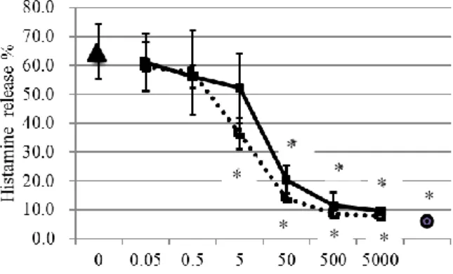
Figure 2 shows the inhibitory curves on histamine release of di-N-acetyl chitobiose and penta-N-acetyl chitopentaose. The IC50 of di-N-acetyl chitobiose and penta-N-acetyl chitopentaose were 18.0 M and 5.3
M, respectively.
2. Importance of N-acetyl residues of the GlcNAc dimers for inhibition.
As di-N-acetyl chitobiose was inhibitory, the effects of various dimers were examined. Ten mM chitosan dimer and 10 mM cellobiose did not inhibit the DSA- induced histamine release (Tables 1 and 2). Table 1 shows there were no significant differences by
12
N-Acetyl chitooligosaccharides (M)
* *
*
*
*
* * *
1
Figure 1. Inhibitory effect of N-acetyl chitooligosaccharides on histamine release induced by DSA.
The mast cells were preincubated with 10 mM di-N-acetyl chitobiose, 10 mM tri-N-acetyl chitotriose, 10 mM tetra-N- acetyl chitotetraose, 5 mM penta-N-acetyl chitopentaose, and 5 mM hexa-N-Acetyl chitohexaose and then were activated by DSA (30g/mL) for 10 min at 37C. Values are given as the mean ± S.E.M. of 3-17 replicate experiments. 1 Spontaneous release (n=16), 2 DSA (n=17), 3 Di-N-acetyl chitobiose (10 mM) + DSA (30
g/mL) (n=4), 4 Tri-N-acetyl chitotriose(10 mM) + DSA (30 g /mL) (n=4), 5 Tetra-N-acetyl chitotetraose (10 mM) + DSA (30 g /mL) (n=4), 6 Penta-N-acetyl chitopentaose (5 mM) + DSA (30 g/mL) (n=4), 7 Hexa-N- acetyl chitohexaose (5 mM) + DSA (30 g /mL) (n=3).
* P<0.01 vs DSA-induced histamine release, by Dunnett test.
Figure 2. Inhibitory curves of di-N-acetyl chitobiose and penta-N-acetyl chitopentaose on histamine release induced by DSA (30 g /mL).
The mast cells were preincubated with various concentrations of di-N-acetyl chitobiose or penta-N-acetyl chitopentaose for 5 min at 37C and then were activated by DSA (30 g /mL) for 10 min. Values are given as the mean ± S.E.M. of 4-6 replicate experiments. Closed squares (―■―) indicate the dose-response curve of di-N- acetyl chitobiose; open squares (•••□•••) indicate the dose- response curve of penta-N-acetyl chitopentaose. Closed triangle and closed circle indicate the histamine release induced by DSA (30 g/mL)(n=17) and spontaneous histamine release (n=16), respectively. * P<0.01 vs DSA- induced histamine release by Dunnett test.
Table 1. Effect of cellobiose on histamine release induced by DSA (30 g /mL).
After preincubation in the absence and presence of cellobiose for 5 min, the mast cells were incubated with DSA (30 g /mL) for 10 min at 37C. Values are given as the mean ± S.E.M. of 4 to 11 replicate experiments. There were no significant differences between the different molarities of cellobiose (Dunnett test). There were no significant differences between the histamine release in the presence of 10mM cellobiose (8.3 ± 4.1%, n=2) and in the absence of cellobiose (8.8 ± 3.8%, n=11).
Table 2. Effect of chitosan dimer on histamine release induced by DSA (30 g/mL).
Chitosan dimer mM Histamine release % n
0 71.4 ± 33.4 10
0.1 67.9 ± 22.4 10
1 75.3 ± 21.4 8
10 67.1 ± 40.1 6
After preincubation in the absence and presence of chitosan dimer for 5 min, the mast cells were incubated with DSA (30
g/mL) for 10 min at 37C. Values are given as the mean ± S.E.M. of 6 to 10 replicate experiments. There were no significant differences between the different molarities of chitosan dimer (Dunnett test). The spontaneous histamine release and the histamine release induced by 10mM chitosan dimer was 8.7 ± 3.7% (n=11) and 21.6 ± 5.4% (n=4), respectively.
Dunnett test between the different molarities of cellobiose. There were no significant differences between the histamine release in the presence of 10mM cellobiose (8.3 ± 4.1%, n=2) and in the absence of cellobiose (8.8 ± 3.8%, n=11)(Table 2).
On the other hand, LacNAc inhibited the histamine release induced by 30g/mL of DSA in dose- dependent manner (Fig. 3).
3. Effects of ECA on mast cell activation.
As LacNAc was inhibitory, the effects of Gal-1, 4- GlcNAc-specific lectin, ECA, were examined. ECA did not release histamine (Table 3) and did not inhibit the histamine release induced by 30 g/mL of DSA (Table 4). There were no significant differences
Cellobiose mM Histamine release % n
0 65.4 ± 37.2 11
0.1 58.3 ± 38.2 6
1 61.4 ± 49.6 8
10 86.9 ± 39.3 4
13 ECA g/mL Histamine release % n
0 5.4 ± 4.4 6
10 6.1 ± 3.6 6
30 5.4 ± 4.3 6
100 3.4 ± 3.5 6
300 4.4 ± 3.9 6
betwe1
Figure 3. Inhibitory curves of N-acetyl Lactosamine on histamine release induced by DSA (30 g /mL).
The mast cells were preincubated with various concentrations of N-acetyl-lactosamine for 5 min at 37C, and then were activated by DSA (30 g/mL) for 10 min.
Values are given as the mean ± S.E.M. of 4-6 replicate experiments. Closed squares (―■―) indicate the histamine release in the presence of DSA; closed triangles (•••▲•••) indicate the histamine release in the absence of DSA. Closed circle and open circle indicate the histamine release induced by DSA (30g/mL) and the spontaneous histamine release, respectively. * P<0.01, Dunnett test.
Table 3. Histamine release in the presence of various amounts of ECA
Purified mast cells were incubated in the presence or absence of ECA for 10 min at 37C. Values are expressed as the mean ± S.E.M. of 6 replicate experiments. There were no significant differences between the presence and absence of ECA (Dunnett test).
Table 4. Effect of ECA on histamine release induced by DSA (30 g/mL).
ECA g/mL Histamine release % n Spontaneous release 5.9 ± 2.9* 8
0 64.4 ± 18.7 8
30 50.5 ± 9.4 6
100 51.5 ± 12.5 6
300 62.6 ± 19.3 6
After preincubation with ECA for 5 min, the mast cells were incubated with DSA (30g/mL) for 10 min at 37C.
Values are given as the mean ± S.E.M. of 6 to 8 replicate experiments. There were no significant differences between the histamine release induced by DSA in the presence and absence of ECA (n=6-8). *P<0.01 vs DSA-induced histamine release by Dunnett test.
between the presence and absence of ECA by Dunnett test (Table 3). And there were no significant differences between the histamine release induced by DSA in the presence and absence of ECA (n=6- 8)(Table 4).
Discussion
Mas-related G protein-coupled receptors are subfamily of orphan G protein-coupled receptors (GPCRs) that are expressed in specific subpopulations of sensory neurons15) and mast cells16). The Mas- related genes comprise a family of many orphan GRCRs. MRGPRX2, one of orphan GRCRs, was detected in primary sensory neurons, several brain areas, mast cells and the adrenal medulla in human.
Activation of MRGPRX2 induces microbial clearance17), neurogenic inflammation and pain18). Human MRGPRX2 and mice Mrgprb2 receptors on mast cells mediate non-IgE dependent allergic and anaphylactic reactions after Ca2+ influx. These receptors are the targets of many small-molecule drugs, e.g. compound 48/80, associated with systemic pseudo-allergic or anaphylactic reactions3). Rat peritoneal mast cells have 6 MrgprB genes, and MrgprB3 is possibly activated by compound 48/80 via IgE-independent pathways. Examining the sugar residues recognized by DSA may give us information about the sugar-specificity of Mrgprb2/MRGPRX2 or the components included in this pathway.
Our group reported that the GlcNAc-specific lectin DSA activates rat mast cells with an IgE-independent and G protein-dependent pathway. GlcNAc sugar residues of the mast cells are important to activate the G protein pathway. WGA inhibits the rat mast cell activation induced by compound 48/80, bradykinin, and substance P6, 7), suggesting that interaction between WGA and GlcNAc sugar residues of the mast cells blocks the G-protein pathway. It was noteworthy that there are differences between DSA and WGA,
14 suggesting that dimeric DSA activates the mast cells, but monomeric WGA does not 6, 7, 9).
In this study, we studied the sugar residues recognized by DSA using a variety of sugars. In Fig.1, the concentrations of di-N-acetyl chitobiose, tri- N-acetyl chitotriose, and tetra-N-acetyl chitotetraose were 10 mM, but that of penta-N-acetyl chitopentaose and hexa-N-acetyl chitohexaose were 5mM, because penta-N-acetyl chitopentaose and hexa-N-acetyl chitohexaose were too sticky to dissolve in the HEPES-buffered Tyrode's solution. The inhibitory effect of di-N-acetyl chitobiose was smaller than that of penta-N-acetyl chitopentaose (Fig.2). The inhibitory effect of GlcNAc-oligomers suggests that GlcNAcβ1-4GlcNAc inhibits the interaction between DSA and mast cells.
Our group previously reported that N-acetyl galactosamine (10 mM), D(+)-galactose (10 mM), D(+)-fucose (10 mM), D(-)-fucose (10 mM), and α- methylmannoside (10 mM) have no effect on the histamine release induced by DSA 9). As dimers of GlcNAc strongly inhibited the histamine release induced by DSA, the effects of various sugar dimers were examined. Both cellobiose without N-acetyl residues and chitosan dimer without acetyl residues were ineffective at inhibiting the histamine release induced by DSA (Tables 1 and 2). This suggests that N-acetyl residues and the 1-4 bond are important in inhibiting the binding of DSA to mast cells.
Our group also previously reported that 10 mM LacNAc inhibited the histamine release induced by DSA, but we did not study the effects of LacNAc at different concentrations. In this study, we examined the inhibition curve of LacNAc, its IC50 being 4.0 mM.
The inhibitory effects of LacNAc were much smaller than those of di-N-acetyl chitobiose. Moreover, Gal
1-4-GlcNAc-specific ECA did not release histamine (Table 3) and did not inhibit the histamine release induced by 30 g/ml of DSA (Table 4), suggesting that
DSA recognizes GlcNAc1-4-GlcNAc rather than Gal1-4-GlcNAc.
The roles of human MrgX1 and MrgX2 have been examined in human mast cells 15- 17). MRGPRX2 likely plays a role in mast cell-mediated host defense, pseudo-allergic drug reactions, and chronic inflammatory diseases18). Monoclonal antibodies of MRGPRX2 will be developed for mast cell IgE- independent anaphylactic reactions4). The sugar specificity we found in this study may show a possible way to control mast cells in order to prevent pseudo- allergic drug reactions.
Conflict of Interest: The authors declare no conflict of interest.
References
1) Orange RP, Stechschulte DJ, Austen KF:
Immunochemical and biologic properties of rat IgE. II.
Capacity to mediate the immunologic release of histamine and slow-reacting substance of anaphylaxis (SRS-A), J Immunol. 105(5), 1087-95 (1970).
2) Nakamura T, Ui M: Islet-activating protein, pertussis toxin, inhibits Ca2+-induced and guanine nucleotide-dependent releases of histamine and arachidonic acid from rat mast cells, FEBS Lett.
173(2), 414-8(1984).
3) McNeil B D, Pundir P, Meeker S, Han L, Undem BJ, Kulka M, Dong X: Identification of a mast cell specific receptor crucial for pseudo-allergic drug reactions, Nature, 519(7542), 237–241(2015).
4) Subramanian H, Gupta K, Ali H: Roles of Mas- related G protein-coupled receptor X2 on mast cell- mediated host defense, pseudoallergic drug reactions, and chronic inflammatory diseases. J Allergy Clin Immunol 138(3), 700-10(2016).
5) Liske R, Franks D: Specificity of the agglutinin in extracts of wheat germ. Nature, 217(5131), 860- 1(1968).
15 6) Suzuki-Nishimura T, Nagaya K, Matsuda K, Uchida MK, Aoki J, Umeda M, Inoue K: Sugar-specific inhibitory effects of wheat germ agglutinin and phytohemagglutinin-E4 on histamine release induced by basic secretagogues from rat peritoneal mast cells and their possible action sites. Jpn J Pharmacol 57(1), 79-90(1991).
7) Matsuda K, Niitsuma A, Uchida MK, Suzuki- Nishimura T: Inhibitory effects of sialic acid- or N- acetylglucosamine-specific lectins on histamine release induced by compound 48/80, bradykinin and a polyethylenimine in rat peritoneal mast cells. Jpn J Pharmacol, 64(1), 1-8(1994).
8) Debray H, Decout D, Strecker G, Spik G, Montreuil J: Specificity of twelve lectins towards oligosaccharides and glycopeptides related to N- glycosylproteins, Eur J Biochem 117(1), 41-55(1981).
9) Matsuda K, Aoki J, Uchida MK, Suzuki-Nishimura T: Datura stramonium agglutinin released histamine from rat peritoneal mast cells that was inhibited by pertussis toxin, haptenic sugar and N- acetylglucosamine- specific lectins: involvement of glycoproteins with N-acetylglucosamine residues, Jpn J Pharmacol 66(2), 195-204(1994).
10) Suzuki-Nishimura T, Furuno T, Uchida MK, Nakanishi M: An initial signal of activation of rat peritoneal mast cells stimulated by Datura stramonium agglutinin: a confocal fluorescence microscopic analysis of intracellular calcium ion and cytoskeletal assembly, Jpn J Pharmacol 66:205- 11(1994).
11) Suzuki-Nishimura T, Uchida MK.: Prostaglandin D2 generation by rat peritoneal mast cells stimulated with Datura stramonium agglutinin and its inhibition by haptenic sugar and wheat germ agglutinin, Jpn J Pharmacol 90(1), 77-80 (2002).
12) Enerbäck L, Svensson I: Isolation of rat peritoneal mast cells by centrifugation on density gradients of Percoll, J Immunol Methods, 39(1-2), 135-45(1980).
13) Shore PA, Burkhalter A, Cohn VH Jr: A method for the fluorometric assay of histamine in tissues, J Pharmacol Exp Ther, 127, 182-6(1959).
14) Sandhu RS, Kamboj SS, Reen RS, Arora JS:
Occurrence and characterization of lympho- agglutinins in Indian plants, Vox Sang, 43(6), 345- 50(1982).
15) Dong X, Han S-k, Zylka MJ, Simon MI, Anderson DJ: A diverse family of GPCRs expressed in specific subsets of nociceptive sensory neurons, Cell, 106,619- 632(2001).
16) Fujisawa D, Kashiwakura J, Kita H, Kikukawa Y, Fujitani Y, Sasaki-Sakamoto T, Kuroda K, Nunomura S, Hayama K, Terui T, Ra C, Okayama Y :Expression of Mas-related gene X2 on mast cells is upregulated in the skin of patients with severe chronic urticarial, J Allergy Clin Immunol 134(3), 622-633(2014).
17) Subramanian H, Gupta K, Guo Q, Price R, Ali H:
Mas-related gene X2 (MrgX2) is a novel G protein- coupled receptor for the antimicrobial peptide LL-37 in human mast cells: resistance to receptor phosphorylation, desensitization, and internalization. J Biol Chem, 286, 44739-49(2011).
18) Kashem SW, Subramanian H, Collington SJ, Magotti P, Lambris JD, Ali H: G protein coupled receptor specificity for C3a and compound 48/80- induced degranulation in human mast cells: roles of Mas-related genes MrgX1 and MrgX2. Eur J Pharmacol 668, 299-304(2011).