1
Second-trimester fetal heart screening using the spatiotemporal image correlation method
Nori Takada, Keisuke Sugimoto, Noboru Inamura
Department of Pediatrics, Kindai University Faculty of Medicine, Osaka-Sayama, Japan
Abstract
Background: The spatiotemporal image correla- tion (STIC) method, an echocardiographic ap- proach to collecting data for offline analysis, can be used to perform fetal heart screenings. How- ever, the utility of this technique for this indica- tion is unknown.
Method: We screened and analyzed the STIC data for all normal pregnancies (n = 405) at 2 obstetrics clinics in Osaka from January 2017 through June 2018. An obstetrician and a labor- atory technician collected the STIC data from a four-chamber view image. A pediatric cardiolo- gist analyzed the data offline. The images ana- lyzed included the four-chamber view, pulmo- nary veins, right and left outflow view, three- vessel view, and three-vessel trachea view. We
classified findings of each image as normal, ab- normal, or unconfirmed, and evaluated the final diagnoses.
Results: There were 324 normal cases (80.0%), 25 abnormal cases (6.2%), and 56 unconfirmed cases (13.8%). Eleven patients had congenital heart disease (CHD) after birth; of these, 9 had severe CHD requiring hospitalization. The sen- sitivity and specificity of the STIC method for detecting CHD was 100% and 95.9%, respec- tively.
Conclusion: The STIC method was useful for performing fetal CHD screenings. However, many images could not be analyzed.
Key words:fetal echocardiography, telemedi- cine, fetal diagnosis, congenital heart disease
Introduction
Congenital heart disease (CHD) affects 3-8 per 1000 live births 1. However, the incidence of CHD in aborted and stillborn infants is 5 times that of live-born infants. Therefore, it is considered to be more prevalent in fetuses 2. Fetal heart screenings are important because 90% of CHD cases begin during gestation in the absence of clear risk fac- tors 3. The spatiotemporal image correlation (STIC) method involves collecting short-term (5- 15 sec) data from a four-chamber view obtained during echocardiography for offline analysis. The purpose of this prospective study was to calculate the sensitivity and specificity of the STIC method for fetal heart screening tests and examine its util- ity and potential drawbacks.
Material and Method
We obtained the STIC data once for each preg- nant woman (n = 405; 19–35 gestational weeks [median 26 weeks]) who visited the offices of one of two obstetricians -(Department of Obstetrics and Gynecology Taniguchi Hospital, Kasamatsu Obsterics and Gynecology)in our hospital neigh- borhood between January 2017 and June 2018.
We explained the “right to know” and the “right not to know” concepts to subjects before the com- mencing screening tests. Furthermore, we ex- plained the purpose of the screening tests and ob- tained informed consent from each subject.
The study protocol was approved by the Ethics Review Board of Kindai University (RH28-07) Heart screening using the STIC method
We used the Voluson E8 and E10 ultrasound systems (GE Healthcare Japan, Tokyo, Japan) to
perform fetal heart screening. A trained labora- tory technician or obstetrician performed the screening during a routine obstetric examination.
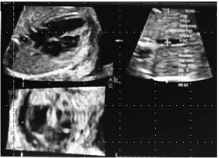
First, we obtained a four-chamber view image and collected the STIC data comprising approxi- mately 10-40 heartbeats (5–15 s). Next, we trans- ferred the STIC data via a virtual private network line. The transferred data were rebuilt as an image on a personal computer by a View PAL system (GE Healthcare Japan, Tokyo, Japan). In the STIC image display, the cross-section (section A)
on the upper left, which is the main part, is a nor- mal B mode tomogram and the cross-section on the upper right (section B) rotates the probe in the A section 90° counterclockwise. At the same time, a cross-sectional image parallel to the base sur- face is displayed on the lower left section (C sec- tion). Sections B and C are displayed by recon- struction of the image by mathematic calculation algorithms. It is possible to rotate around three mutually orthogonal axes or translate it and ob- tain an arbitrary tomogram of the collected data (Figure 1).
Figure 1 Spatiotemporal image correlation images (normal case)
A pediatric cardiologist (N.I.) analyzed the re- built images.
The analyses confirmed the following points sequentially (Table 1): 4
1) Abdominal cross-section: position of the fetal apex of the heart and the stomach;
2) Four-chamber view: heart size, laterality, heart axis, ventricular septum, and connection of the pulmonary vein to the left atrium;
3) Left and right outflow tract view: size of the great vessels and crossing over of the great vessels;
4) Three-vessel view: major vascular balance and position; and
5) Three-vessel trachea view: size of the aortic
arch and the ductal arch and the bloodstream direction.
We confirmed each image point in the evalua- tion and classified the analysis results as normal, abnormal, or unconfirmed. The analysis method is based on "fetal echocardiography guidelines"
Level I and II. 5 A second examination was con- ducted by a pediatric cardiologist in cases in which the analytical results were abnormal. Neo- nates with suspected CHD within 1 month after birth were identified as having CHD, while those with CHD that required hospitalization and treat- ment within 1 month after birth were identified as having severe CHD.
Table 1 Heart screening checklist
Section Check item
Upper abdomen View
Position of stomach and apex ○Same, ○Opposite, ○Unconfirmed Four-chamber View ○Normal, ○Abnormal, ○Unconfirmed Heart size
Cardiac axis
Balance of both ventricles ventricular septum
Pulmonary vein ○Normal, ○Abnormal, ○Unconfirmed Left ventricular outflow view ○Normal, ○Abnormal, ○Unconfirmed Right ventricular outflow view ○Normal, ○Abnormal, ○Unconfirmed Three vessels view ○Normal, ○Abnormal, ○Unconfirmed Three vessels trachea view ○Normal, ○Abnormal, ○Unconfirmed Analysis results ○Normal, ○Abnormal, ○Unconfirmed
Results
Of the 405 prenatal diagnoses, 324 (80.0%) were classified as normal, 25 (6.2%) as abnormal, and 56 (13.8%) as unconfirmed.
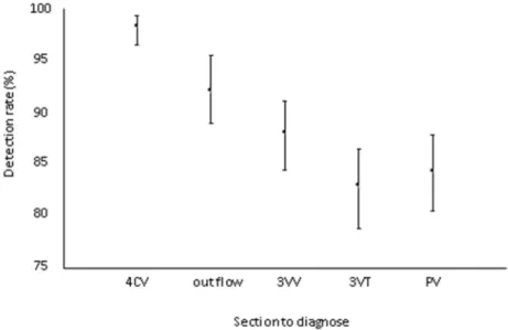
The detection rate by view was 98.3% for the four-chamber, 92.1% for the right and left out- flow, 88.1% for the three-vessel, 83.0% for the three-vessel trachea, and 84.4% for the pulmo- nary vein views (Figure 2). The 95% confidence intervals were calculated using the exact method based on binomial distribution.
Table 2 shows the pre- and postnatal diagnosis results of CHD using the STIC method. Among the 324 patients with a normal prenatal diagnosis, none were diagnosed with CHD postnatally (i.e., a false-negative rate of 0%). In contrast, we diag- nosed 14 normal heart cases postnatally among 25 cases of abnormal prenatal diagnosis (i.e., a false- positive rate of 56.0%); of these, a ventricular septal defect (VSD) was identified in 6. The sen- sitivity and specificity of the STIC method for CHD screening was 100% and 95.9%, respec- tively.
Figure 2 Detection rate by telemedicine using the spatiotemporal image correlation method
4CV, four-chamber view; 3VV, three-vessel view; 3VT, three-vessel trachea view; PV, pulmonary vein
Table 2 Results of CHD Fetal diagnosis
Neonatal diagnosis
Normal Abnormal
CHD(+) 0 11 11
CHD(-) 324 14 338
324 25 349
Sensitivity of CHD: 11/11 = 100%
Specificity of CHD: 324/338 = 95.9%
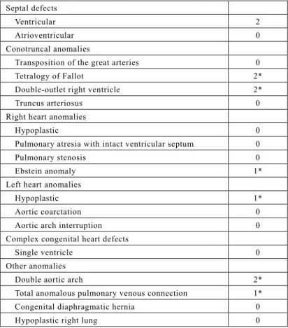
Table 3 shows the postnatal CHD diagnosis re- sults. A total of 11 infants had CHD, 2 had VSD with a small defect not requiring hospitalization, and 9 had severe CHD requiring hospitalization.
There were 2 cases each of Tetralogy of Fallot, a double-outlet right ventricle, and a double aortic arch. There was 1 case each of Ebstein anomaly,
hypoplastic left heart anomaly, and total anoma- lous pulmonary venous connection. Other cardiac abnormalities including great vessel abnormality were noted in 3 cases. These findings show that fetal heart screening using the STIC method was able to detect congenital heart diseases with high efficiency and accuracy.
Table 3 Details of congenital heart defect (n = 11) confirmed postnatally Septal defects
Ventricular 2
Atrioventricular 0
Conotruncal anomalies
Transposition of the great arteries 0
Tetralogy of Fallot 2*
Double-outlet right ventricle 2*
Truncus arteriosus 0
Right heart anomalies
Hypoplastic 0
Pulmonary atresia with intact ventricular septum 0
Pulmonary stenosis 0
Ebstein anomaly 1*
Left heart anomalies
Hypoplastic 1*
Aortic coarctation 0
Aortic arch interruption 0
Complex congenital heart defects
Single ventricle 0
Other anomalies
Double aortic arch 2*
Total anomalous pulmonary venous connection 1*
Congenital diaphragmatic hernia 0
Hypoplastic right lung 0
* Severe congenital heart disease requiring hospitalization
Discussion
The utility of telemedicine in the diagnosis of fetal heart disease was recently reported. 6-9 Here, we prospectively examined the usefulness of the
STIC method for fetal heart screening. Of the im- ages included in this study, 14% could not be an- alyzed. This, of course, is the main disadvantage of the STIC method. Its detection rate varies de- pending on the different characteristics of the
mothers, including obesity and presence of poly- hydramnios and oligohydramnios, and fetuses, in- cluding presentation, size, and presence of hy- drops, characteristics. 6,10 These were the reasons that we could not analyze a subset of the images.
Short screening times result in little influence of fetal movements. The Voluson E10 can complete screening within 5 seconds. Thus, as the use of the Voluson E10 becomes more popular, we can expect to see a concomitant reduction in the rate of unconfirmed diagnoses.
However, it is difficult to increase the diagnosis rate of VSD without increasing image resolution.
This is one of the drawbacks of the STIC method.
Another drawbacks is that the detection rate differs for the images taken from different views. The de- tection rate with the four-chamber view was 98.3%, while that with the three-vessel trachea view was 82.9%. The STIC data included approximately 10–
40 heartbeats (5–15 sec). After loading the four- chamber view, we collected the STIC data. For this reason, the detection rate with the four-chamber view, which was scanned first, was high, whereas that of the three-vessel trachea view, which was scanned last, was low. Fetal respiration and body movements influenced the detection rate despite the short data collection time. 6 We think that the aforementioned problems can be solved by further shortening the scanning time.
According to our study, the sensitivity and spec- ificity rates of the STIC method for detecting CHD were 100% and 95.9%, respectively. The STIC method is useful for screening for CHD. Our method was performed by obstetricians and ultra- sound technicians using only the four-chamber view and a cardiologist analyzed the images. For this reason, no expert knowledge or special train- ing is required for obstetricians or ultrasound tech- nicians. 7 In other words, screening can once the device becomes available. The rate of false-posi- tive VSD diagnosis was high with this technique.
We believe that the spontaneous closure of the VSD at birth could be a reason for this finding. 11
The high specificity of 95.9% can be attributed to the fact that the pediatric cardiologist was able to use multiple views to arrive at a diagnosis. Be- cause there were no false-negative cases, our method is suitable for cardiac screening. Severe CHD is aggravated after birth. Complete transpo- sition of the great arteries, total anomalous pul- monary venous drainage, and coarctation of the aorta are representative of CHD that worsens in
normal in the four-chamber view. Thus, making a diagnosis based on a four-chamber view is diffi- cult. A report from the Nevada State Clark County concluded that the sensitivity of screen- ing using only the four-chamber view, was 36%.
However, the sensitivity increased to 71% when the right and left ventricular outflow views were included.12 The sensitivity of the CHD screening described here also improved if a second view was added. 12,14 However, the detection rates var- ied with the use of different views. The detection rate with the four-chamber view was 98.3%. The detection rate using views other than the four- chamber was high using the STIC method as well.
The detection rate using the right and left outflow view was 92%; three-vessel view, 88%; and three-vessel trachea view, 83%. Our method had higher detection rates than the traditional four- chamber view because a specialist made the diag- noses. Therefore, a double aortic arch could be screened. The STIC method, that can easily re- ceive expert diagnosis is useful for CHD.
Study limitation
In this study, several echocardiographic labor- atory technicians and obstetricians collected im- ages that were analyzed by a single cardiologist.
Because there was only 1 analyst, there were no inter-analyst differences. This fact may have in- fluenced the sensitivity and specificity of the study. In a future study, we will include multiple examiners and examine the differences in their di- agnoses.
Conclusion
In this study, we attempted to diagnose prena- tal CHD using the four-dimensional STIC method in 405 pregnant women who underwent fetal heart screening at an obstetrics and gynecology depart- ment. A total of 56 cases (13.8%) could not be analyzed. There were 14 false-positive and 0 false-negative diagnoses. The sensitivity and specificity of the STIC method for identifying CHD were 100% and 95.9%, respectively. Thus, our data suggest that the STIC method is useful for screening for fetal CHD.
Acknowledgement
The authors express gratitude to Yasutaka Chiba, PhD (Clinical Research Center, Kindai
comments.
Conflicts of interest
The authors declare no conflicts of interest.
References
1.Allan L, et al (2001) Isolated major congenital heart disease. Ultrasound Obstet Gynecol 17: 370-379.
2.Smythe JF, Copel JA, Kleinman CS (1992) Outcome of prenatally detected cardiac malformations. Am J Cardiol 69: 1471-1474.
3.Achiron R, Glaser J, Gelerntter I, Hegesh J, Yagel S (1992) Extended fetal echocardiographic examination for detecting cardiac malformations in low risk preg- nancies. BMJ 304: 671-674.
4 .ISUOG (2013) ISUOG Practice Guidelines (up- dated): sonographic screening examination of the fetal heart. Ultrasound Obstet Gynecol 41: 348-359, 2013 5.Fetal heart echocardiography guideline creation com-
mittee. (2006) Fetal echocardiography guidelines. Ped Cardiol Card Surg 22(5):591-613.
6.Zhao L, et al. (2016) Feasibility study on prenatal cardiac screening using four-dimensional ultrasound with spatiotemporal image correlation: a multicenter study. PloS ONE 11(6): e0157477.
7.Bennasar M, et al. (2010) Accuracy of four-dimen- sional spatiotemporal image correlation echocardiog-
raphy in the prenatal diagnosis of congenital heart de- fects. Ultrasound Obstet Gynecol 36: 458-464.
8.Juliana Y, et al. (2015) Screening of congenital heart diseases by three-dimensional ultrasound using spatio- temporal image correlation: influence of professional experience. Echocardiography 33: 99-104.
9.Bennasar M, et al. (2009) Feasibility and accuracy of fetal echocardiography using four-dimensional spatio- temporal image correlation technology before 16 weeks, gestation. Ultrasound Obstet Gynecol 33: 645- 651.
10.De Vore G, et al. (1993) Fetal echocardiography: fac- tors that influence imaging of the fetal heart during the second trimester of pregnancy. J Ultrasound Med 12:
659-663.
11.Mosimann B, Zidere V, Simpson JM, Allan LD (2014) Outcome and requirement for surgical repair following prenatal diagnosis of ventricular septal defect. Ultra- sound Obstet Gynecol 44(1): 76-81.
12.Evans W, et al. (2015) Moving towards universal pre- natal detection of critical congenital heart disease in southern Nevada: a community-wide program. Pediatr Cardiol 36: 281-288.
13.Pinto NM, et al. (2012) Barriers to prenatal detection of congenital heart disease: a population-based study.
Ultrasound Obstet Gynecol 40: 418-425.
14.Carvalho JS, Mavrides E, Shinebourne EA, Campbell S, Thilaganthan B (2002) Improving the effectiveness of routine prenatal screening for major congenital heart defects. Heart 88: 387-391.